


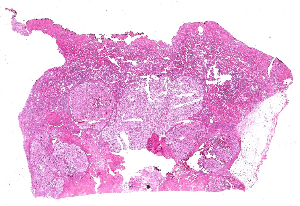
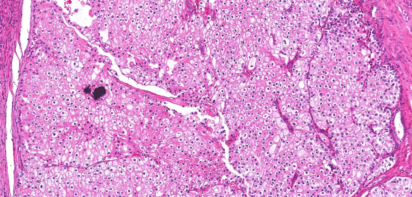
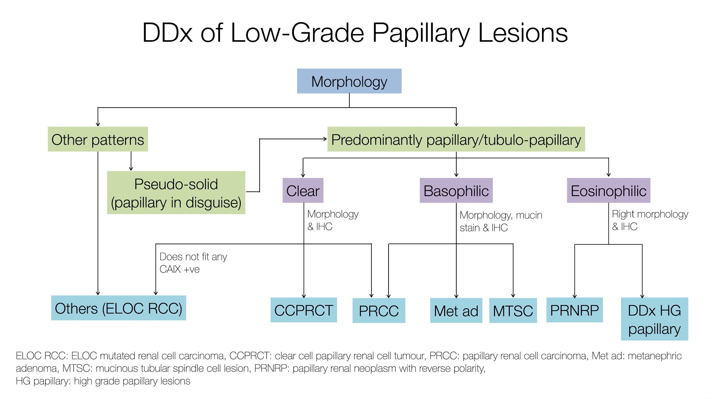
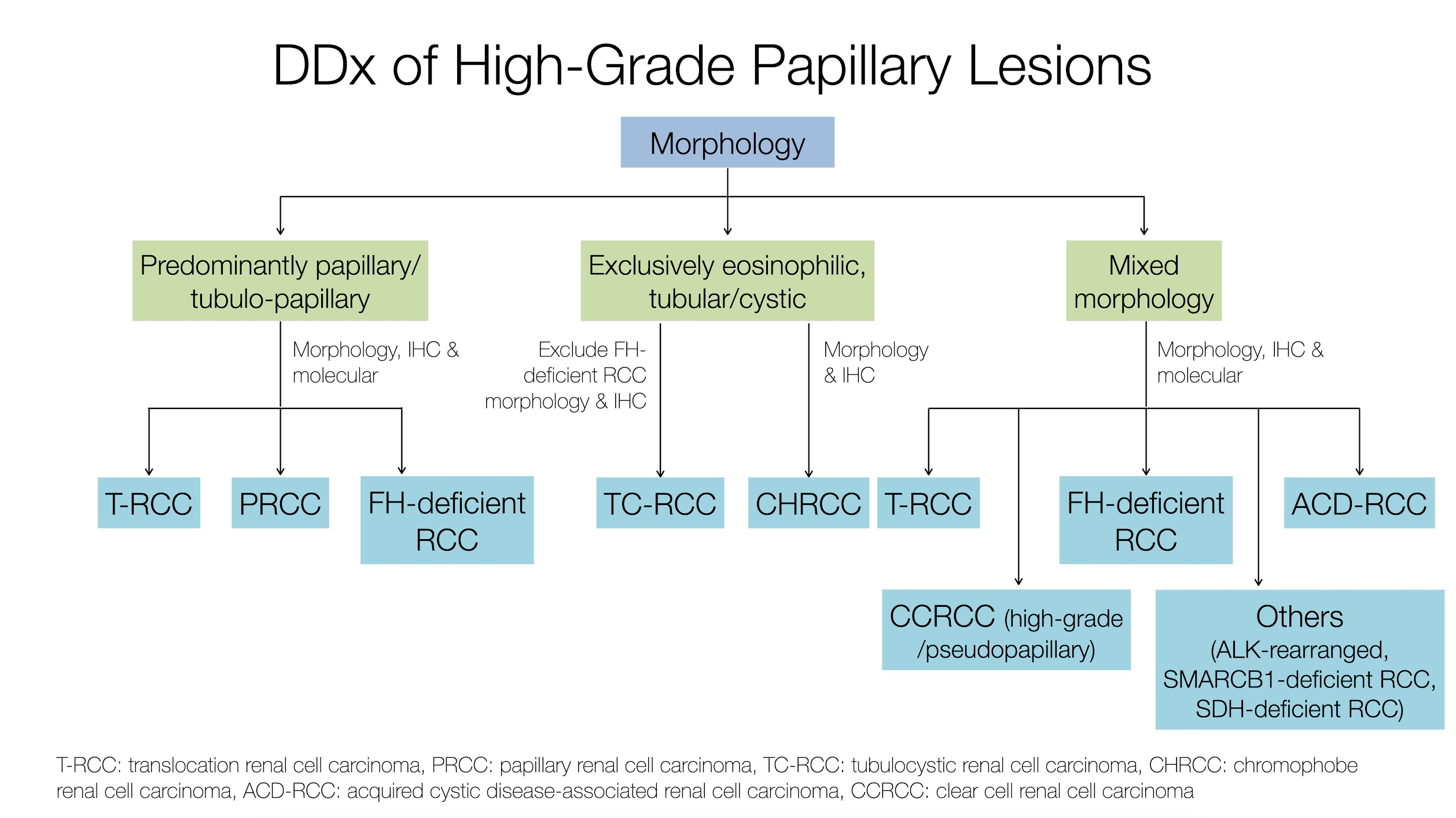
Images hosted on other servers:
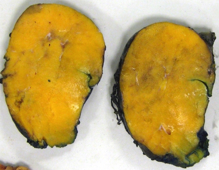
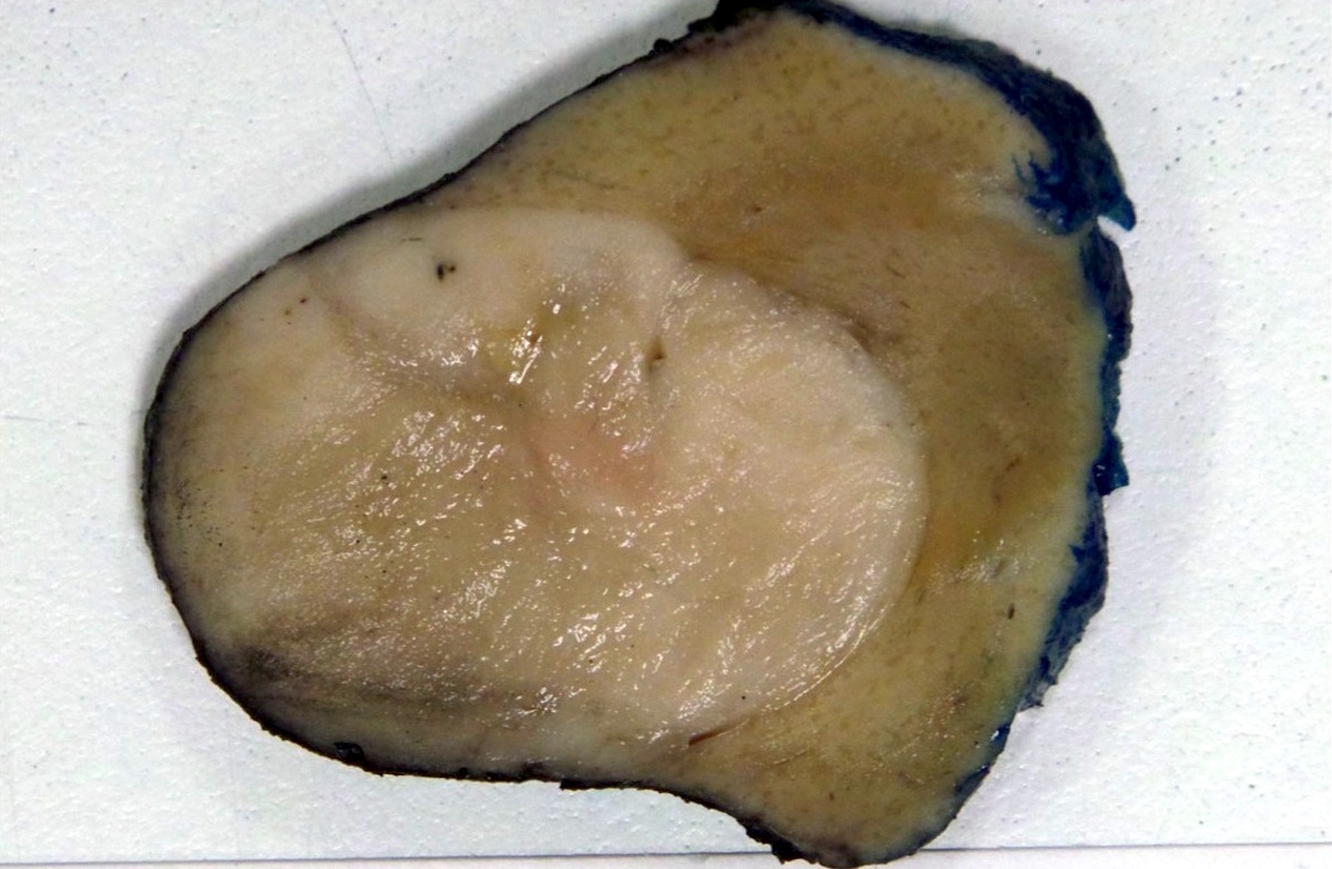
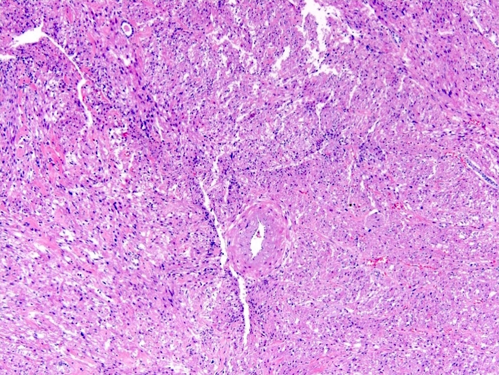
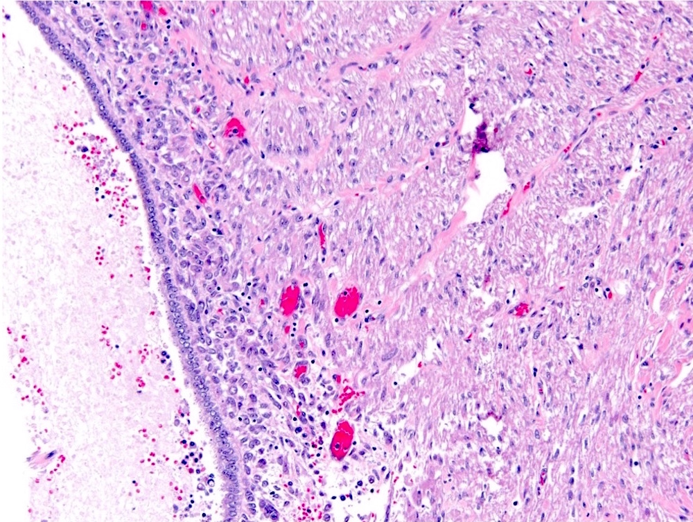
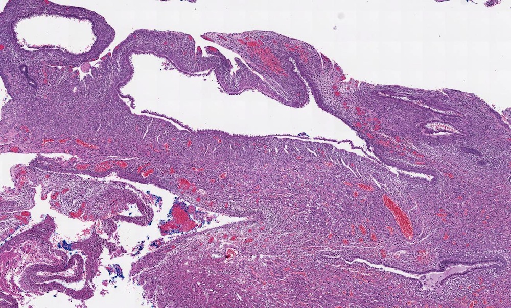
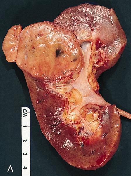
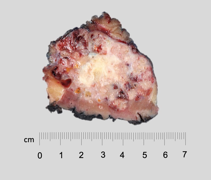
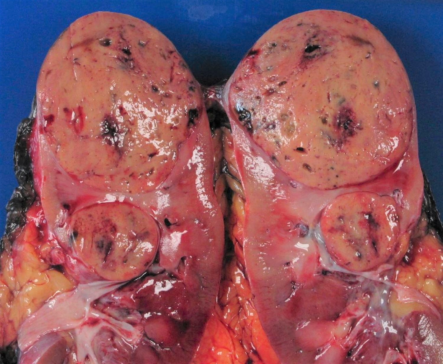
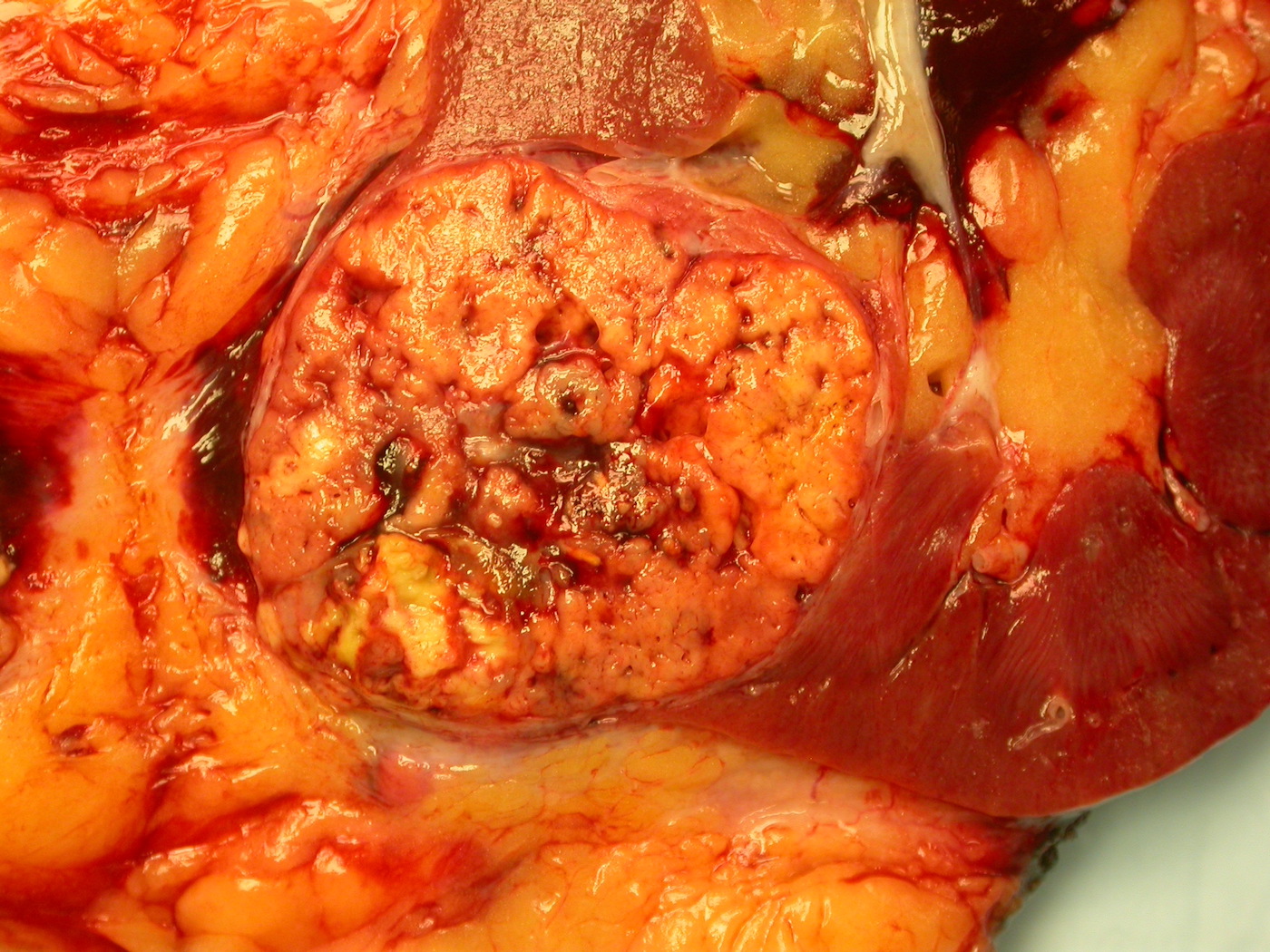
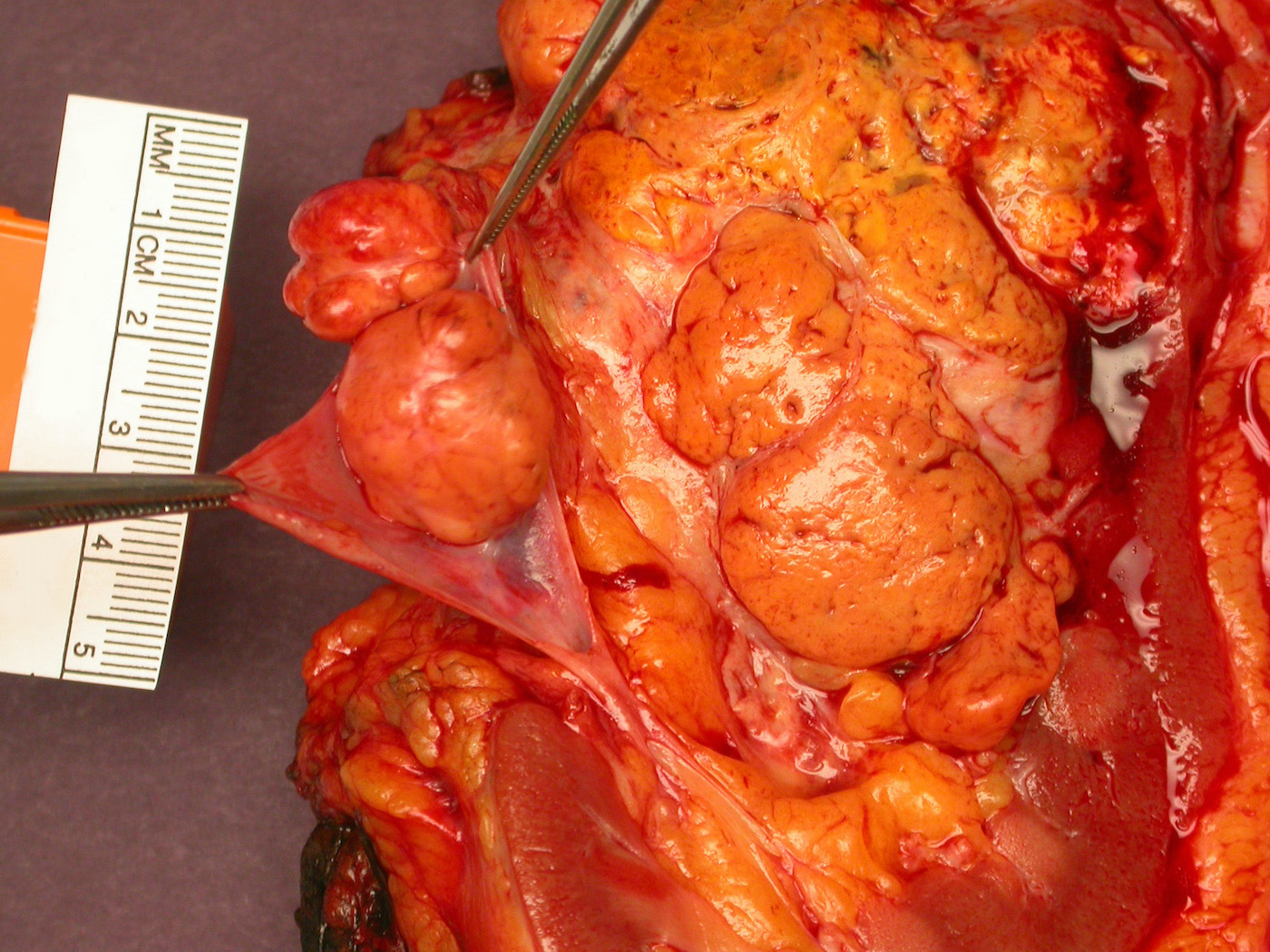
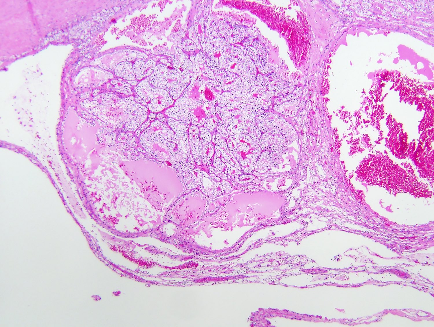
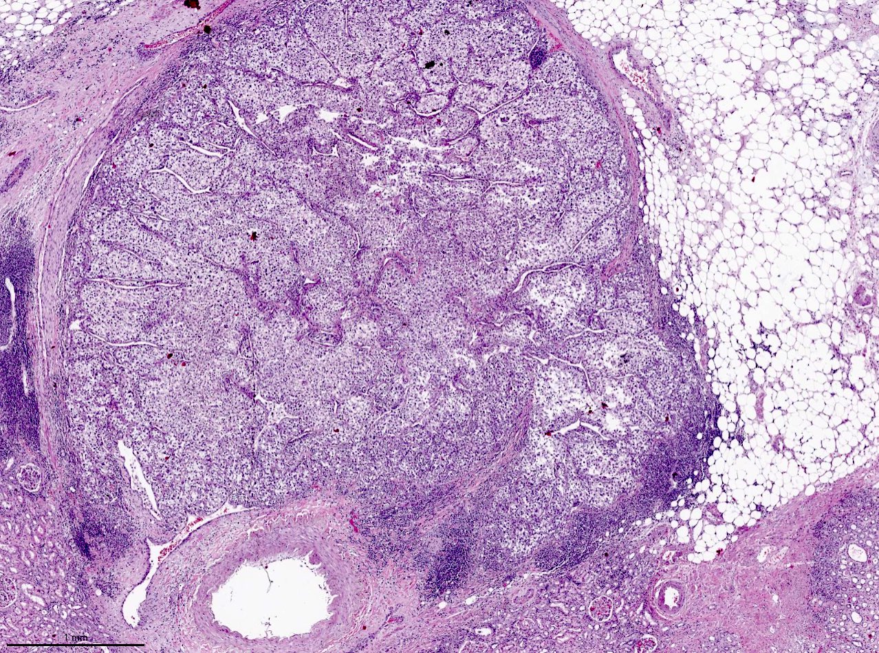
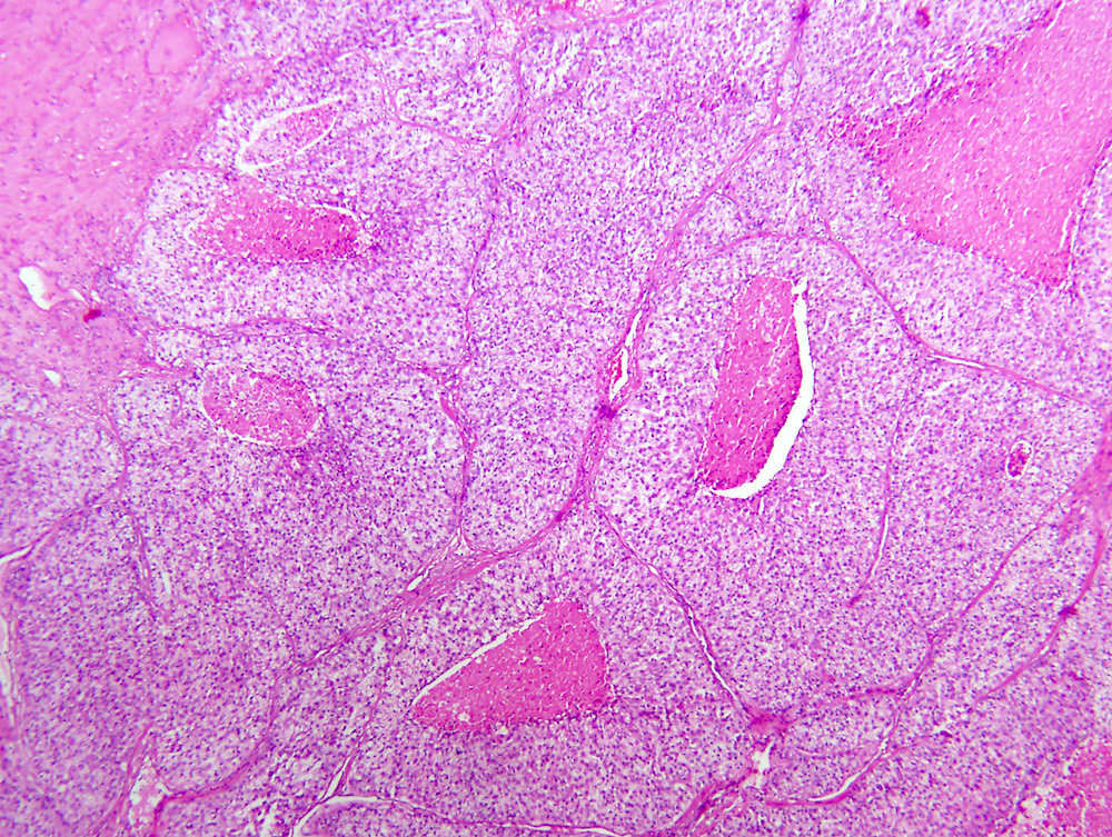
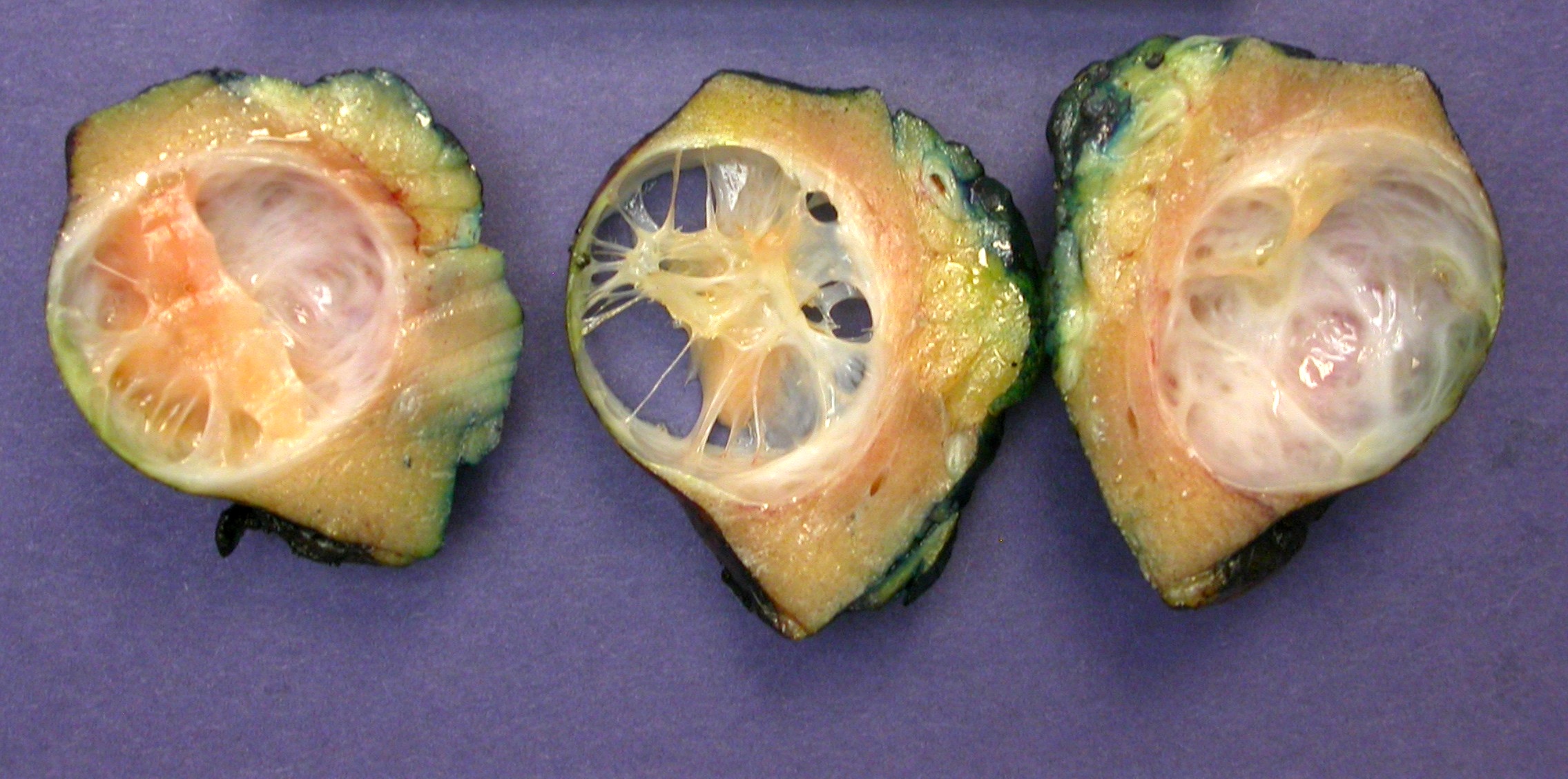
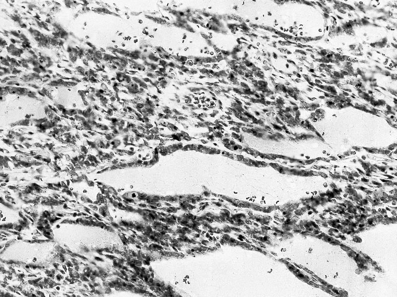
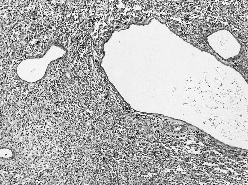
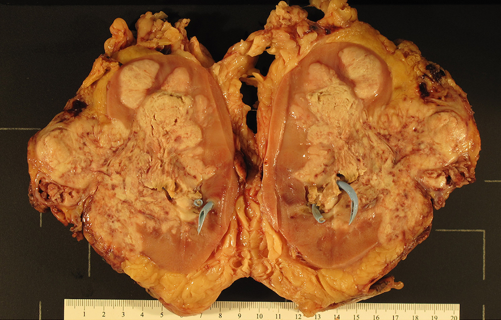
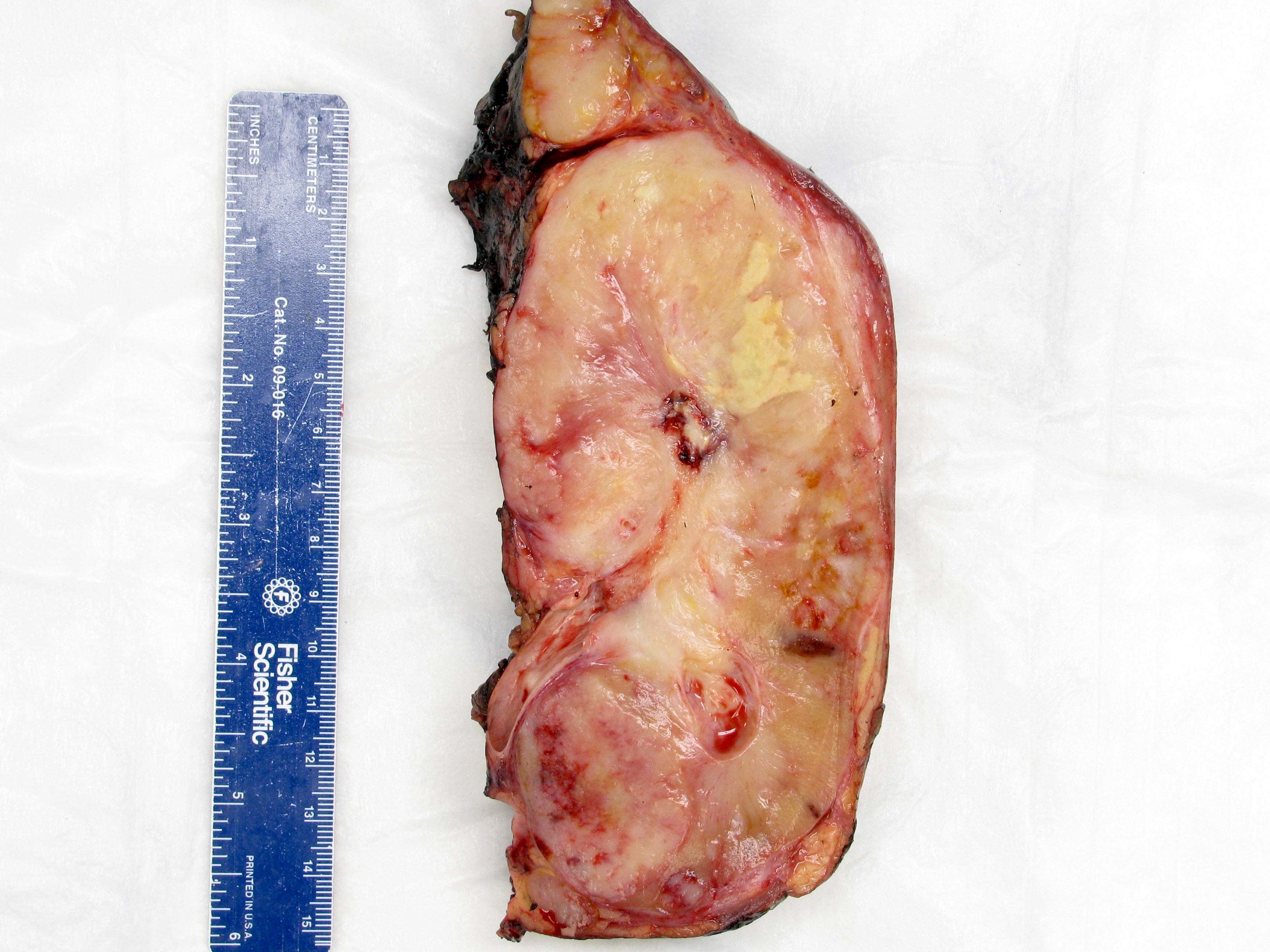
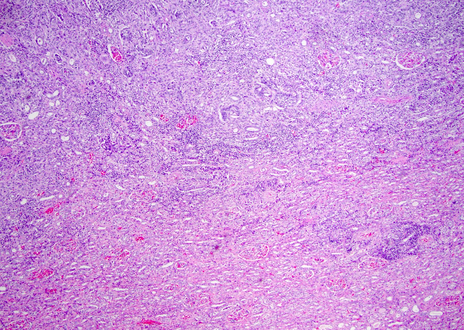
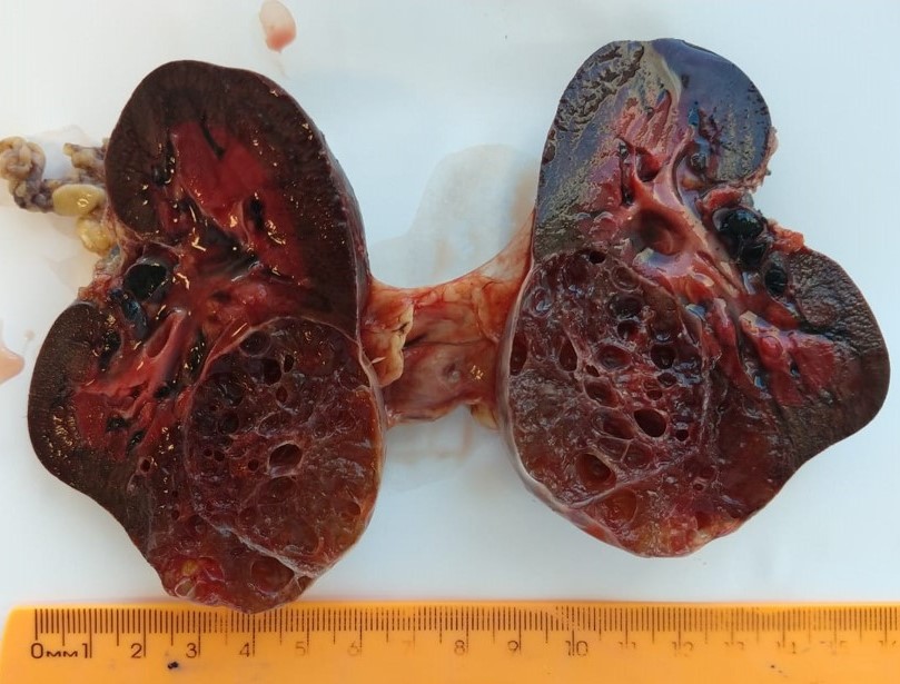
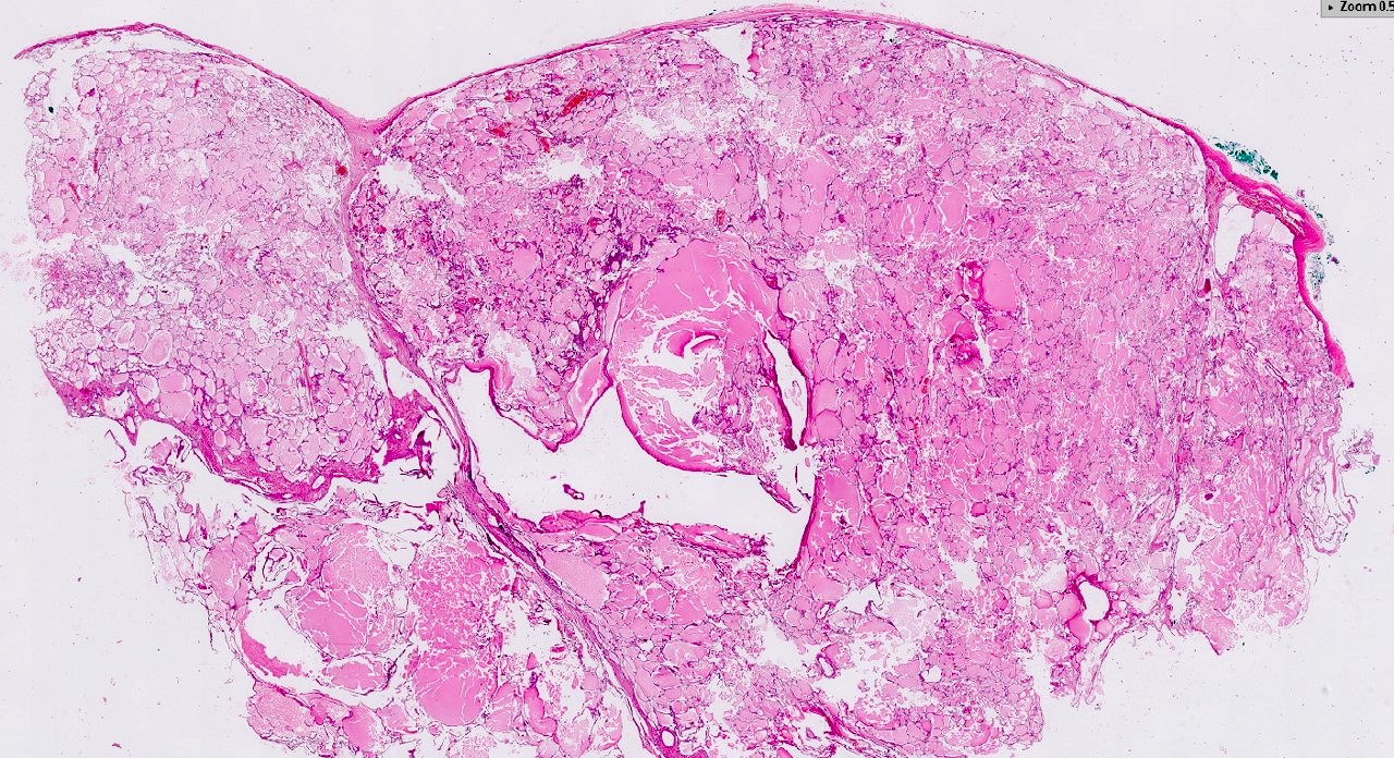
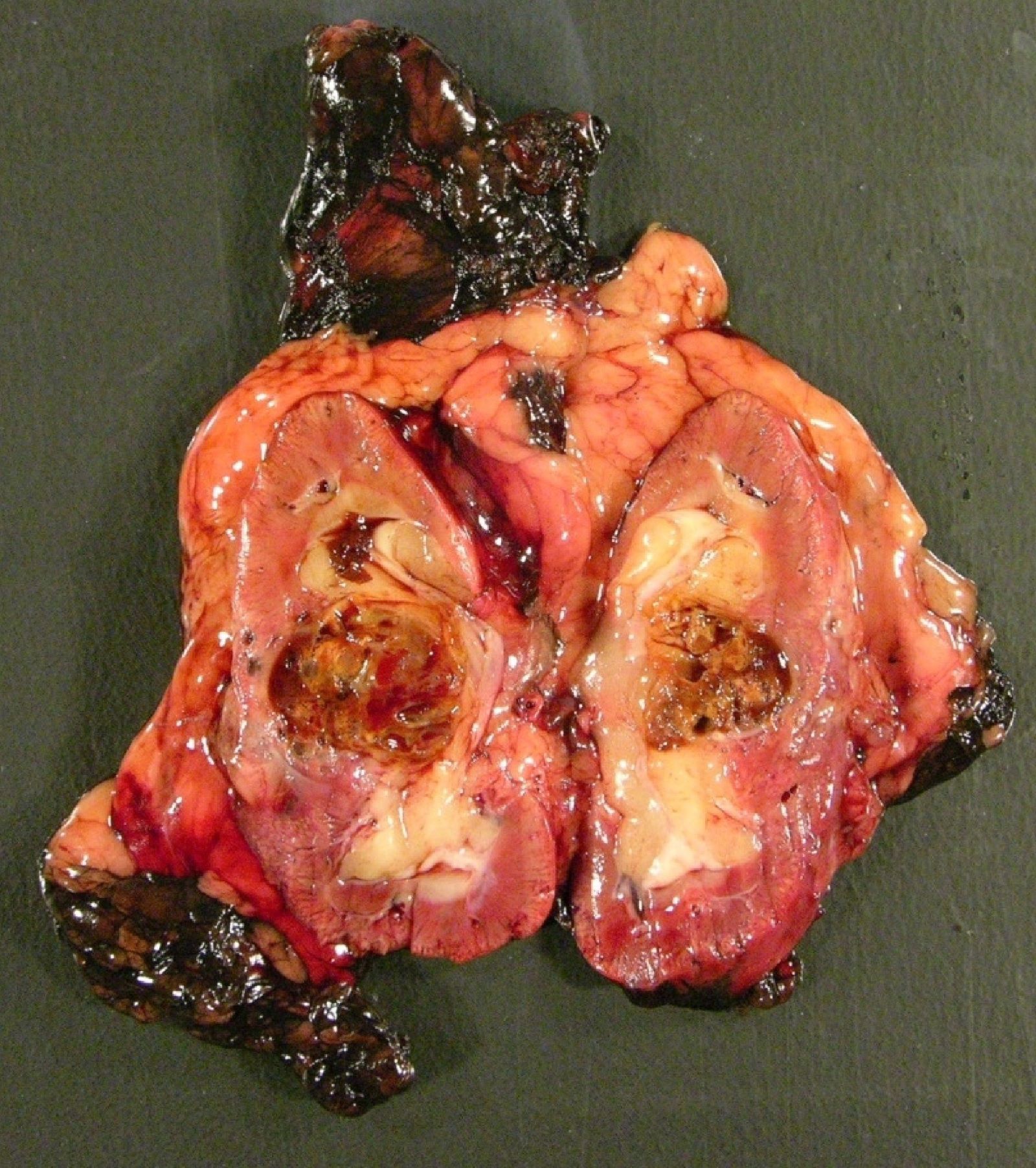
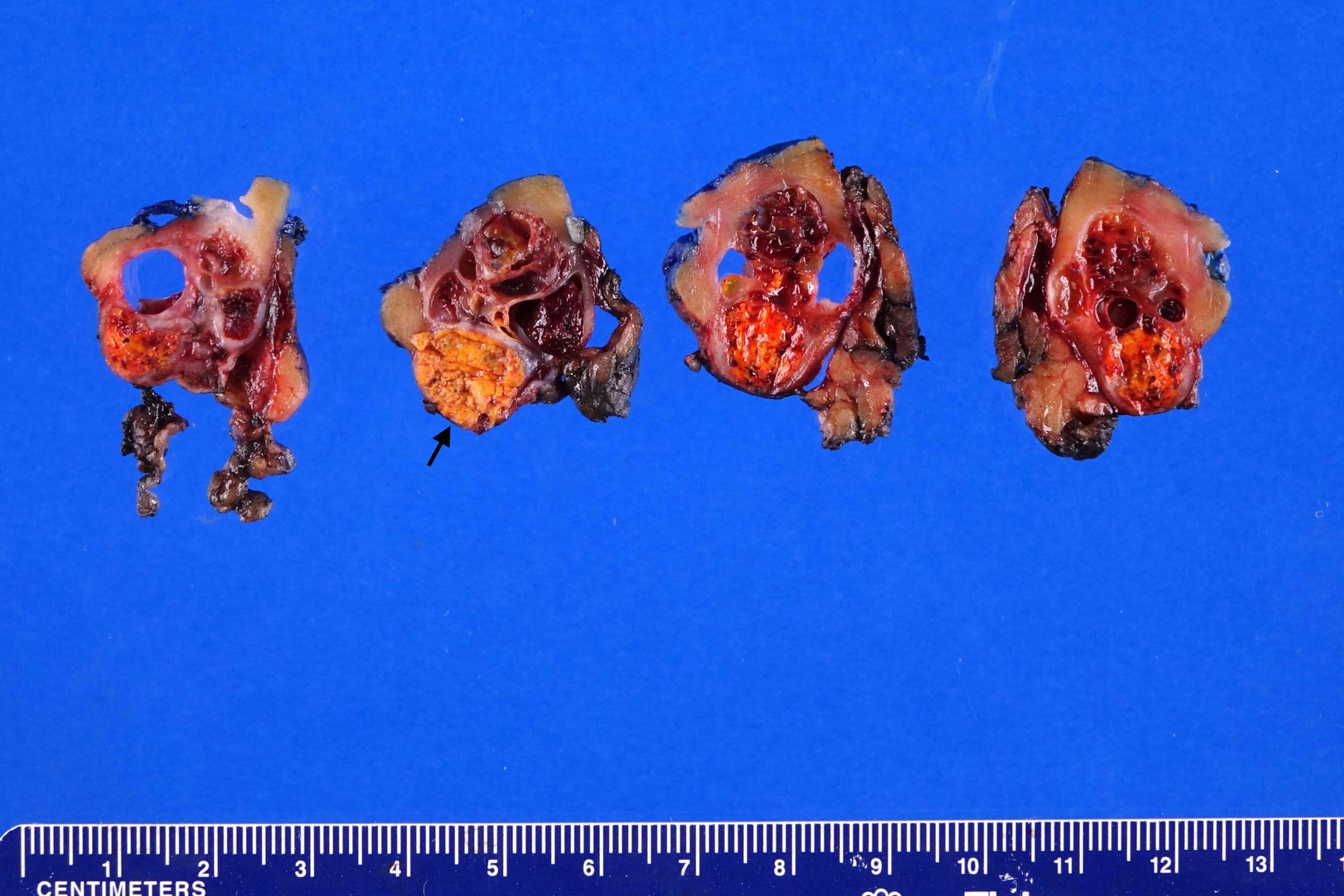
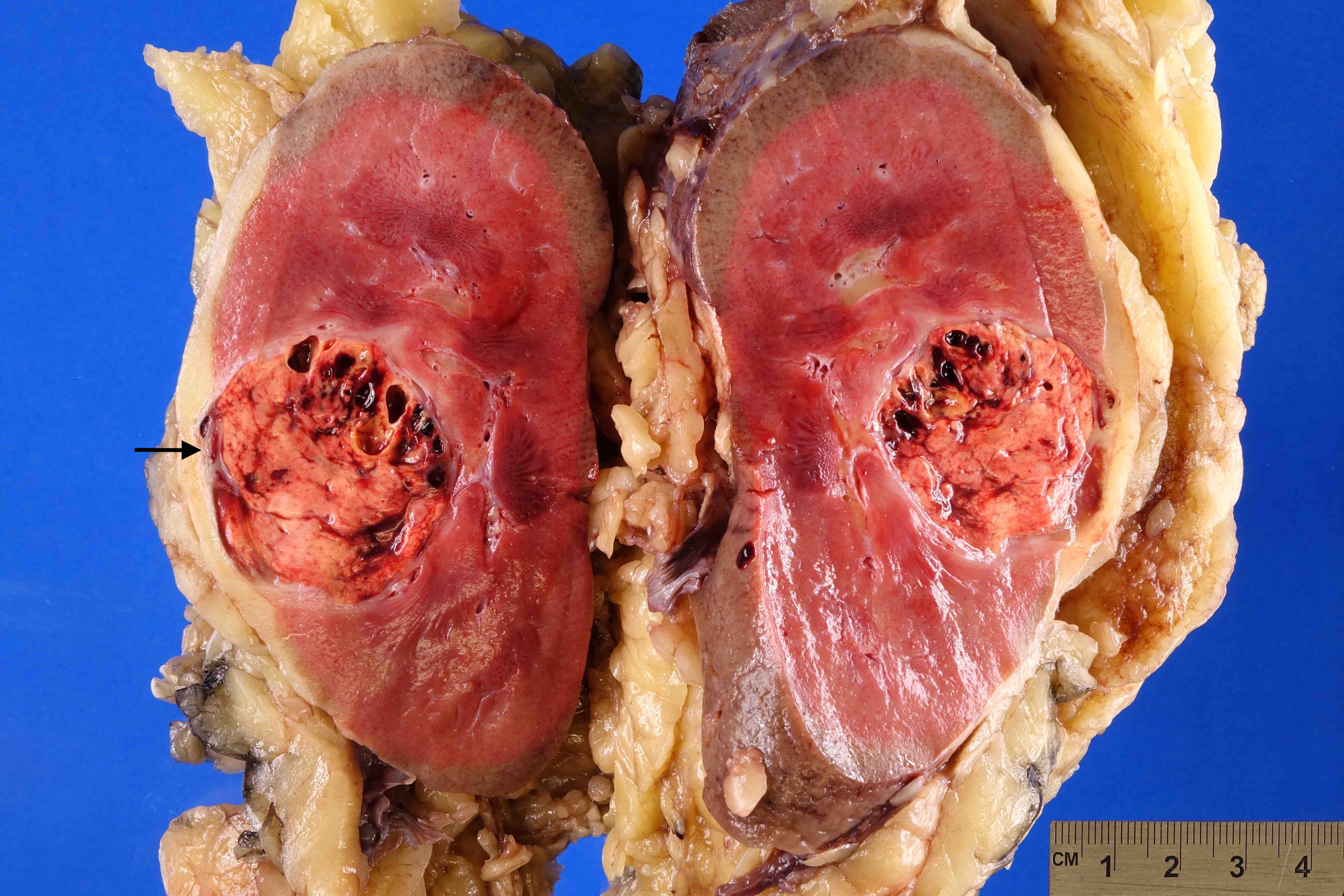
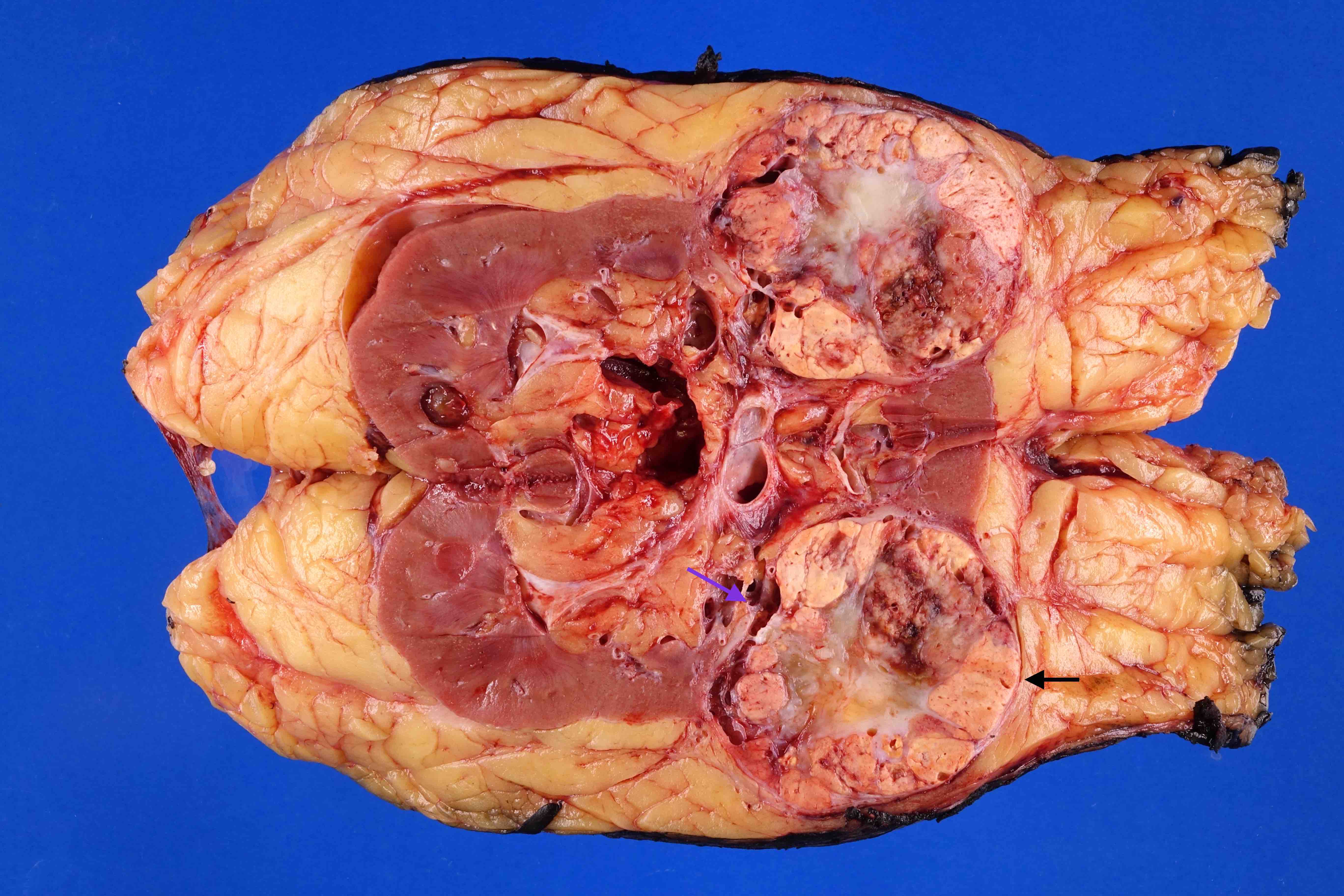
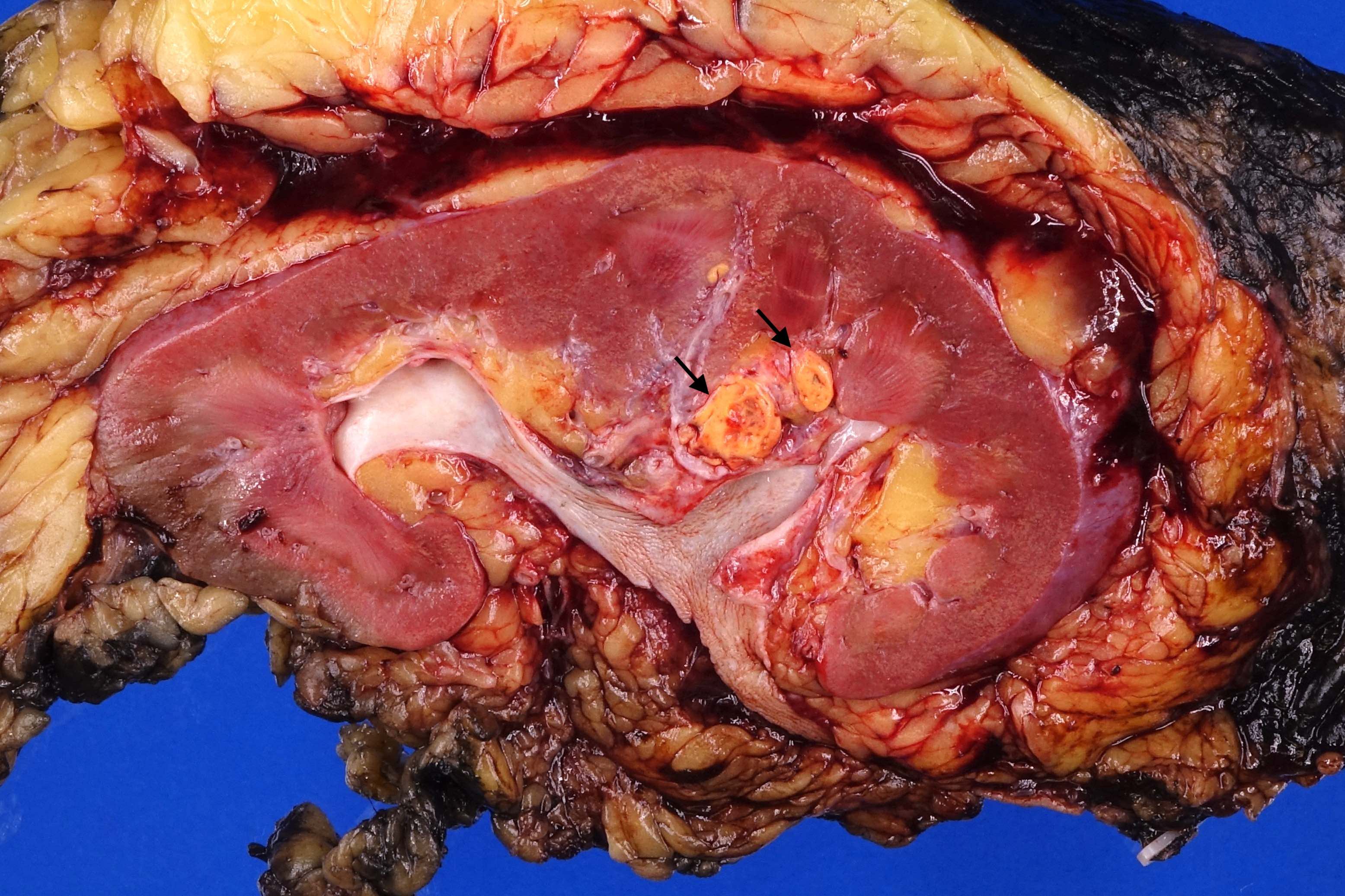
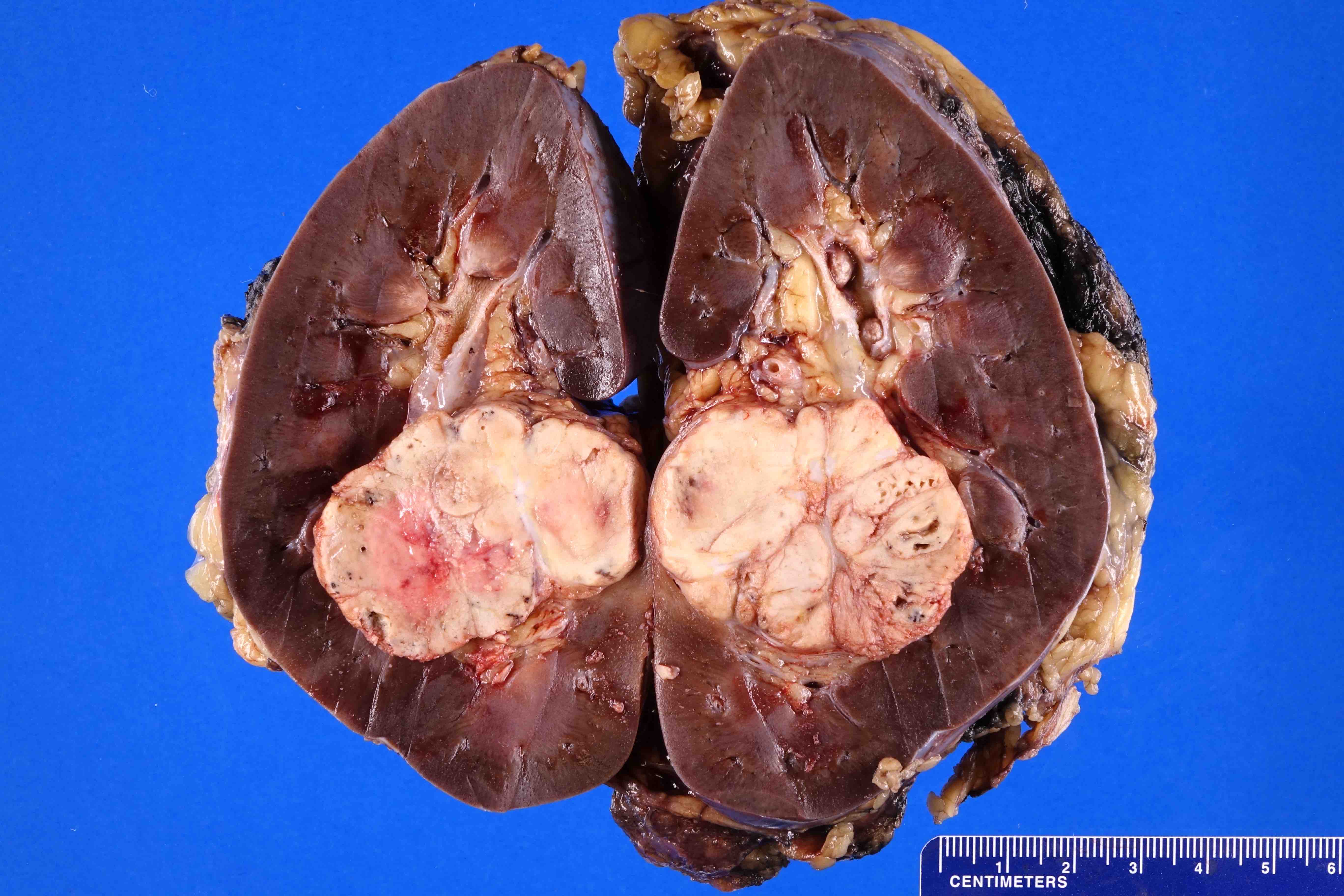
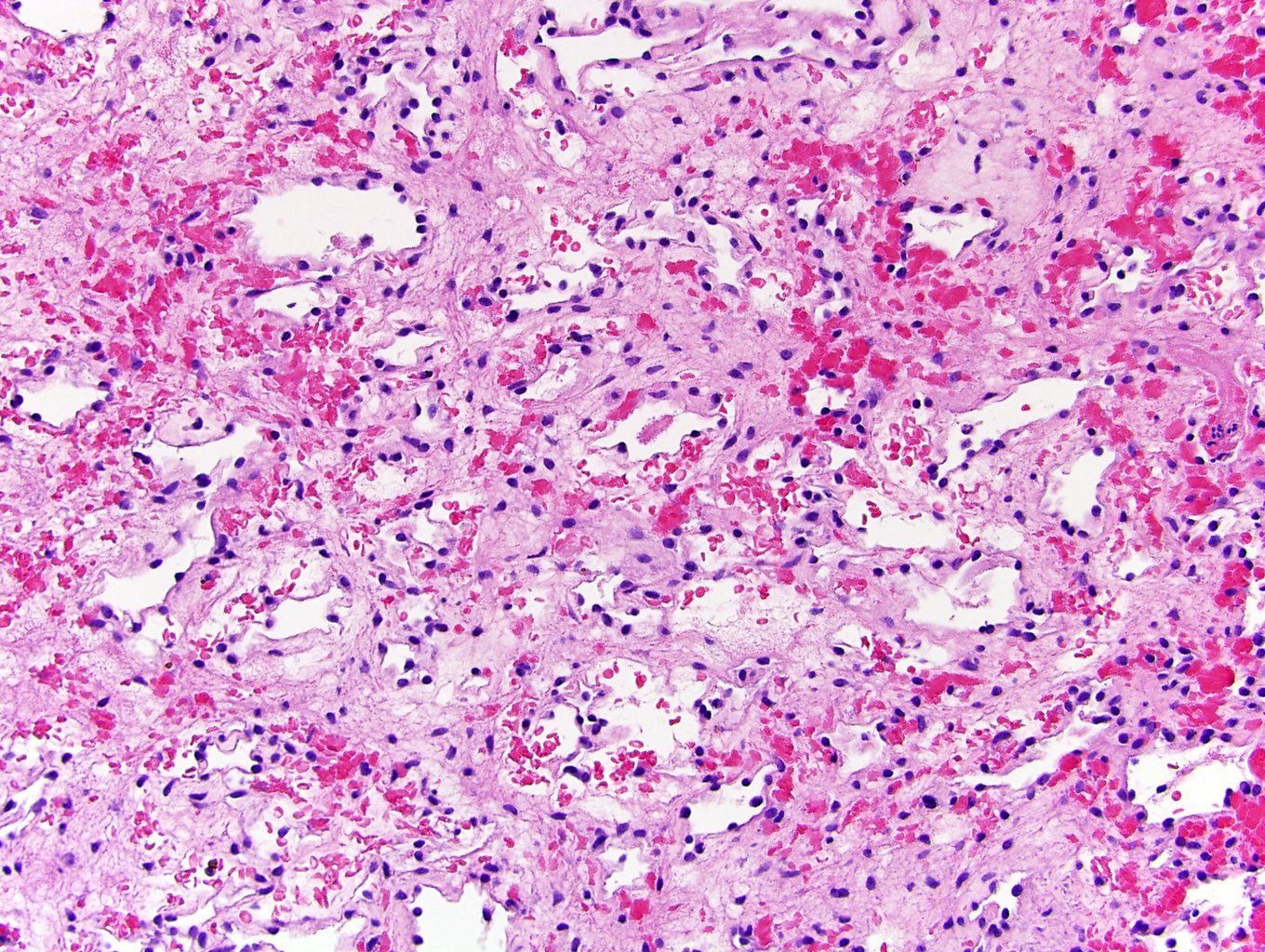
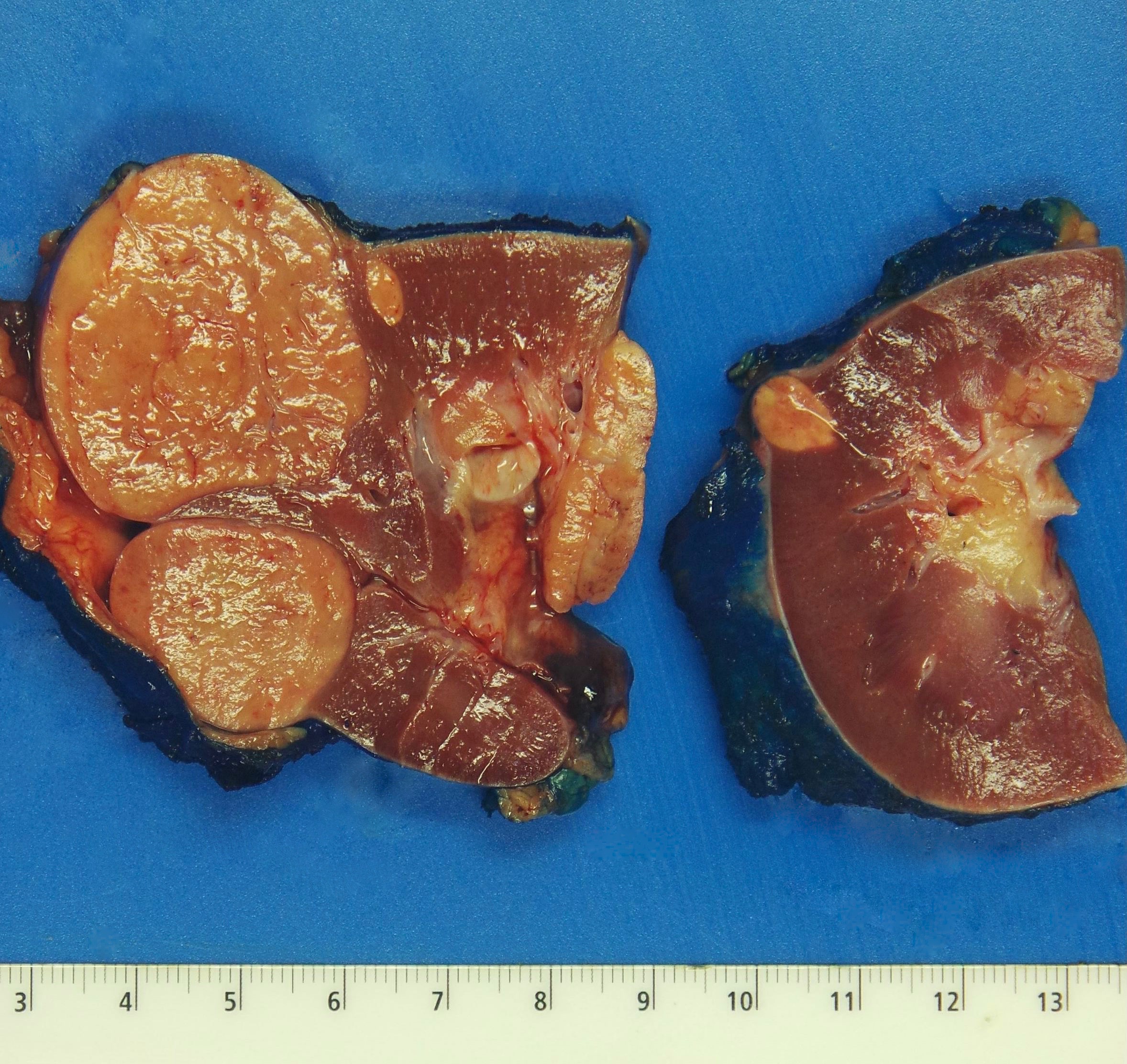
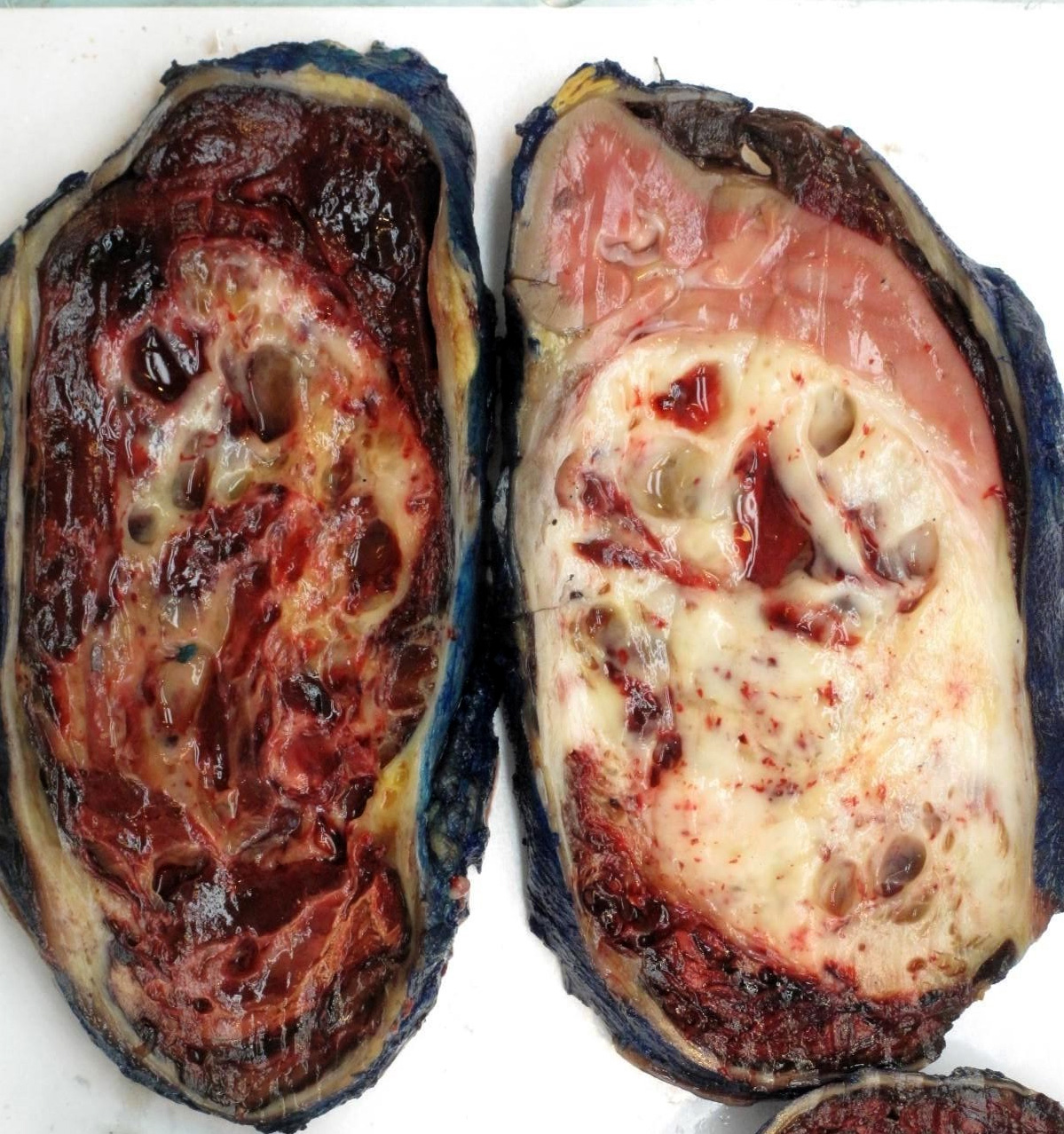
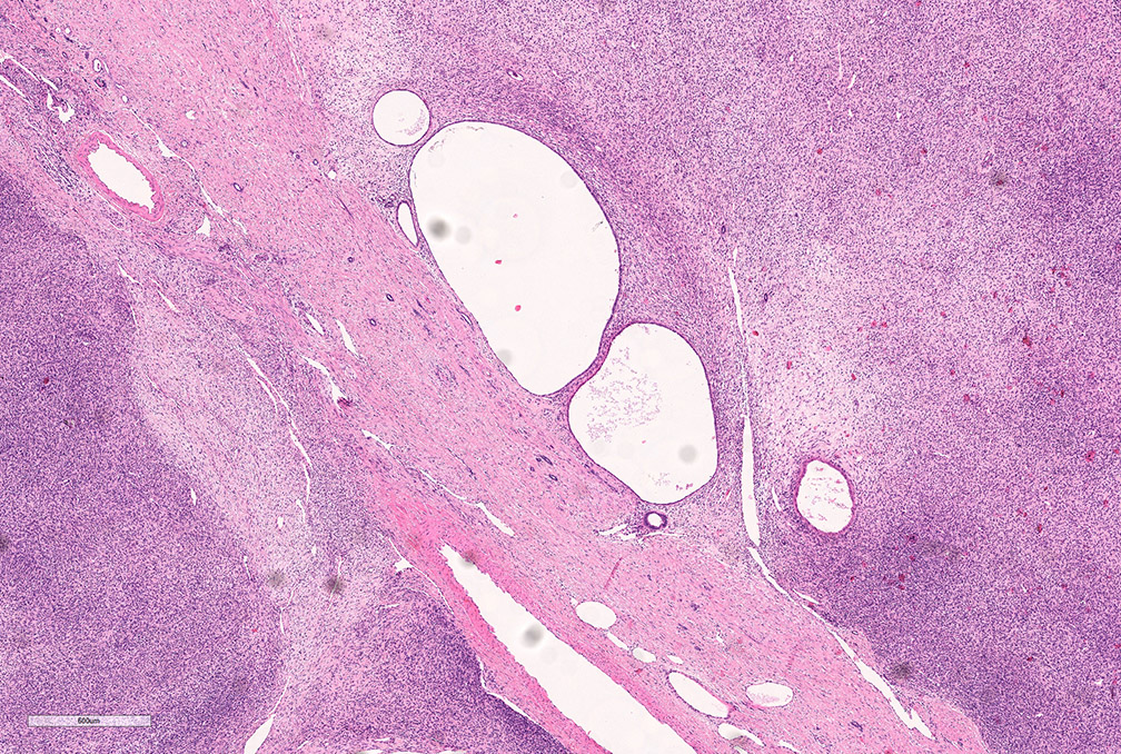
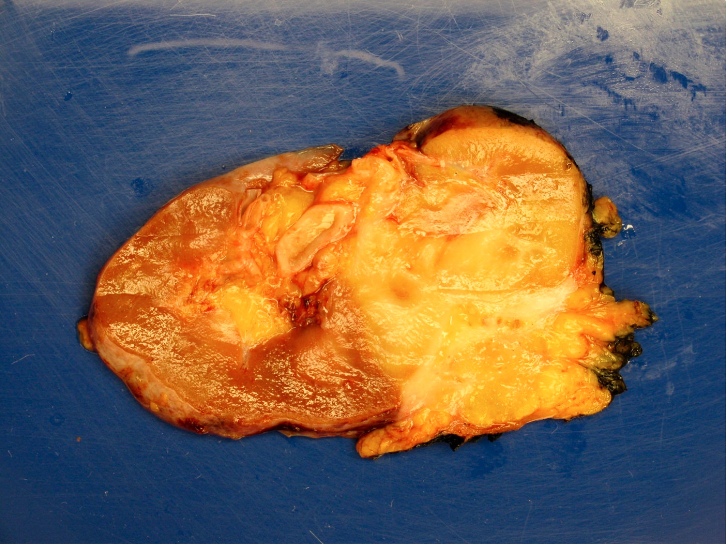
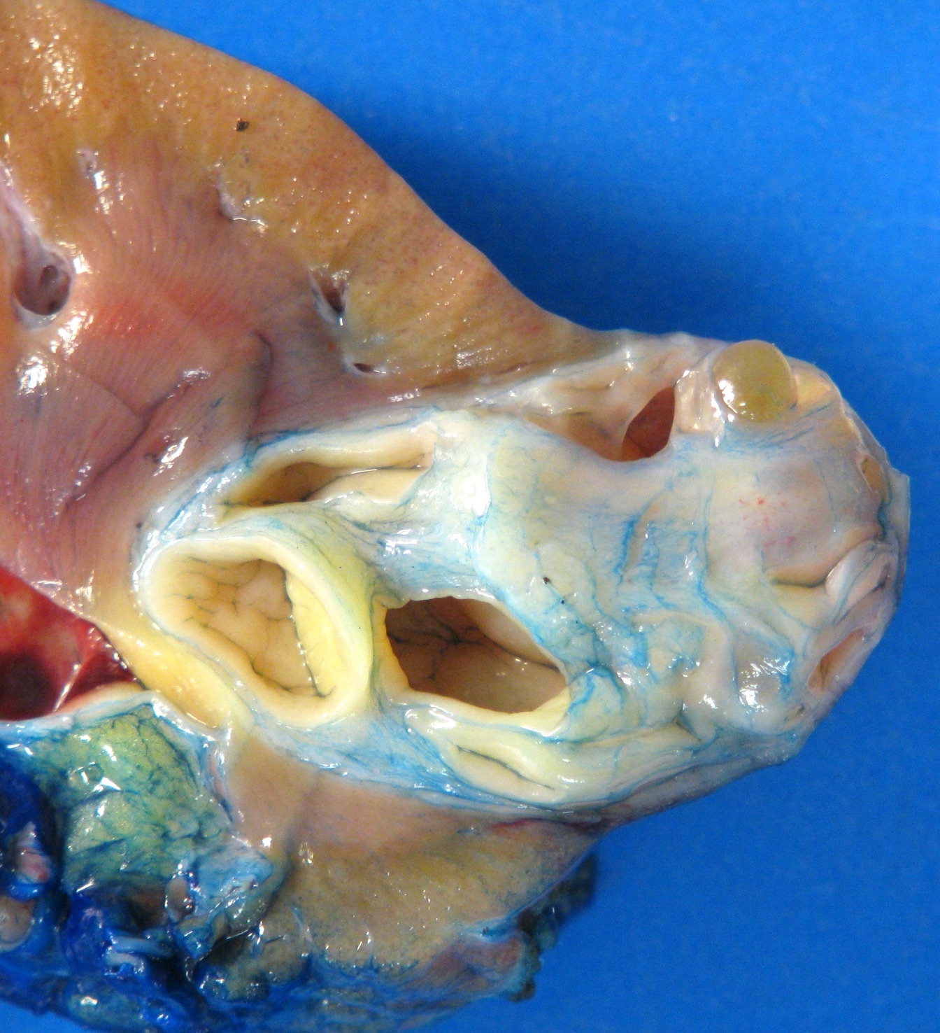
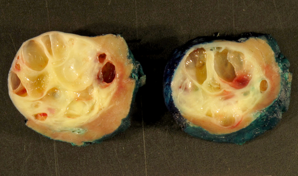
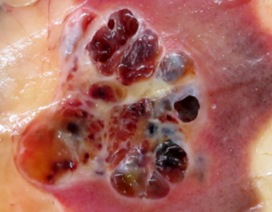
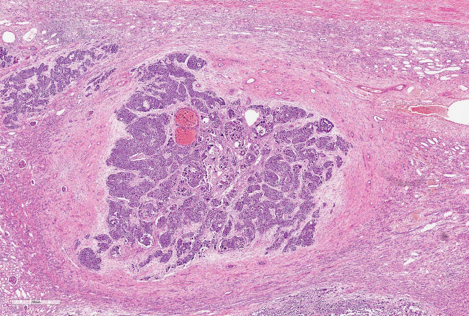
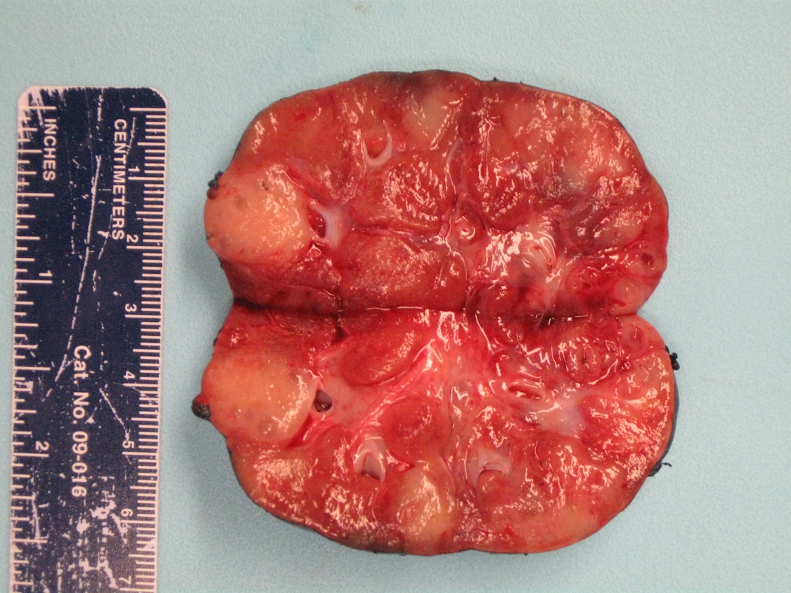
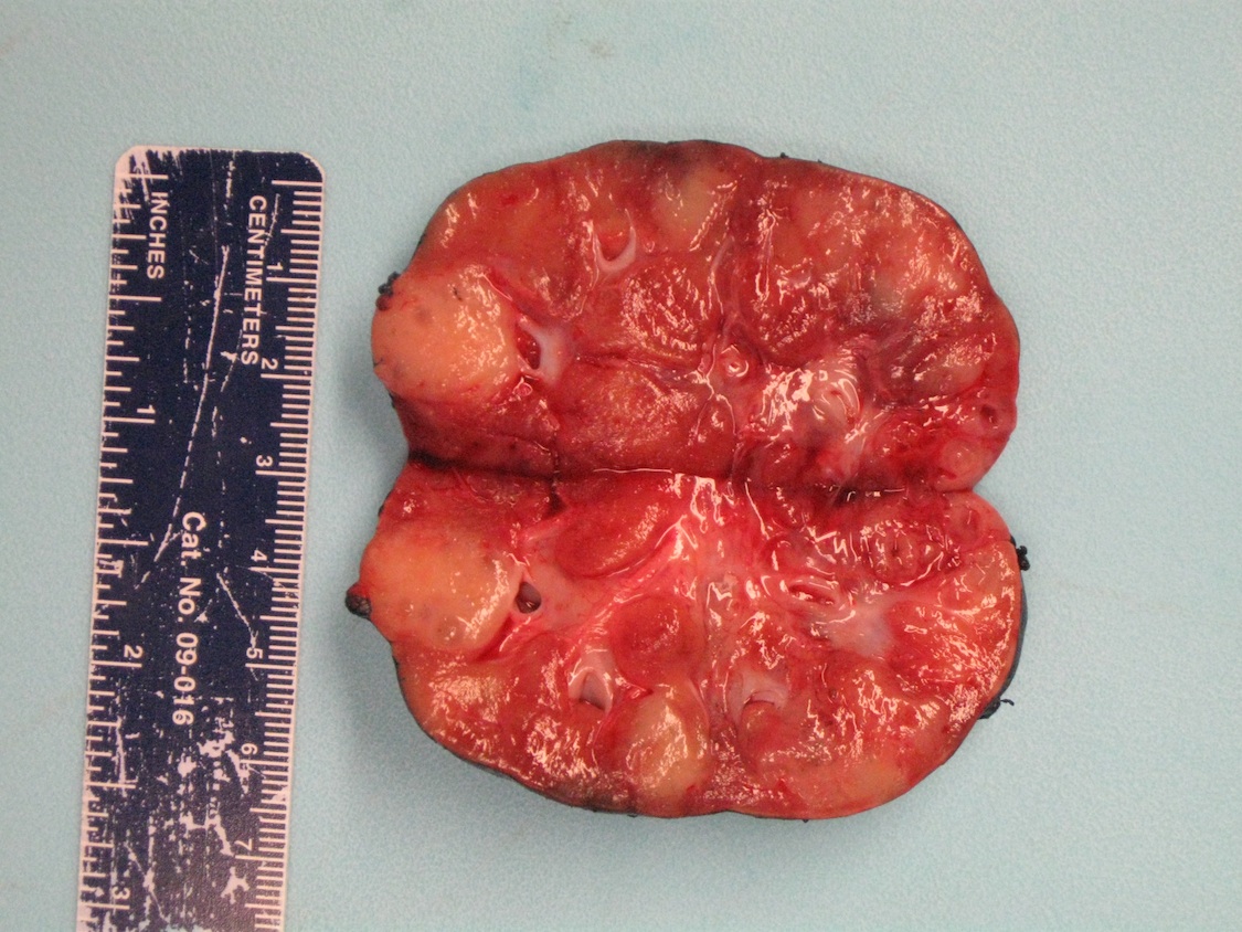
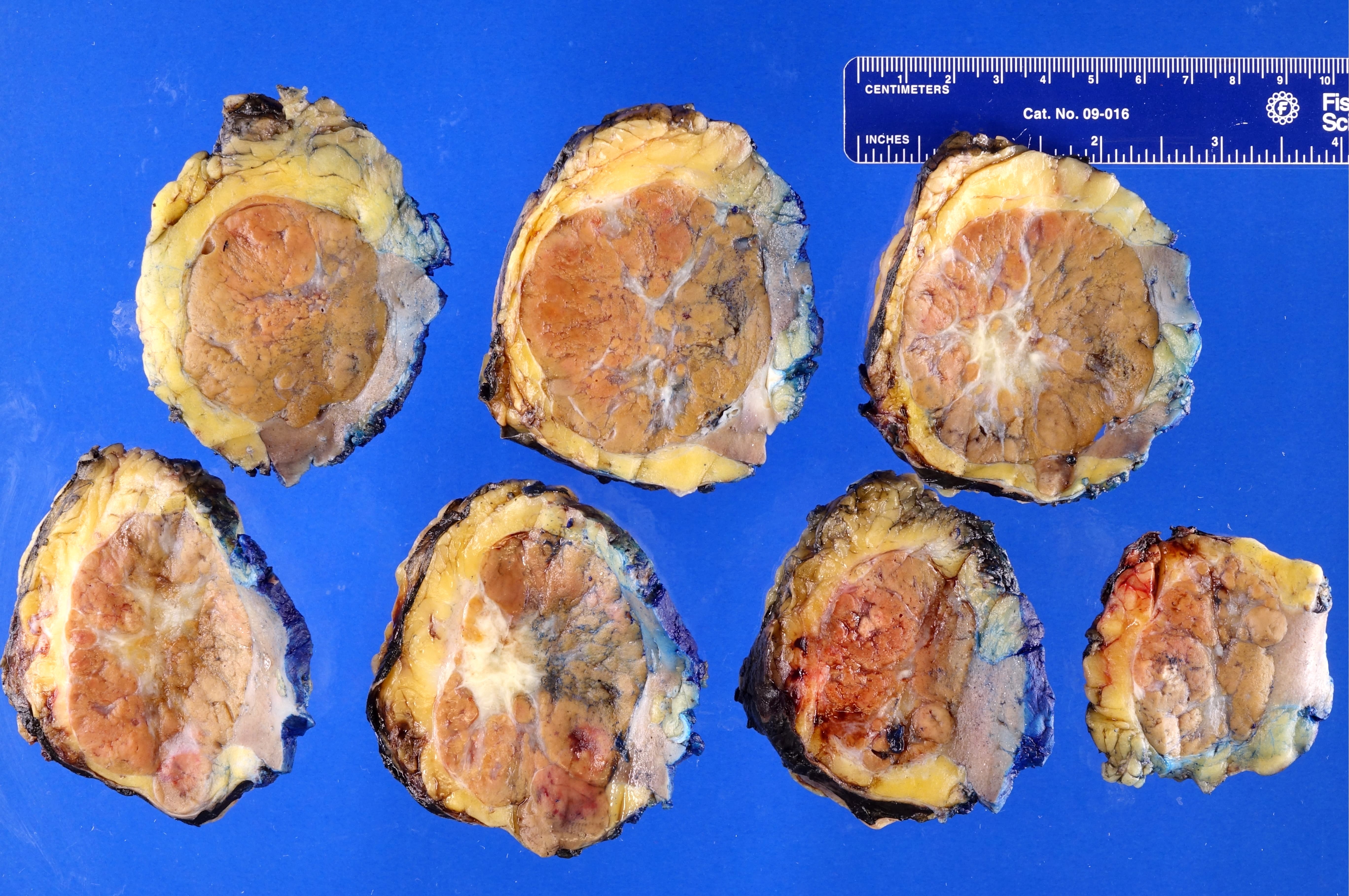
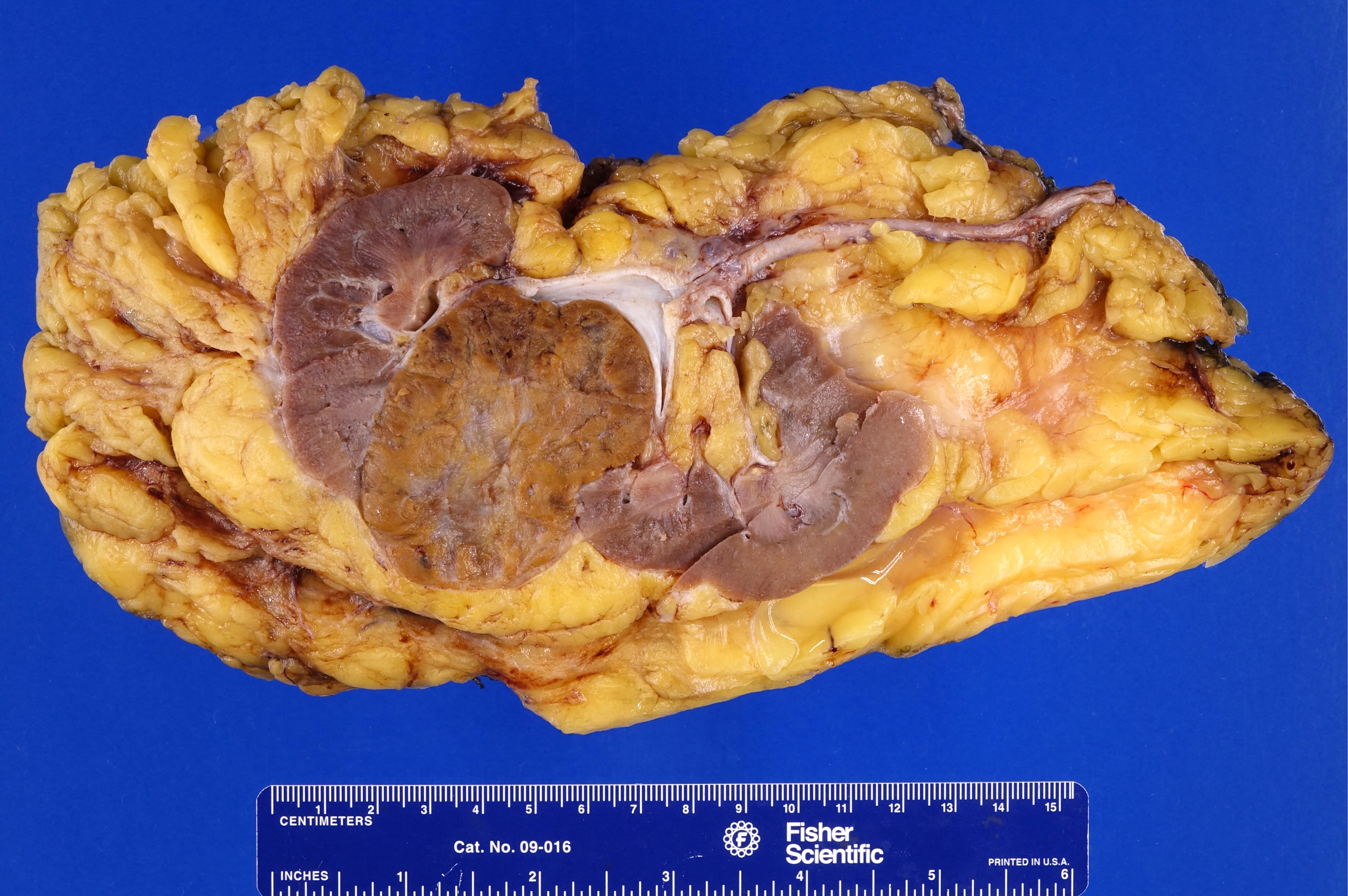
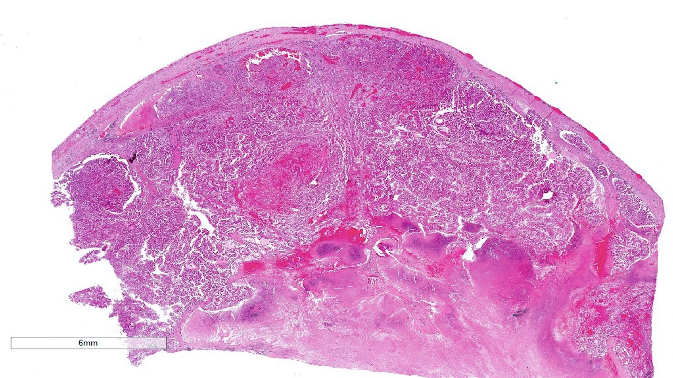
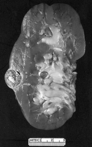
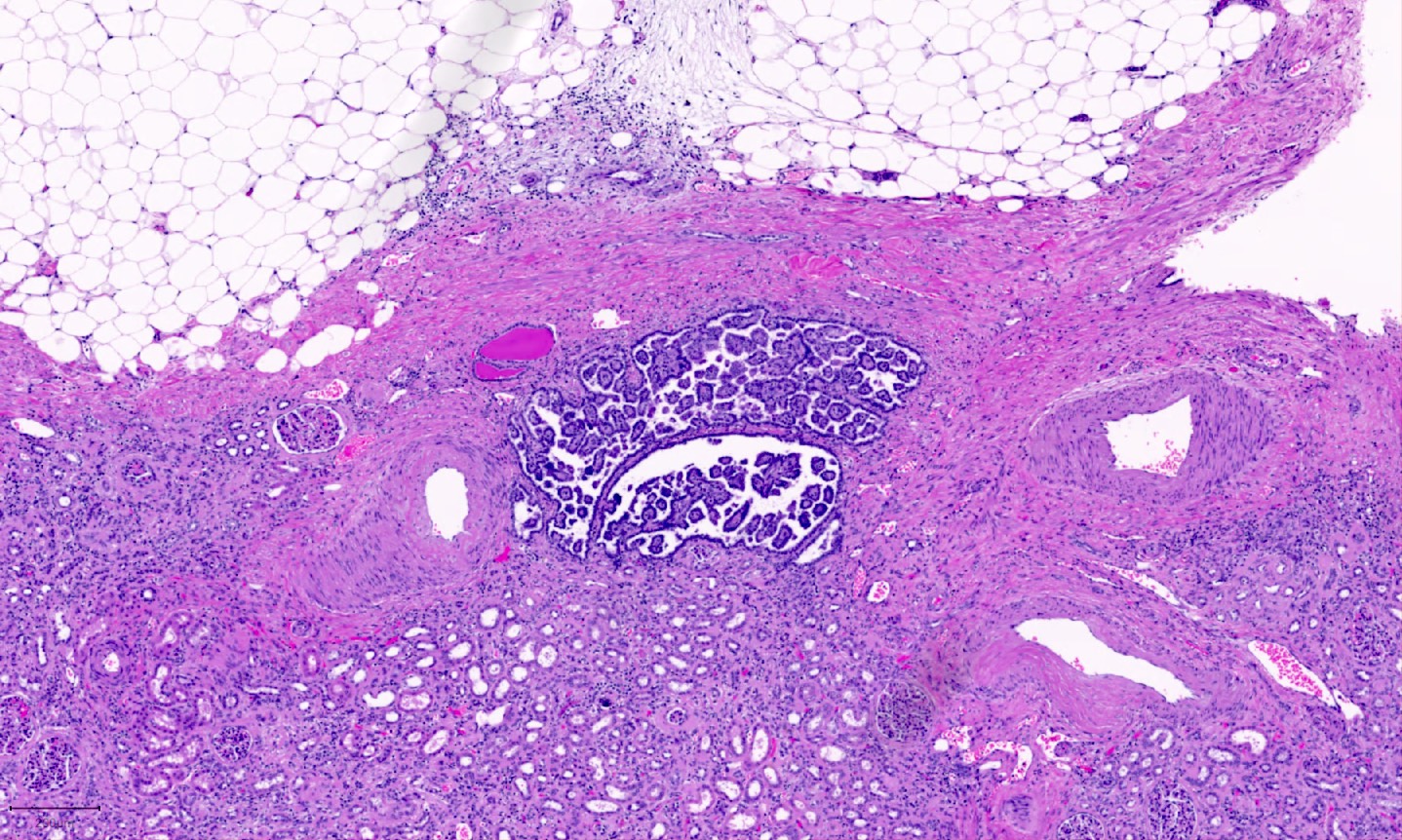
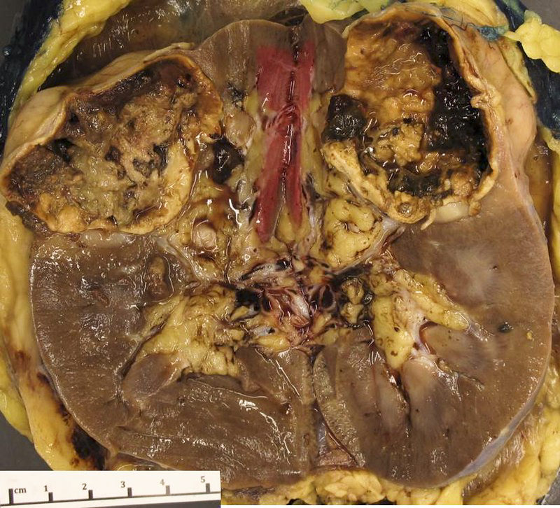
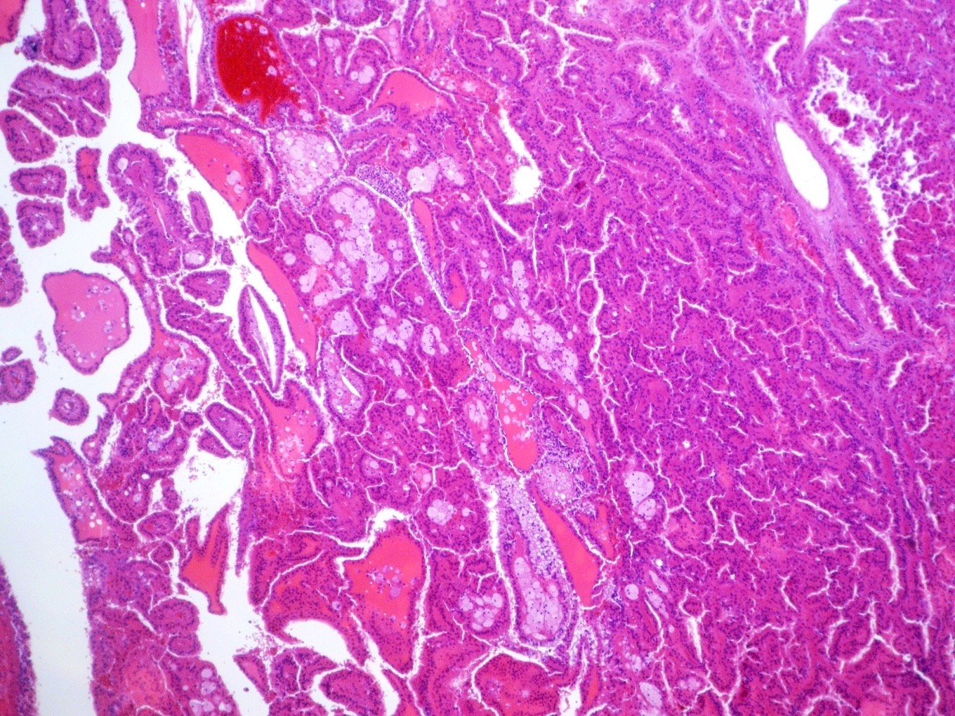
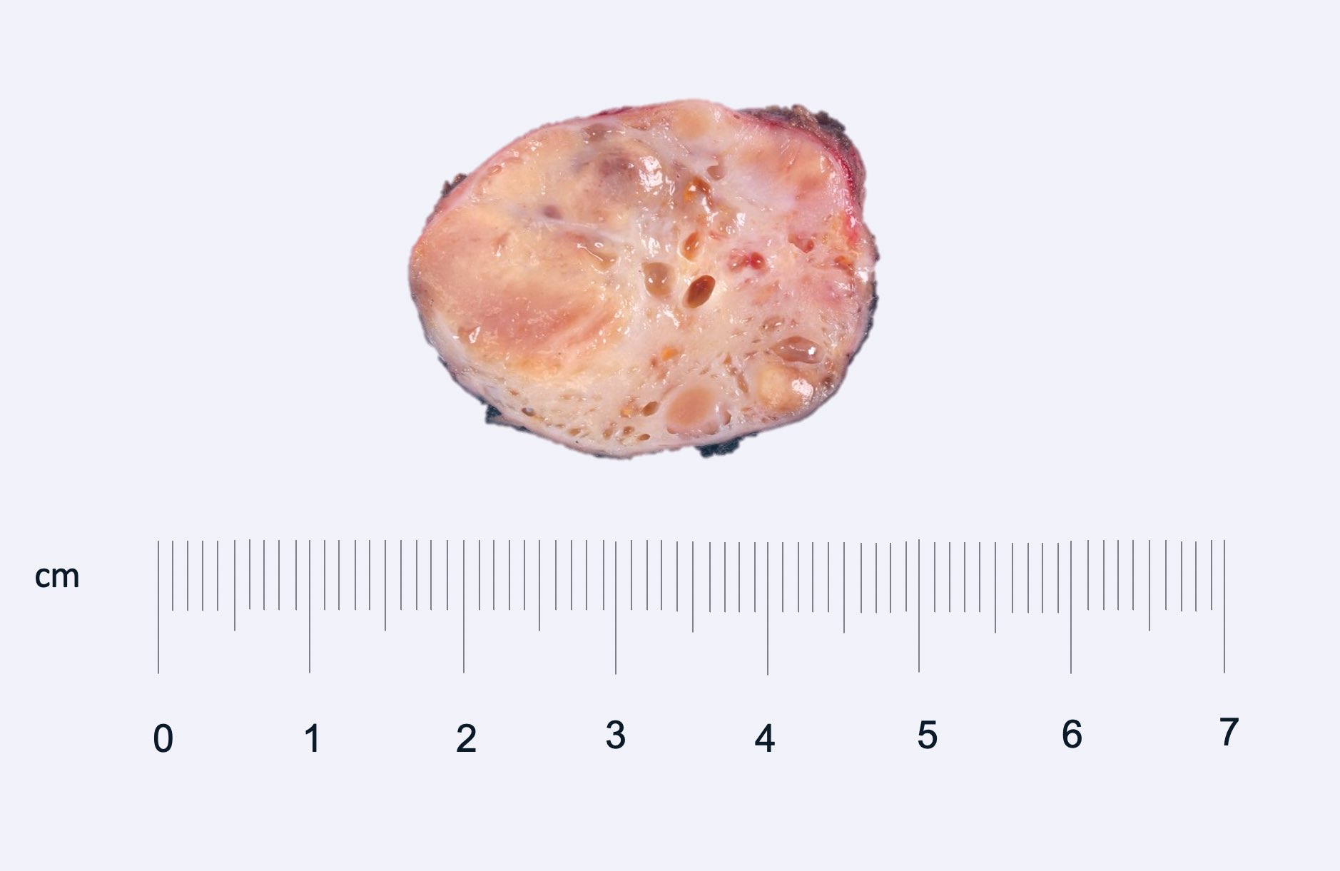
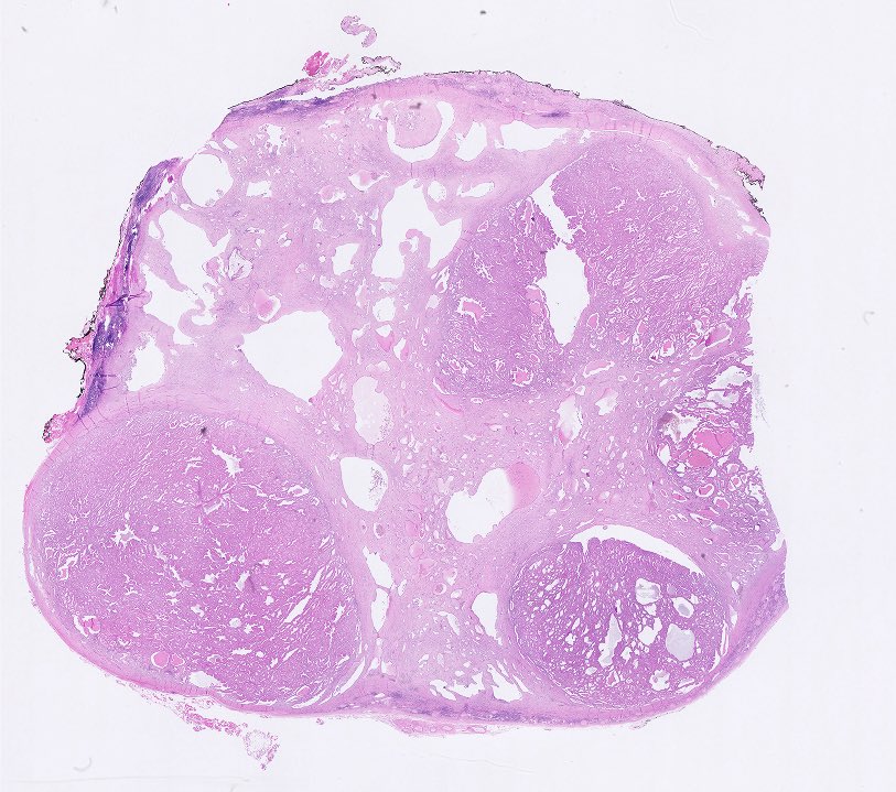
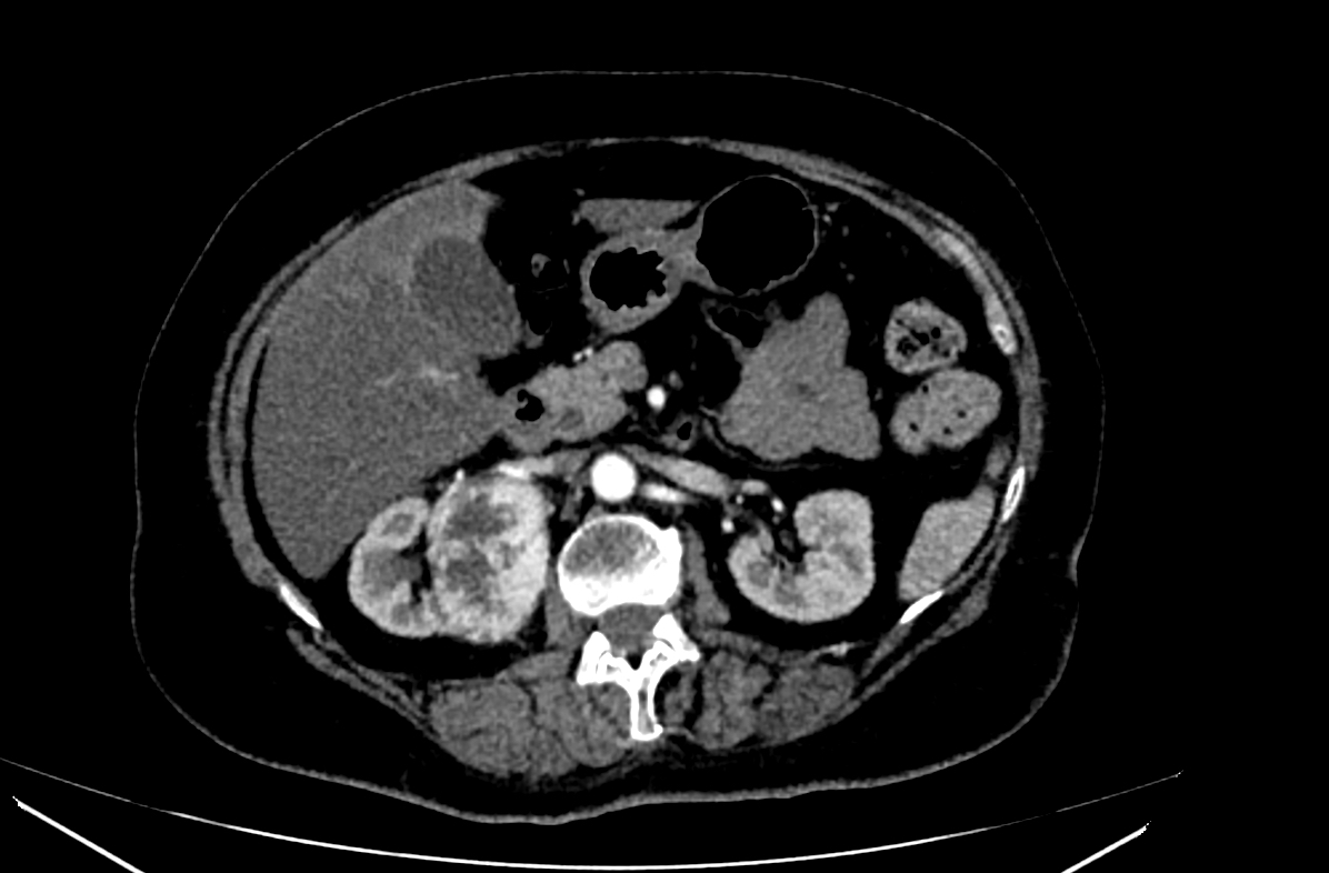
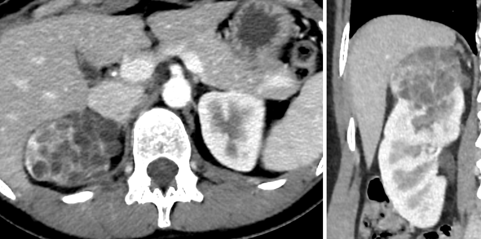
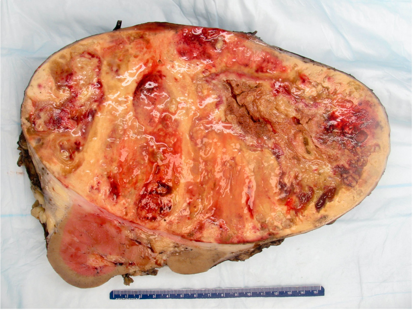
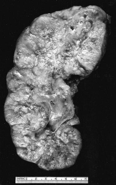
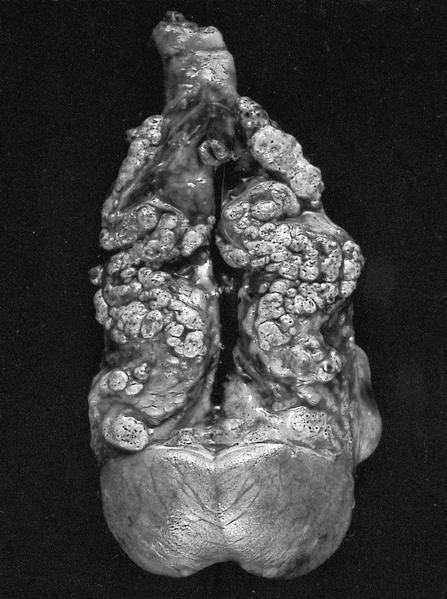
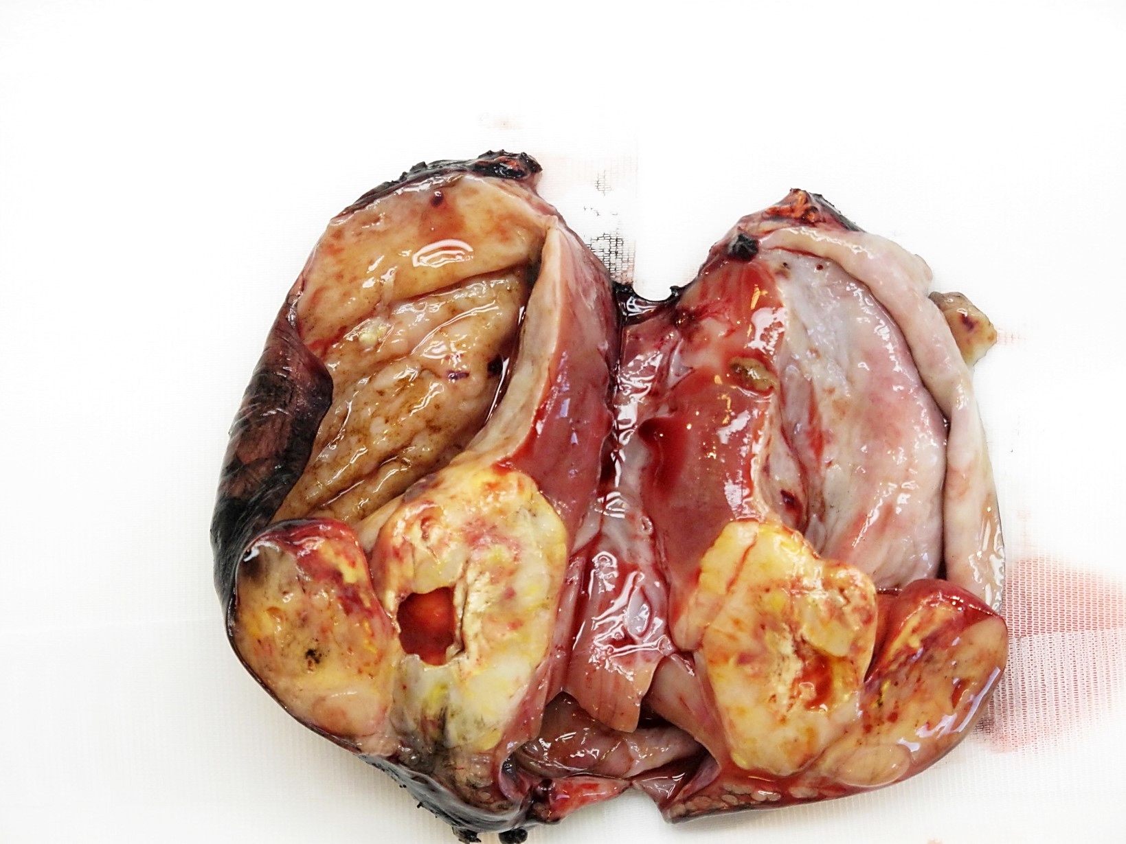
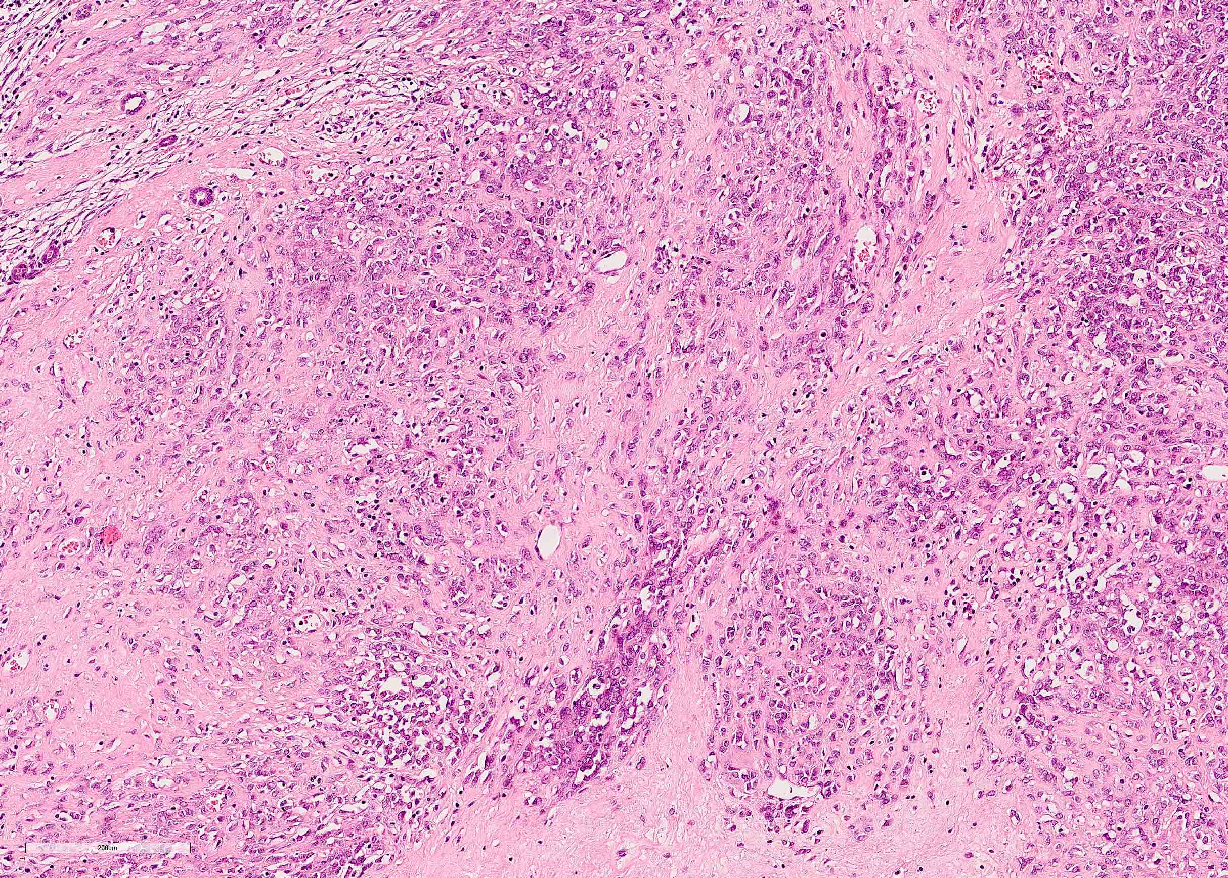
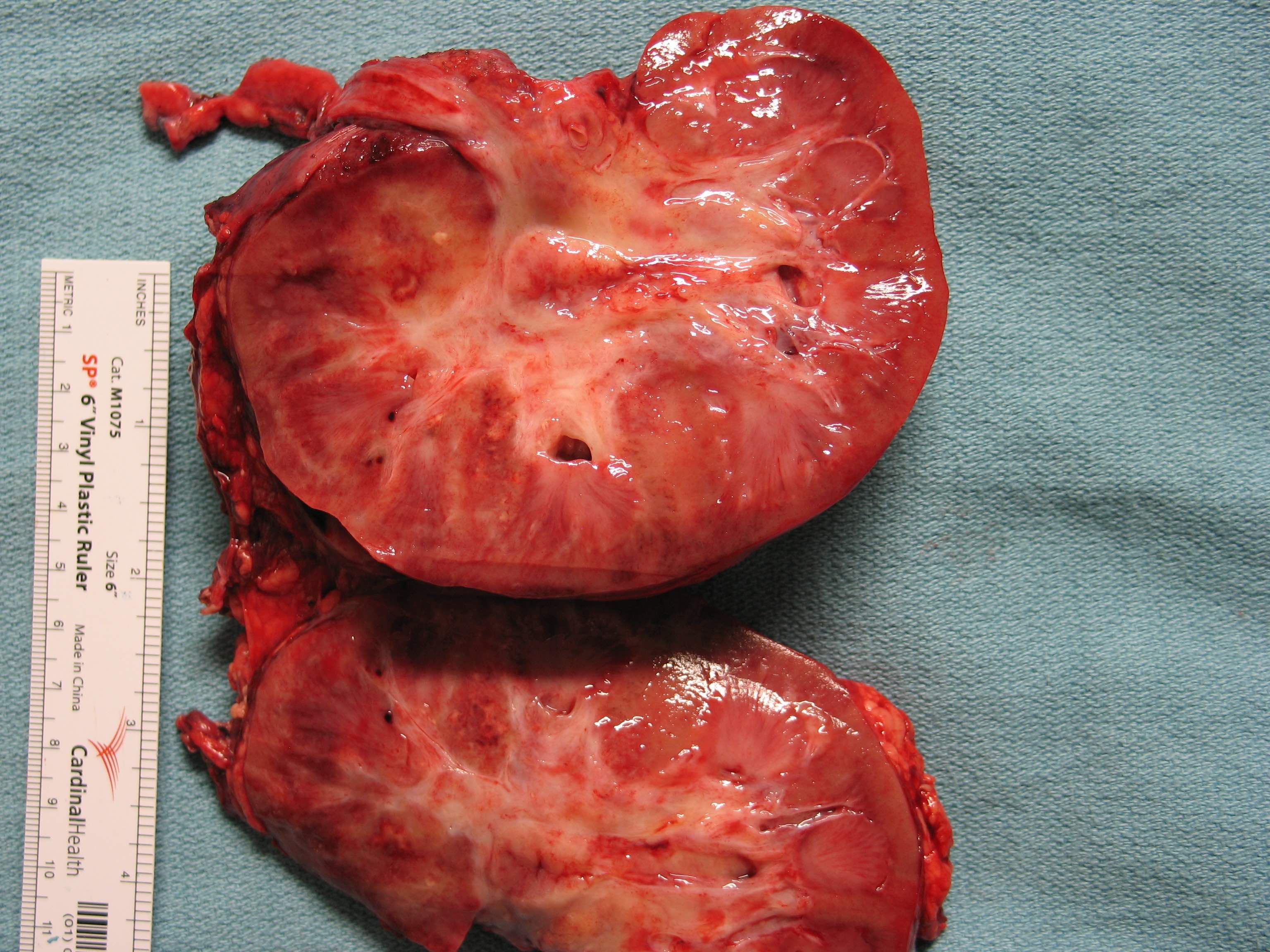
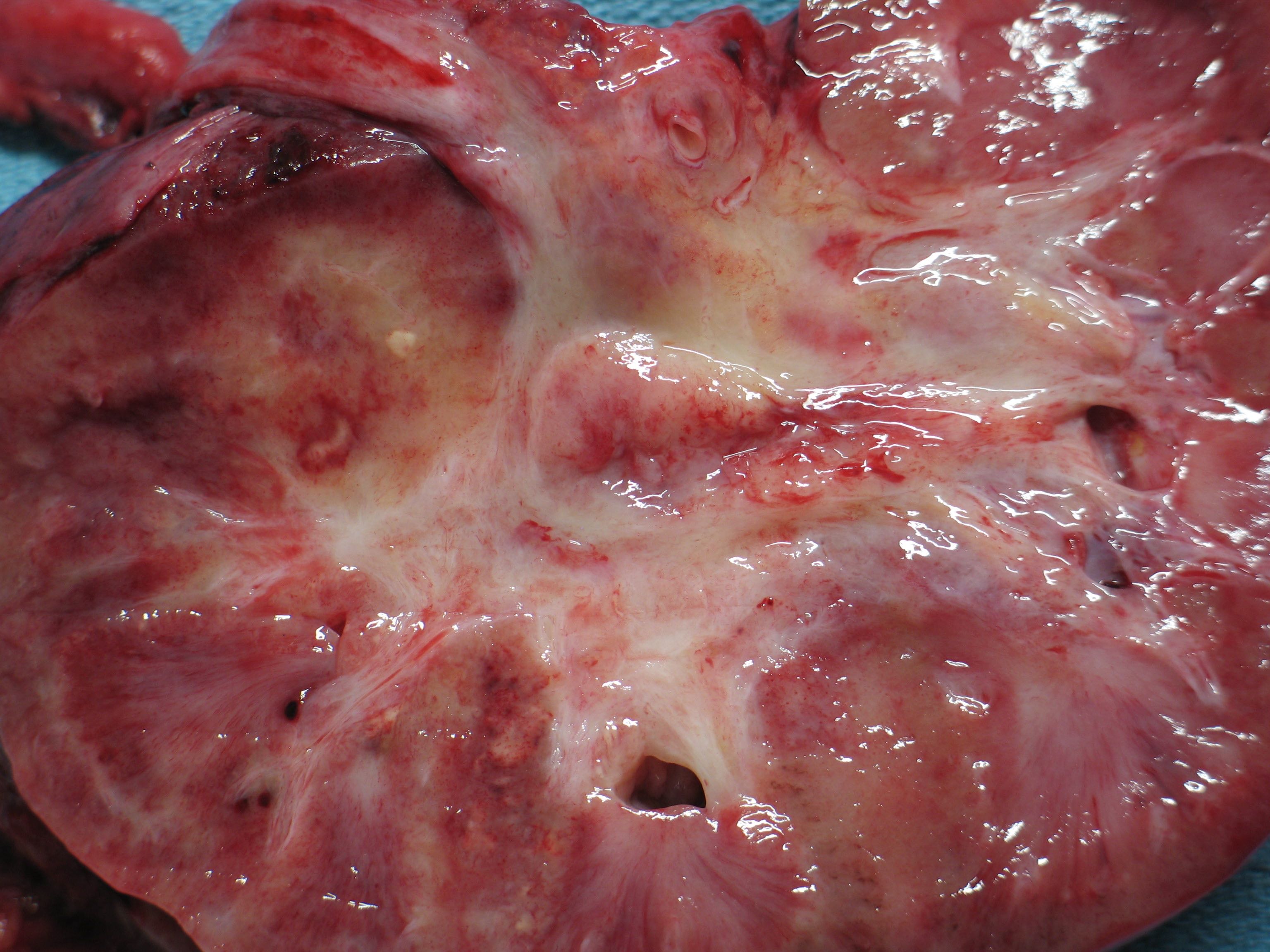
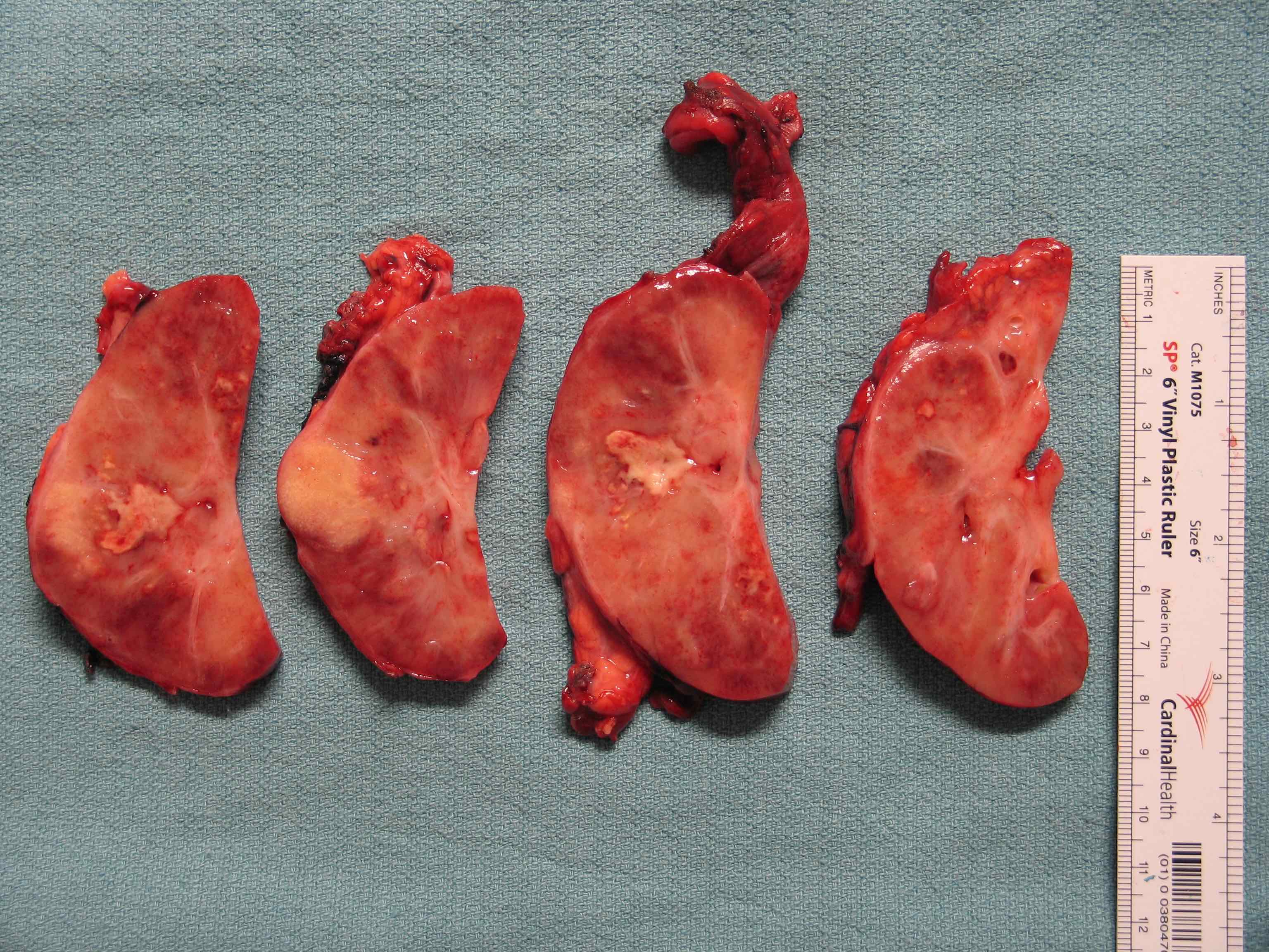
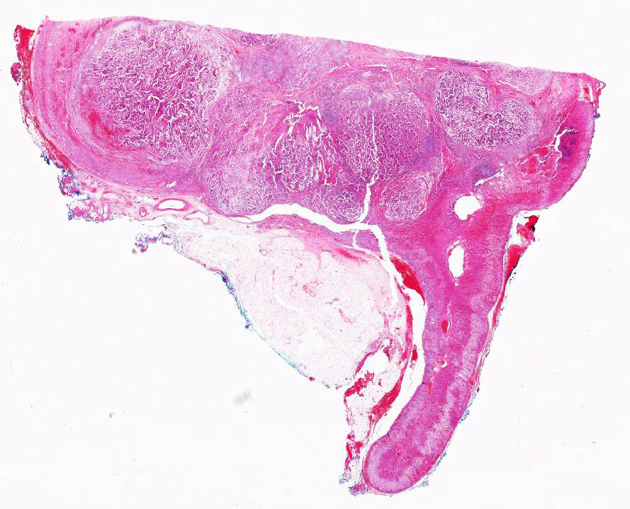
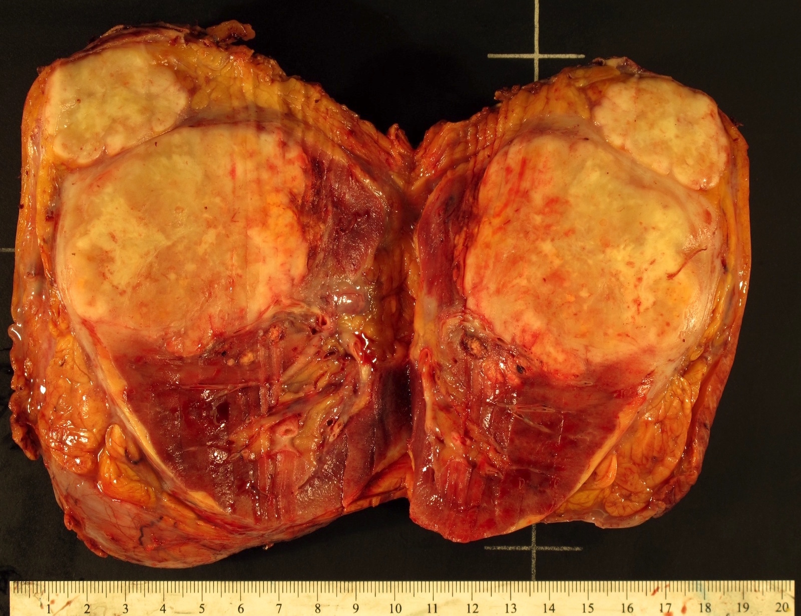
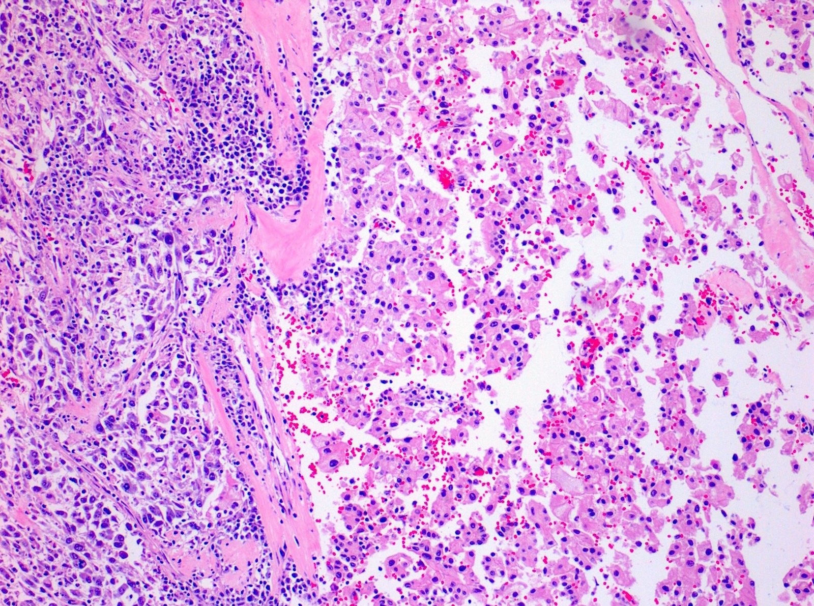
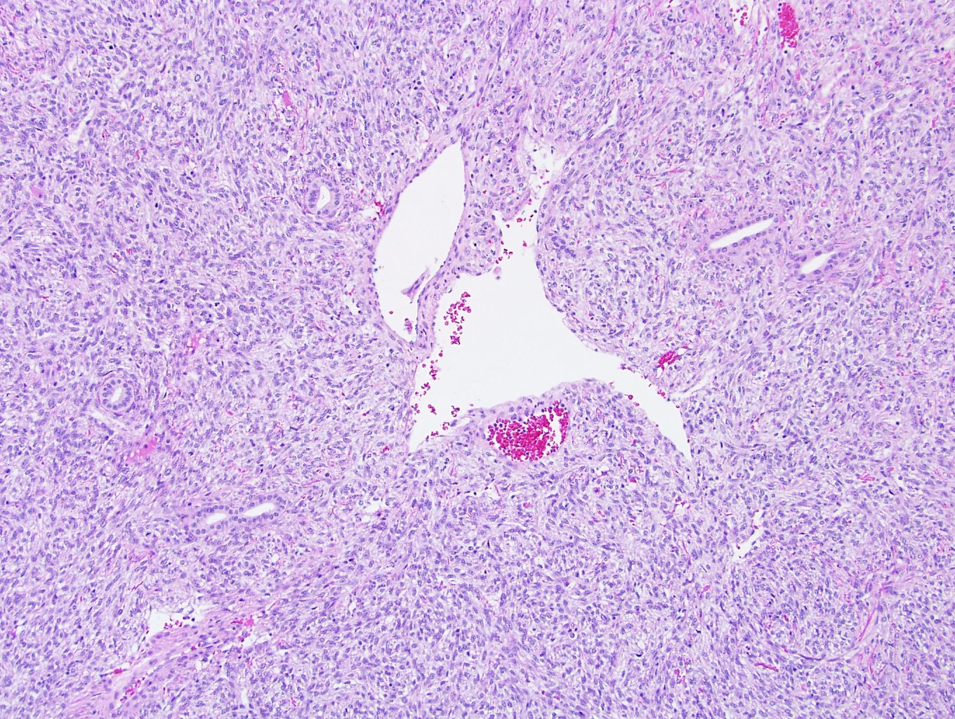
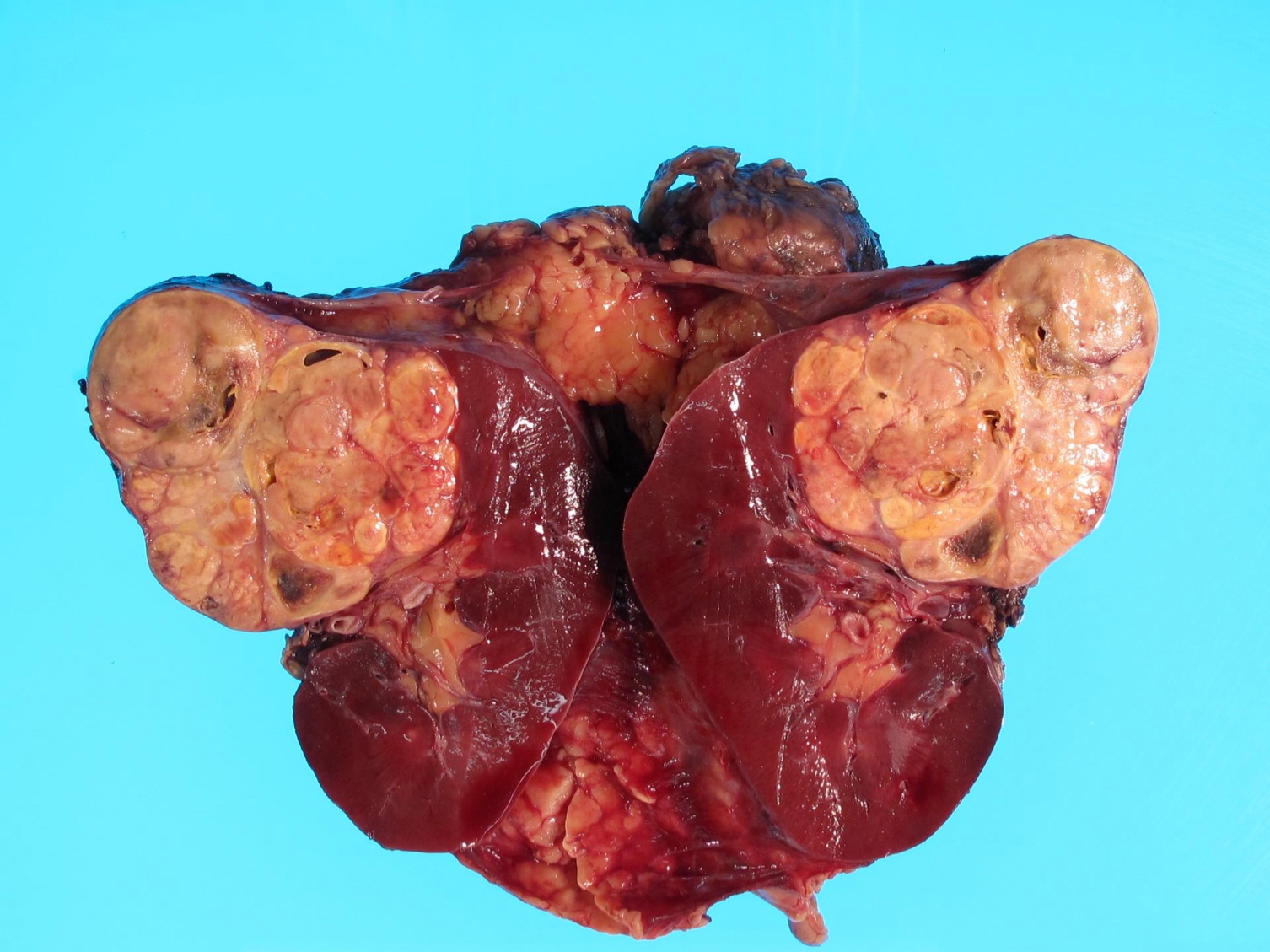
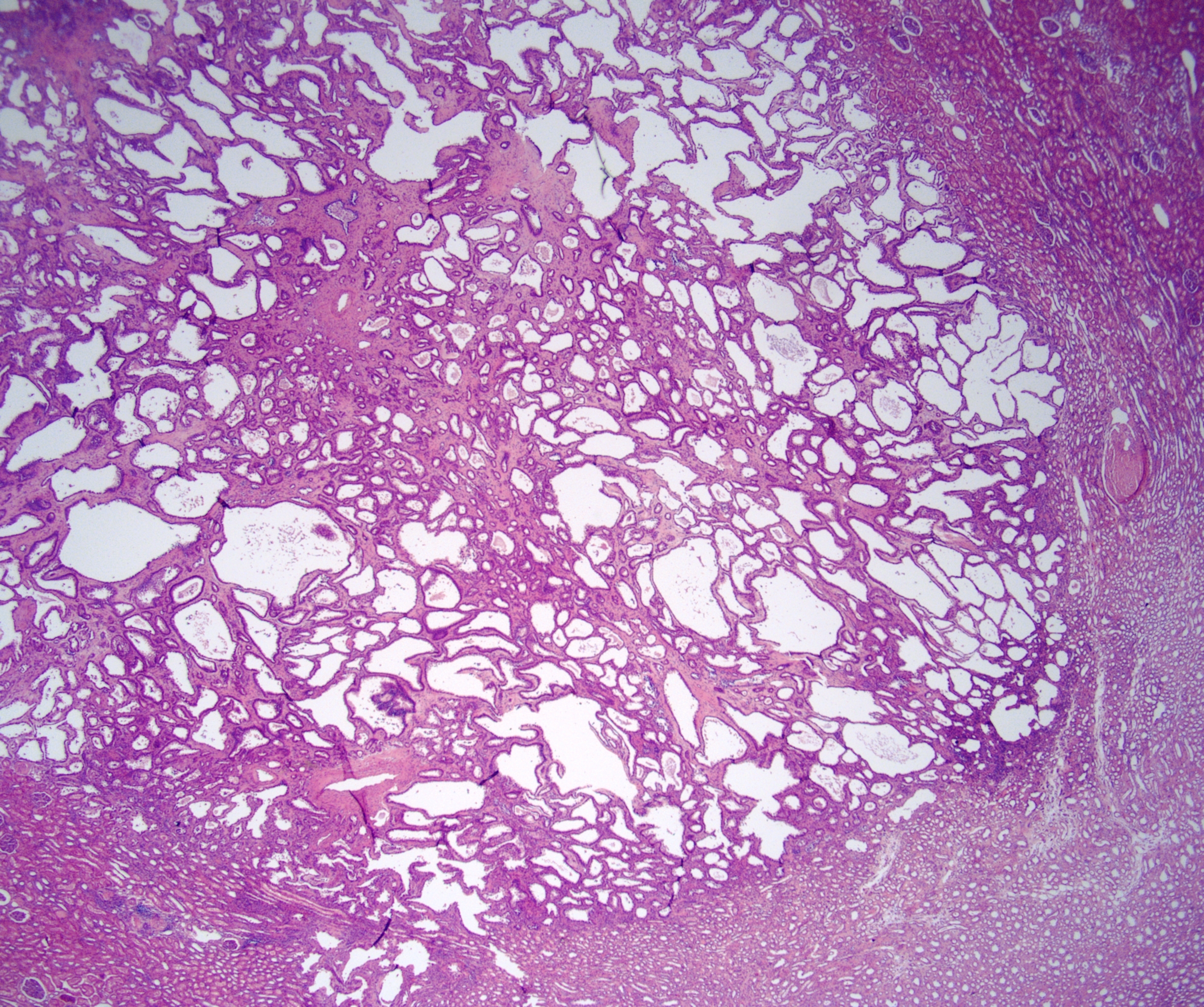
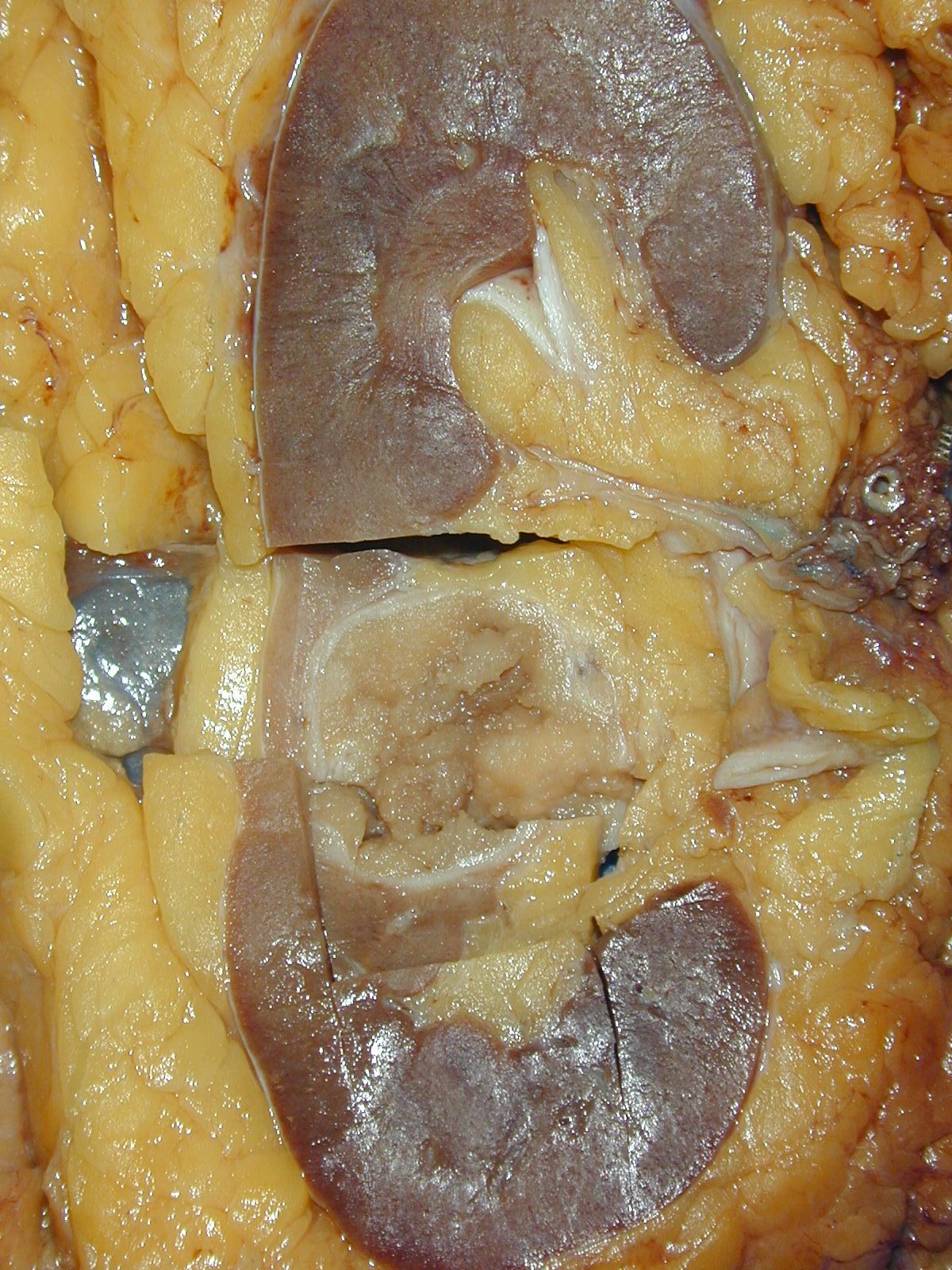
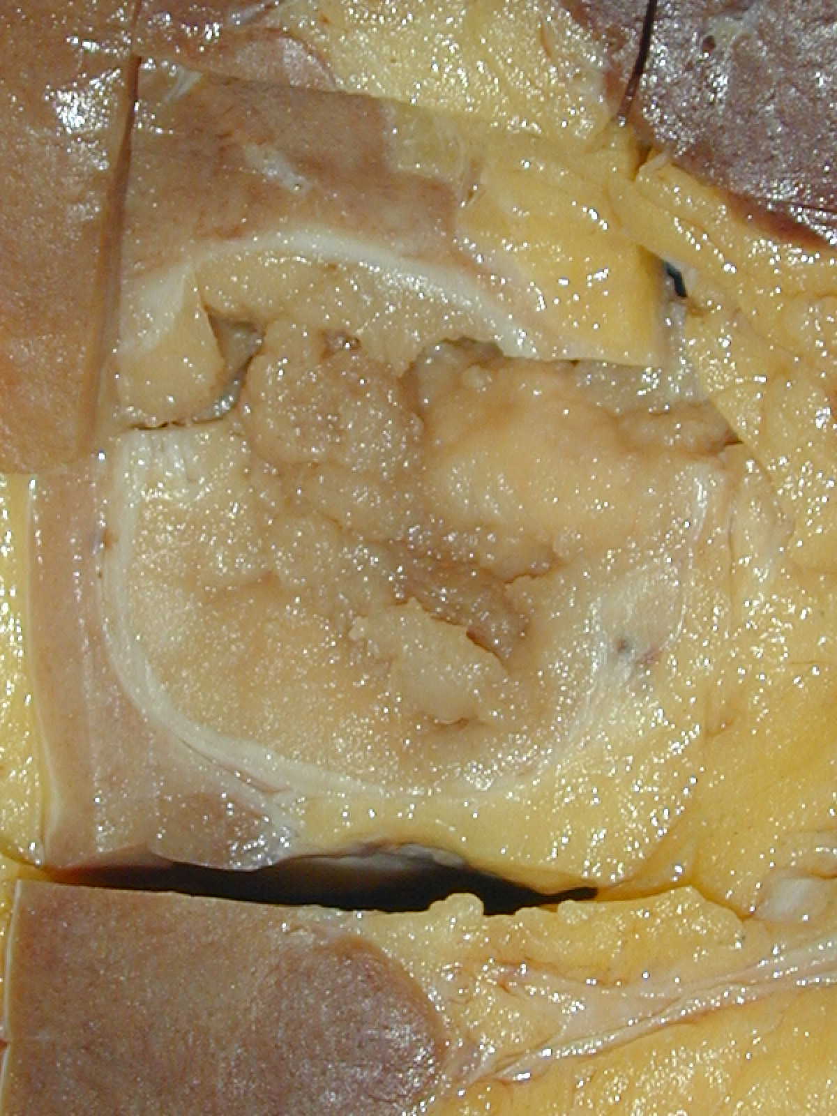
Solid mass
expanding to
collecting system

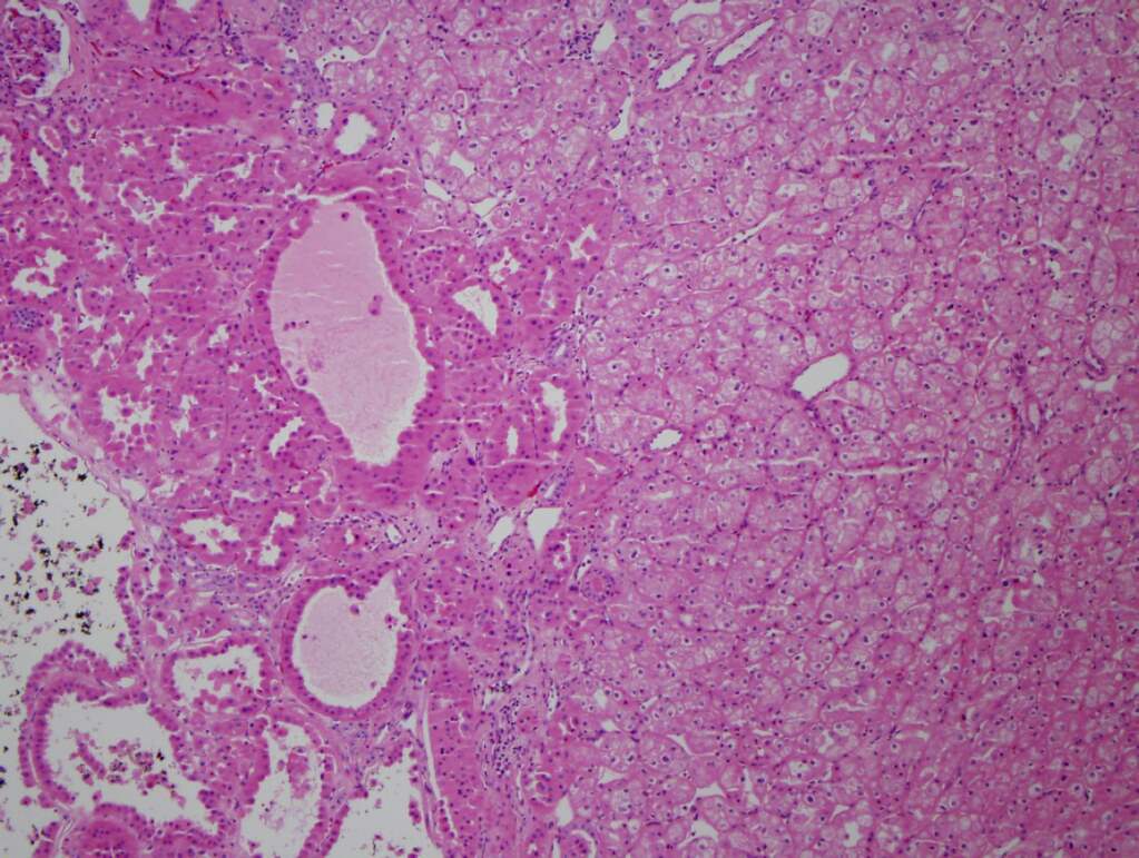
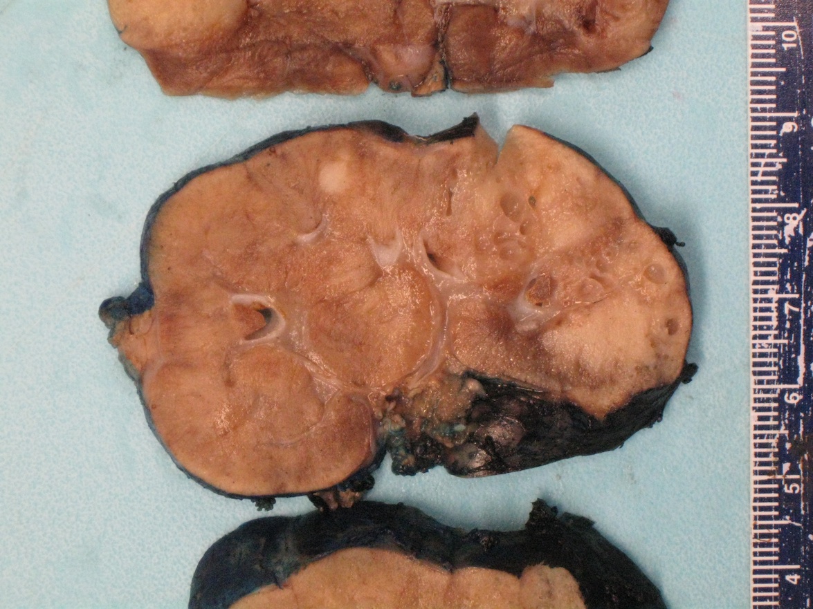
Mid kidney
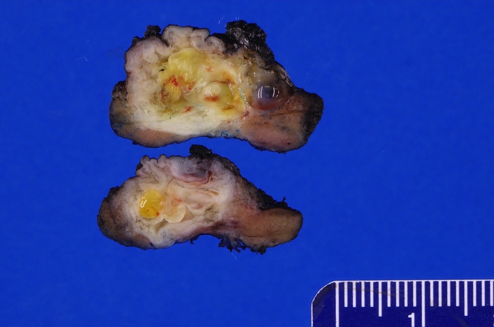
gray-white mass
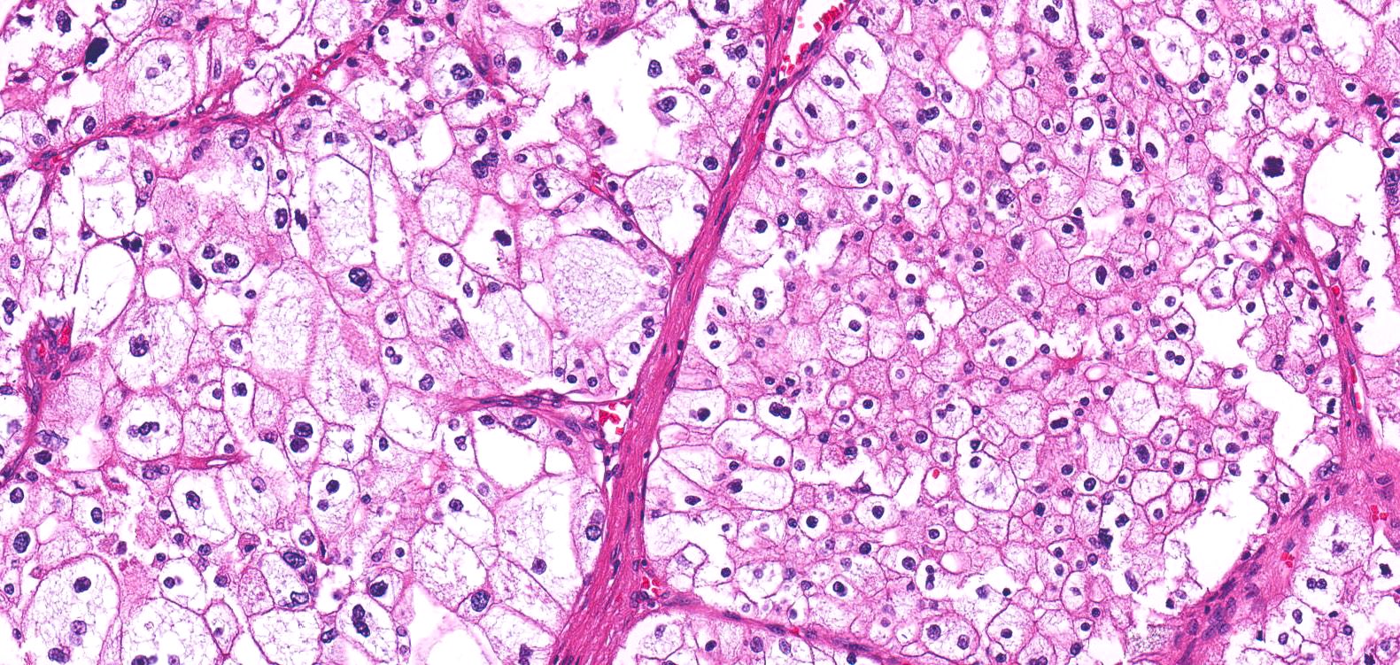
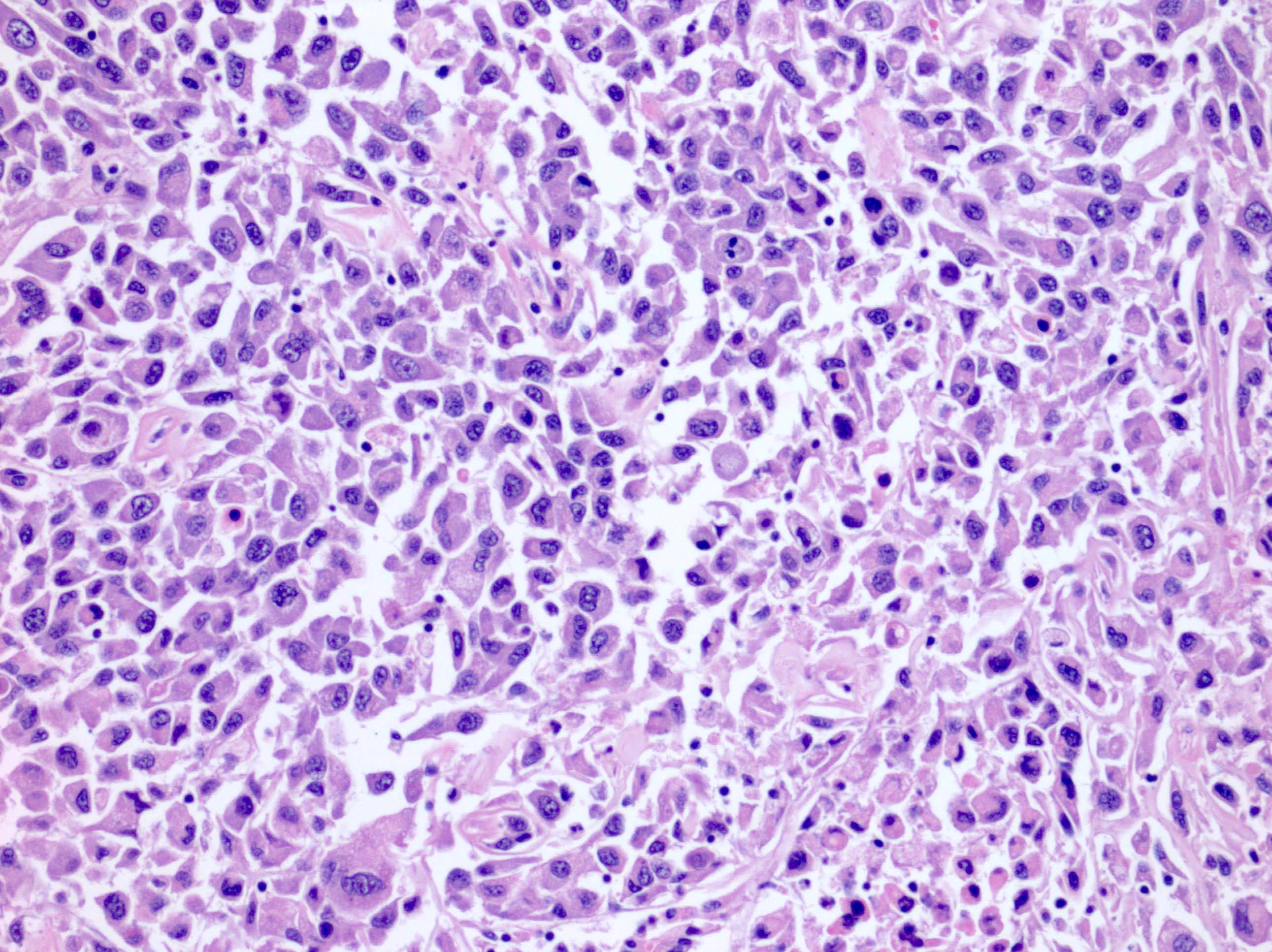
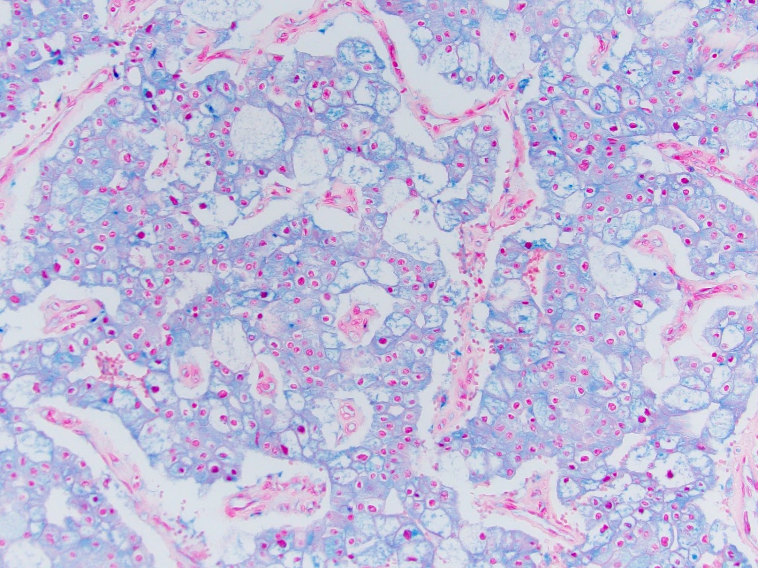
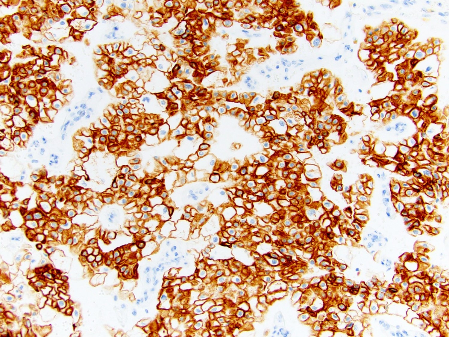
Contributed by Maria Tretiakova, M.D., Ph.D.
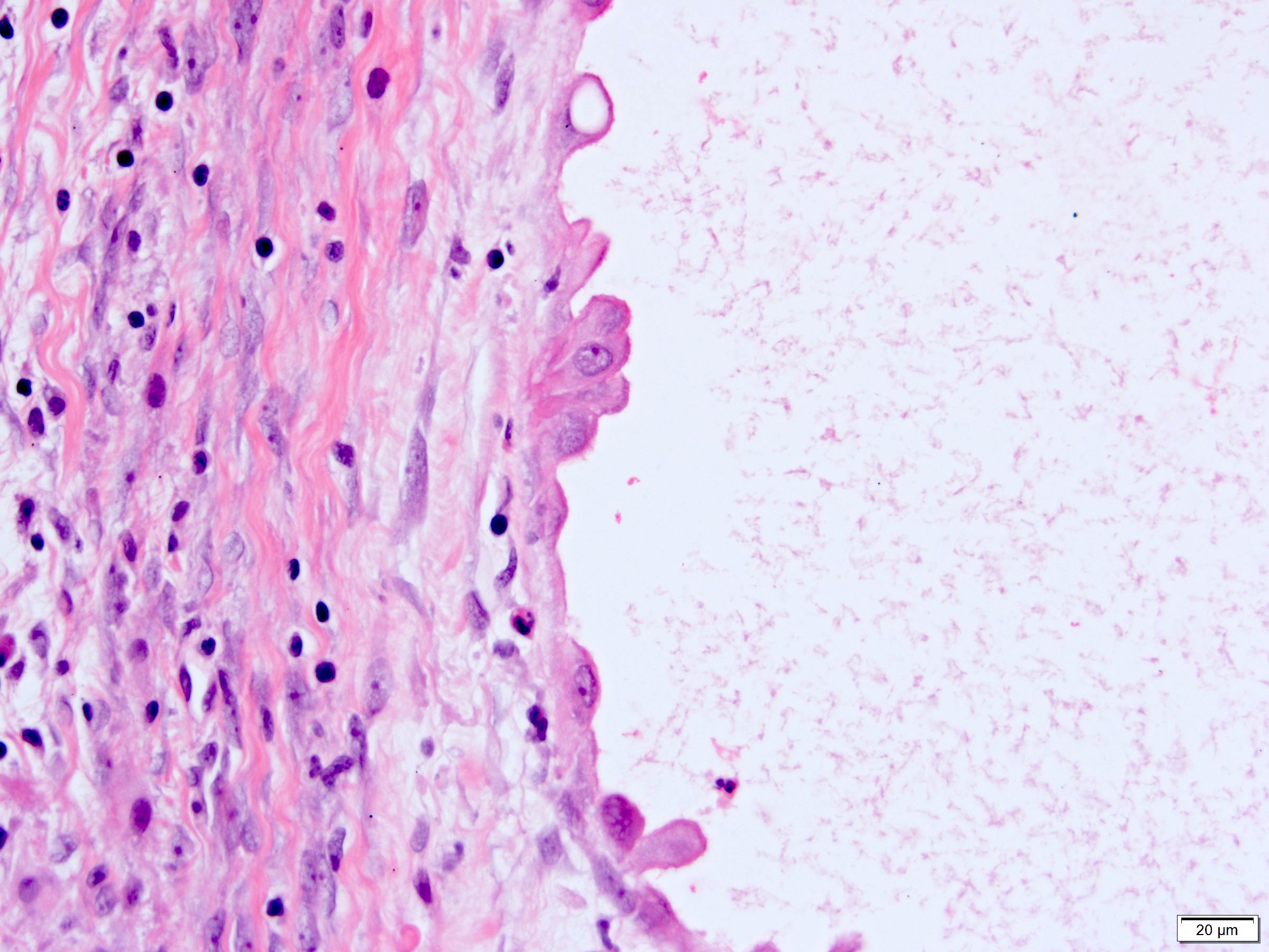
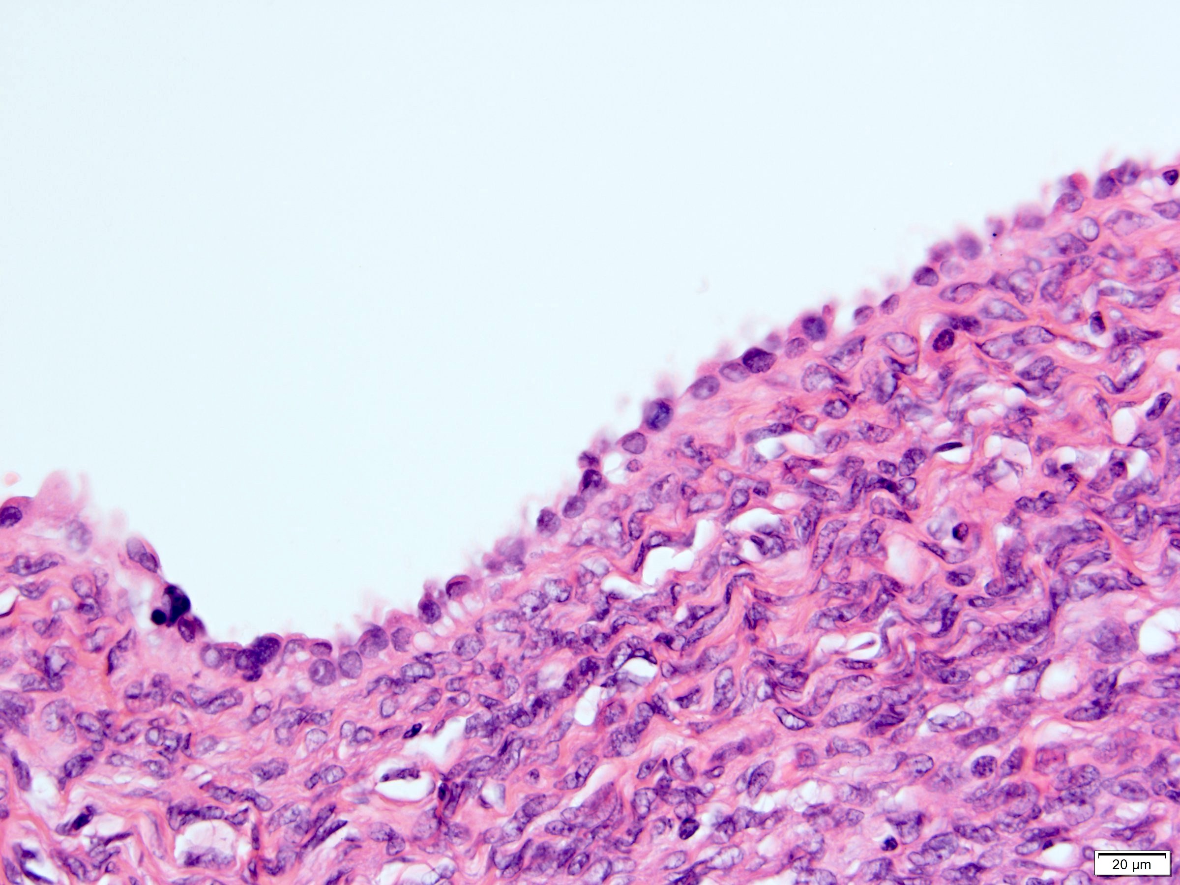
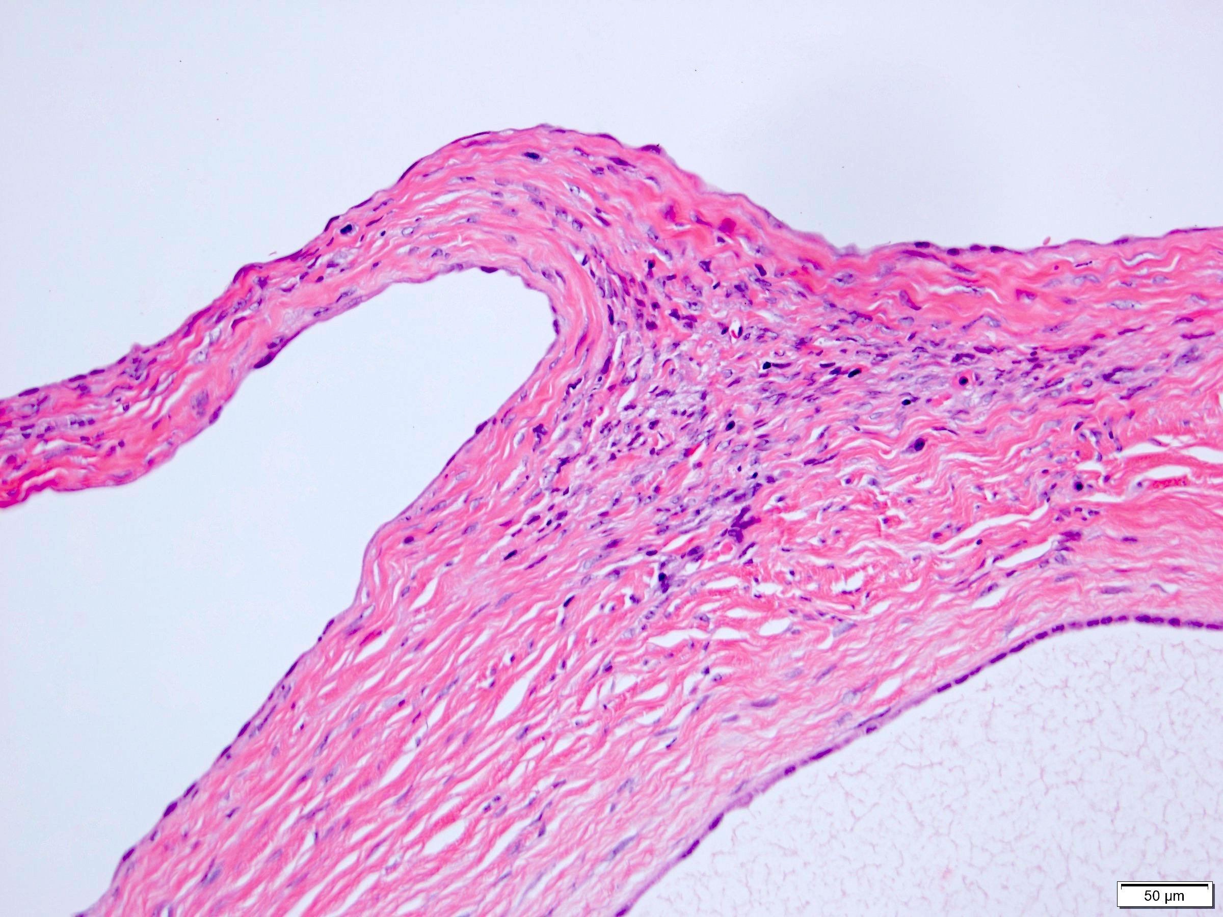
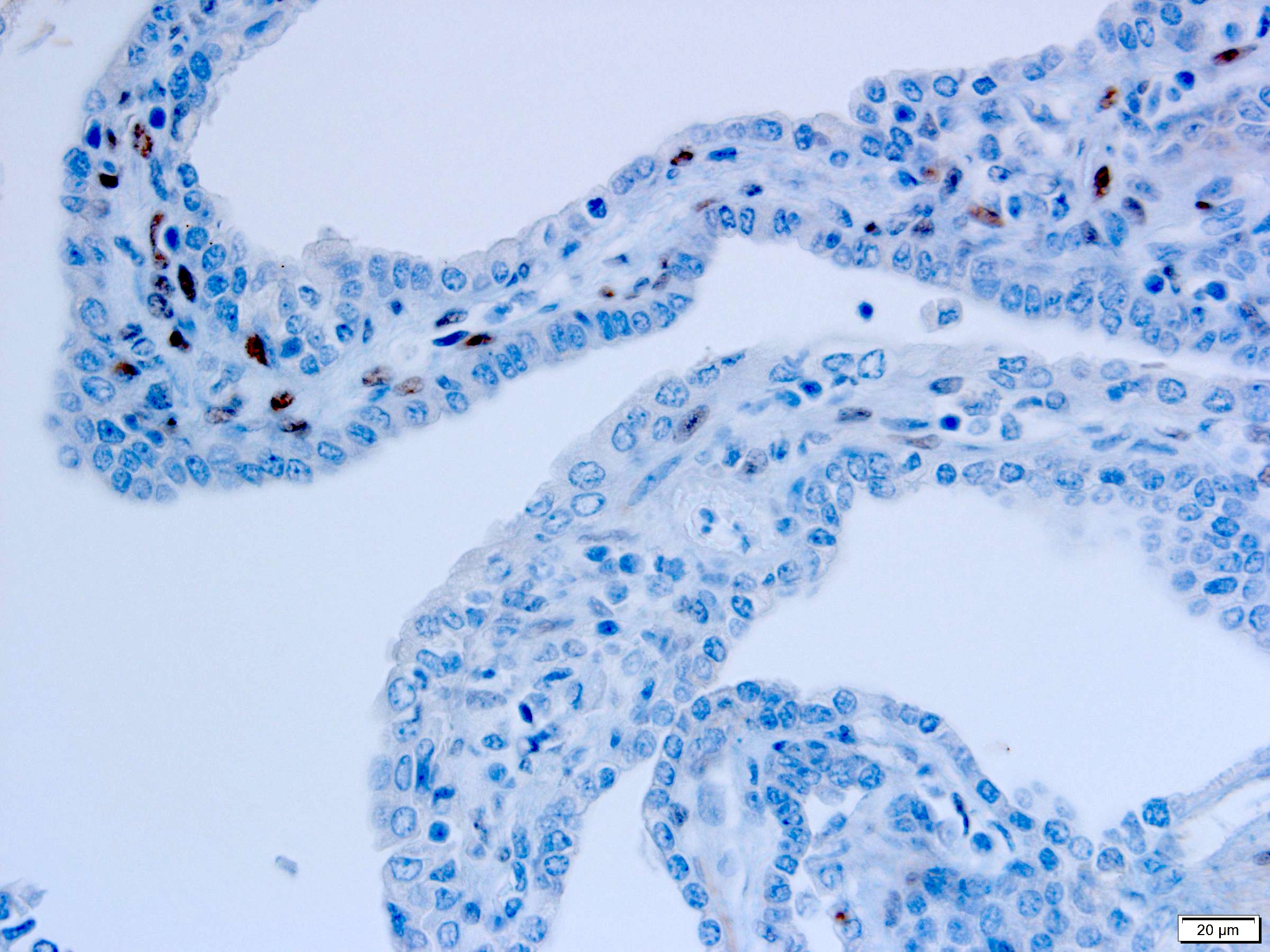
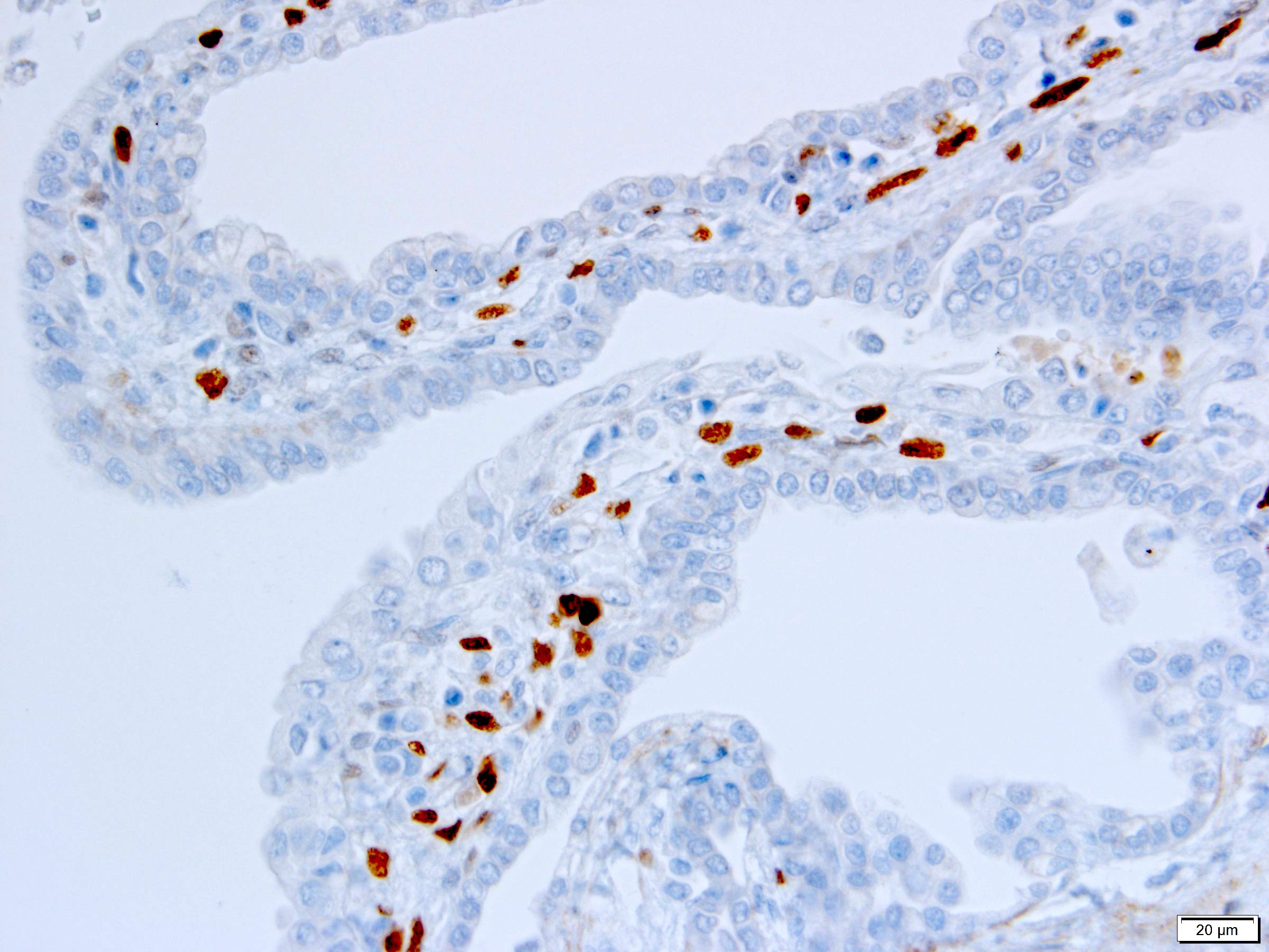
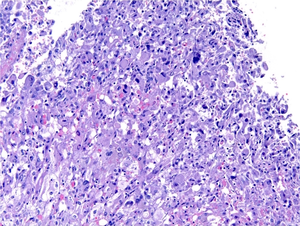
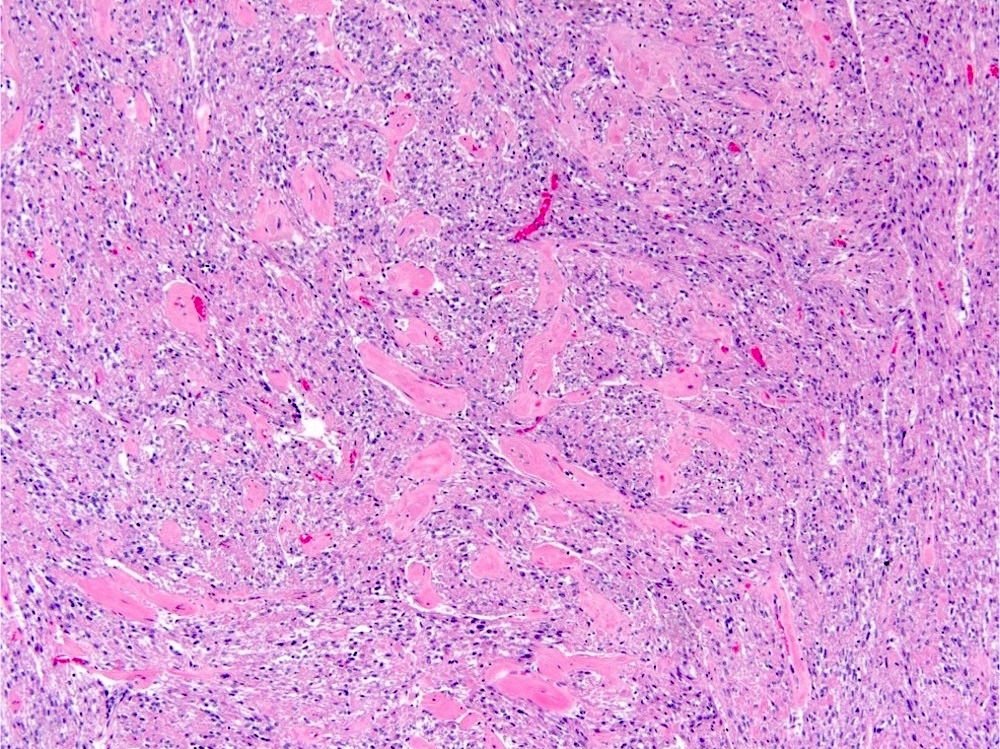
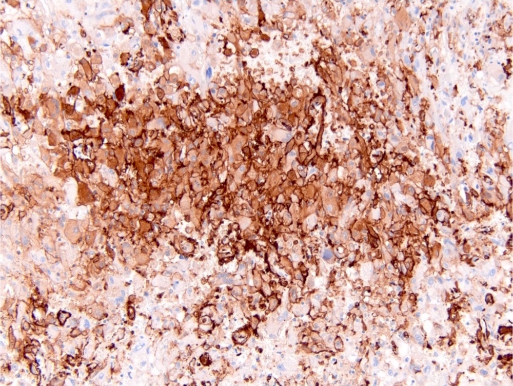
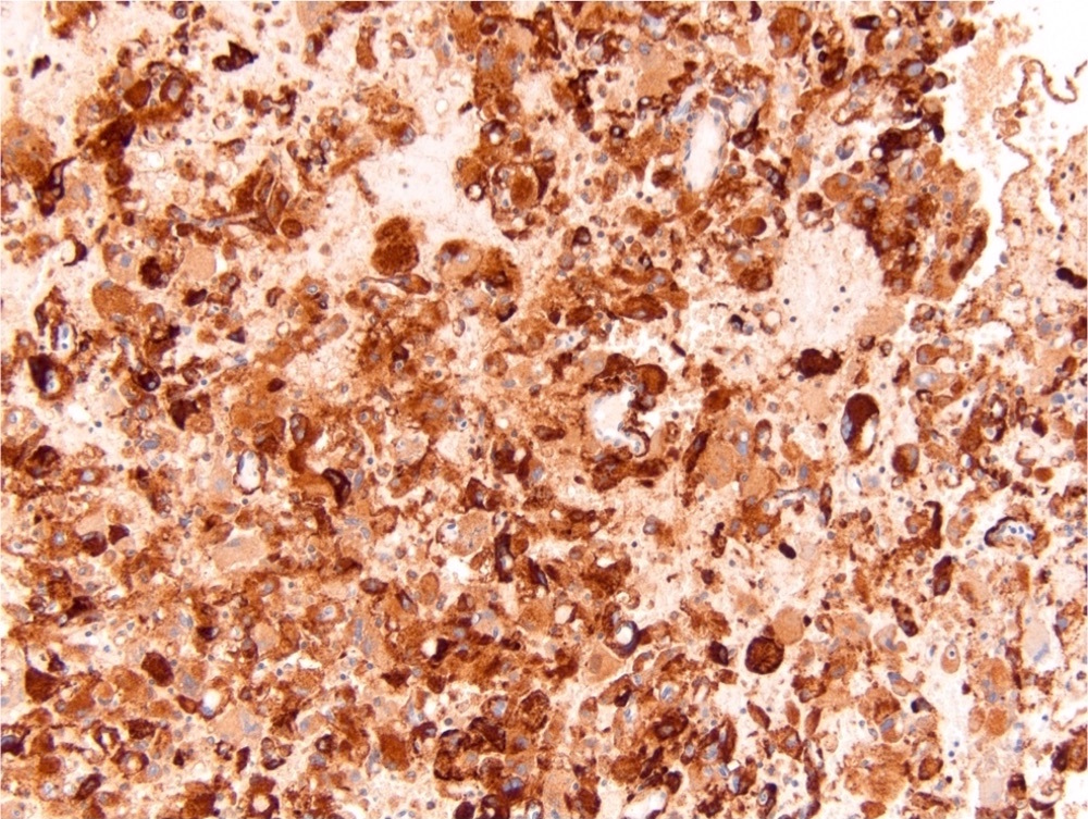
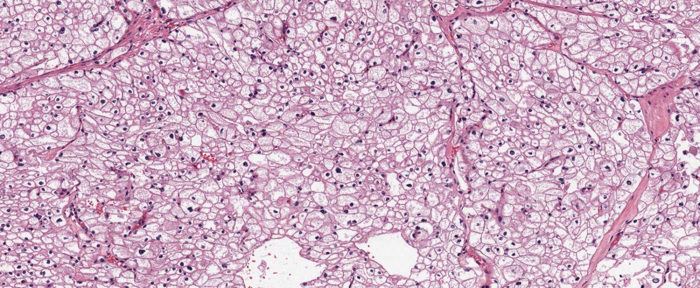
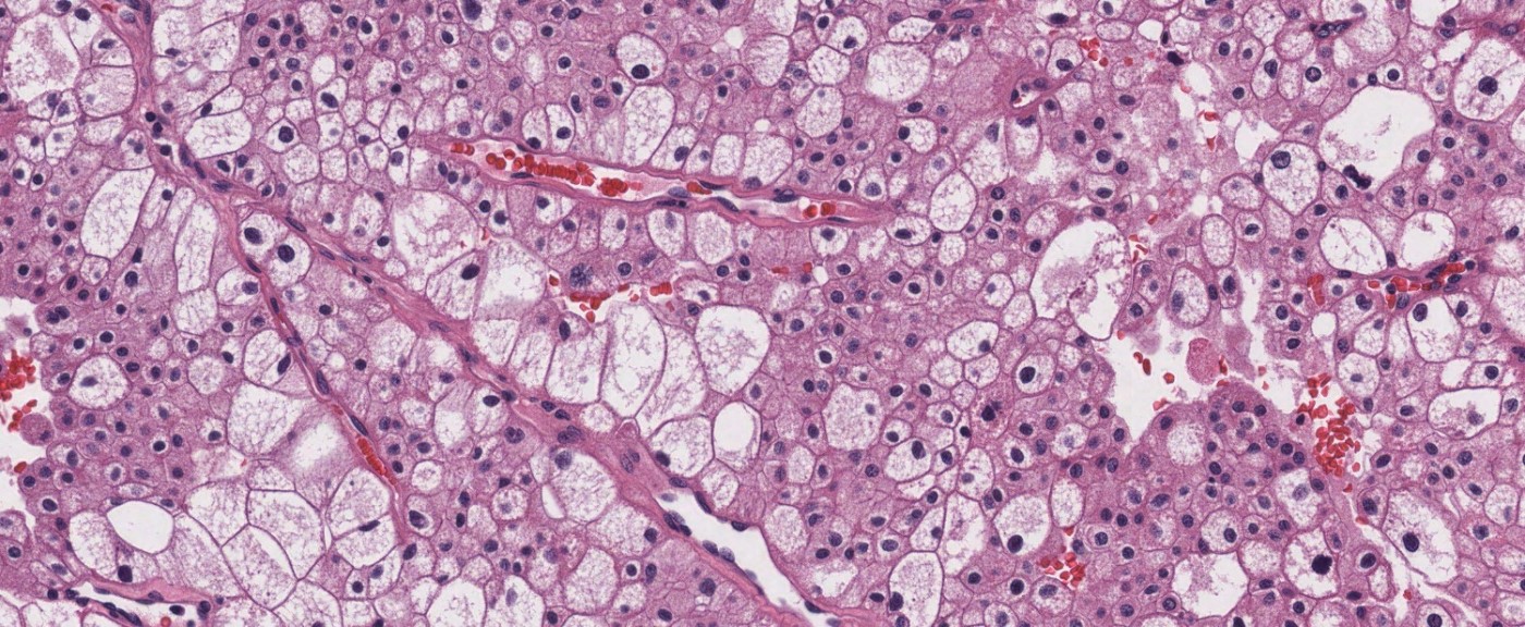
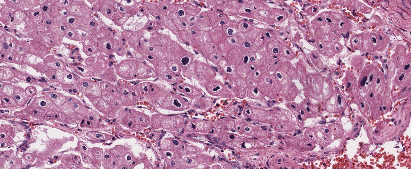
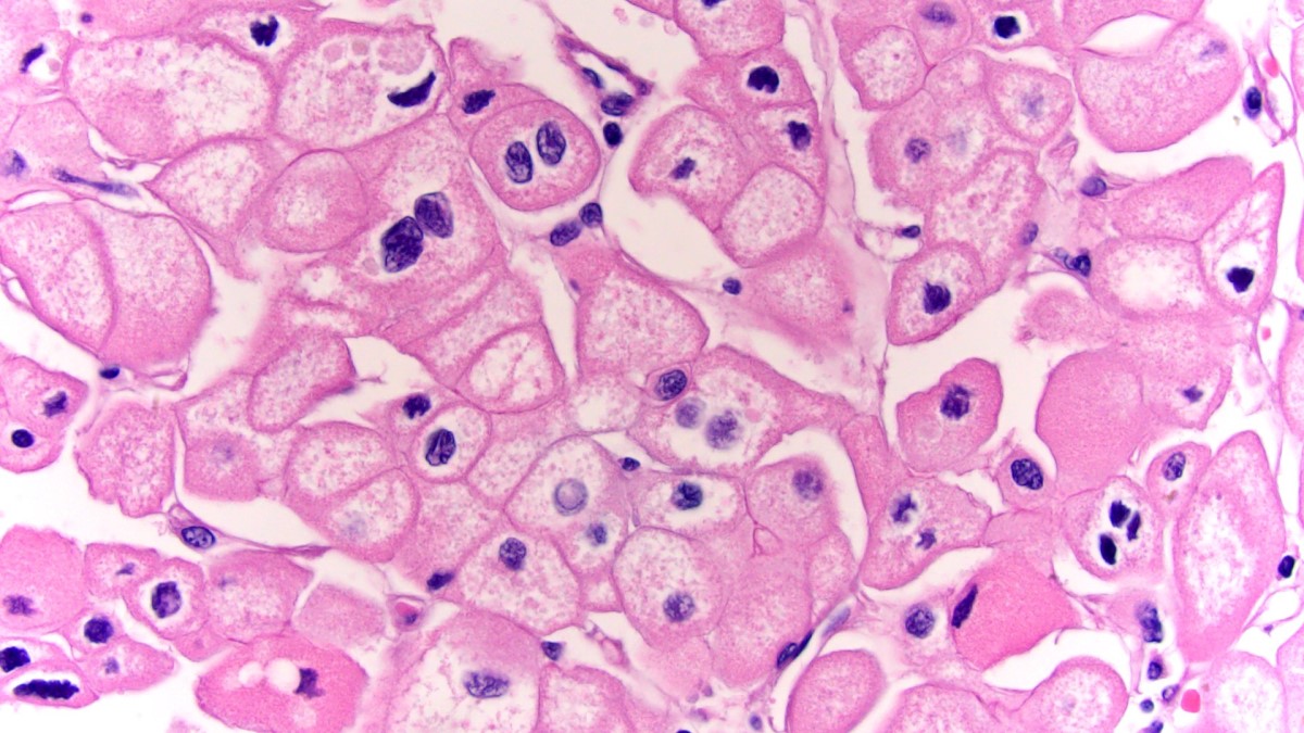
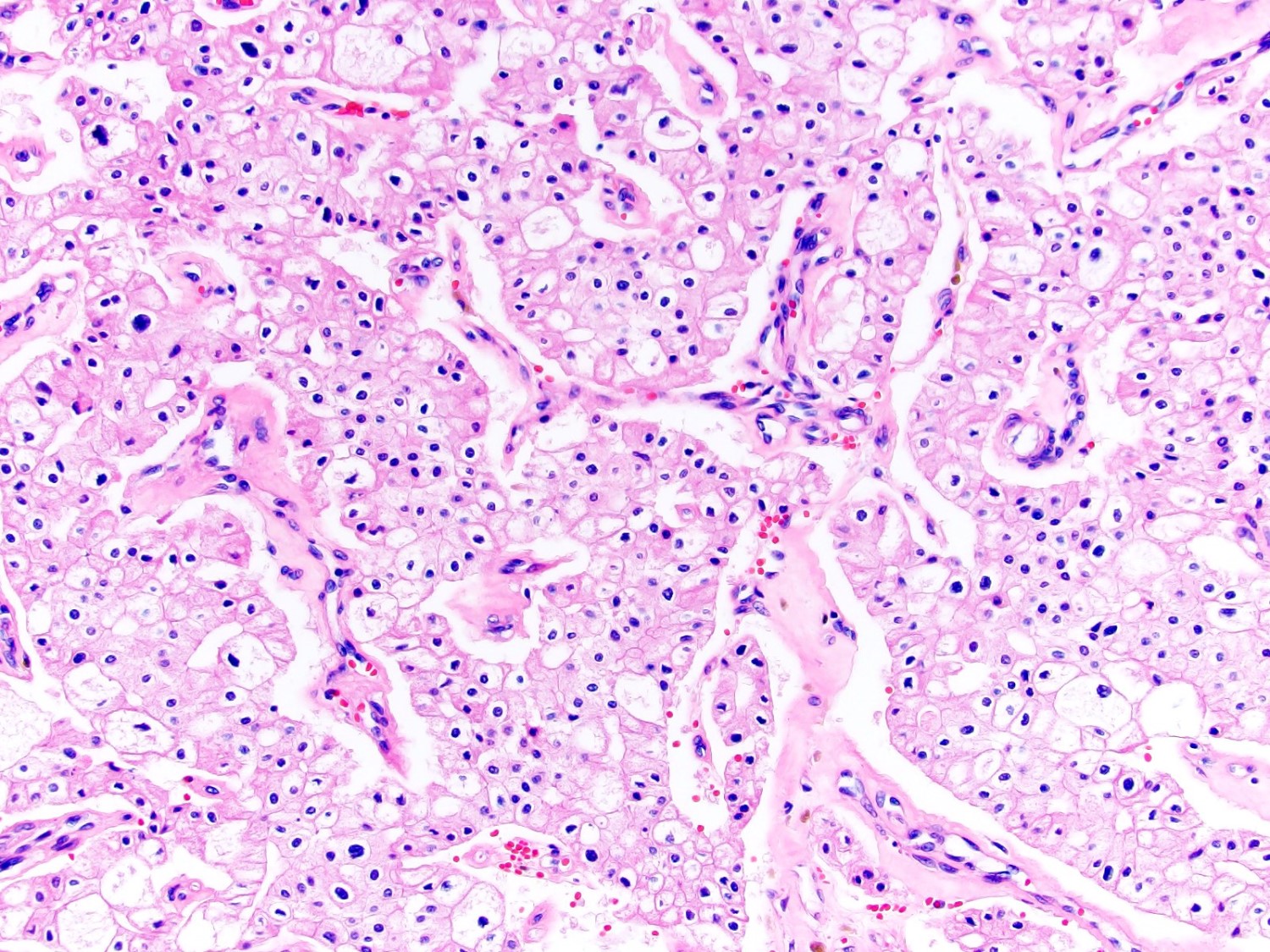
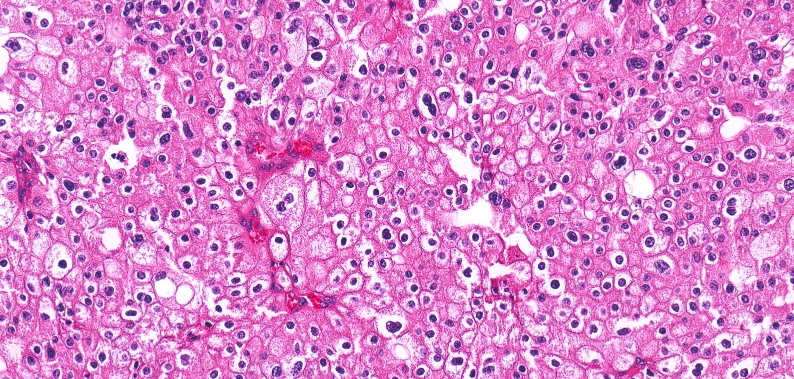
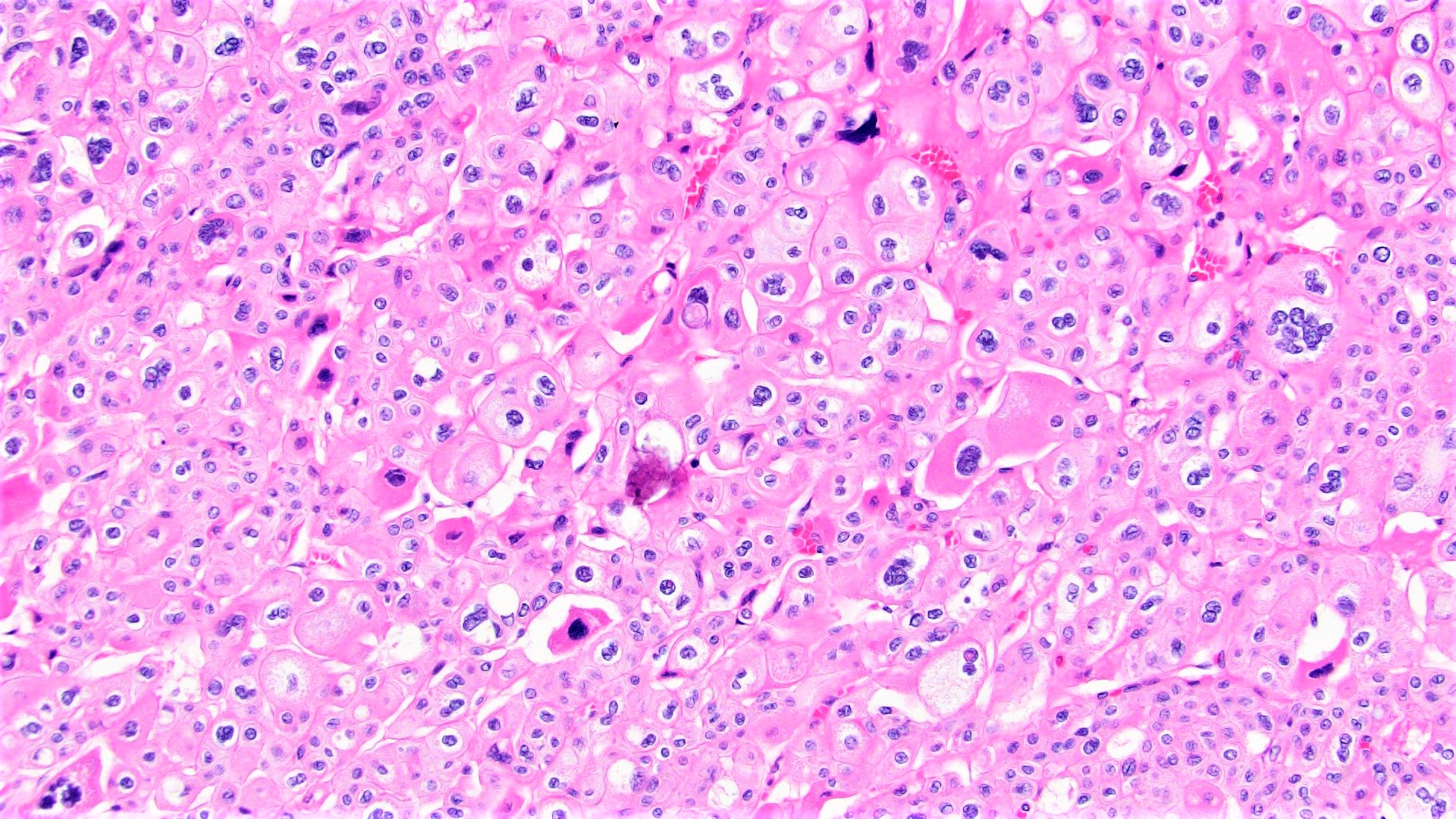
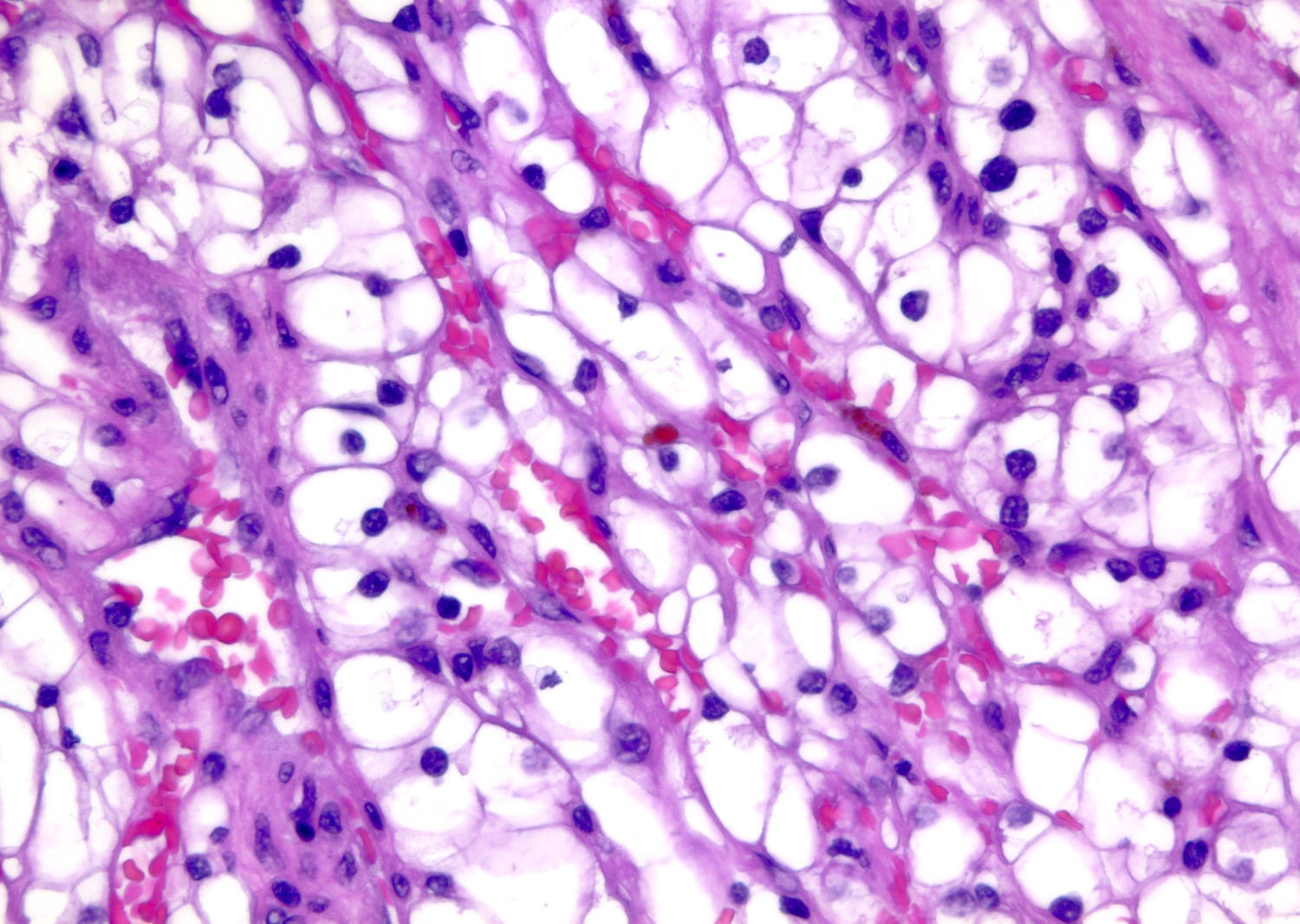
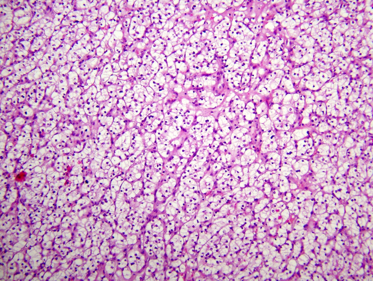
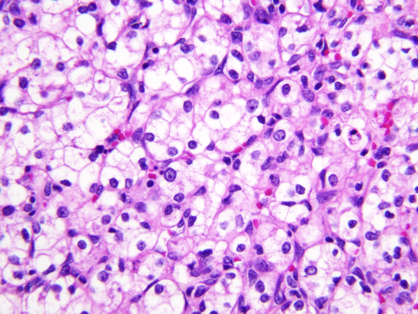
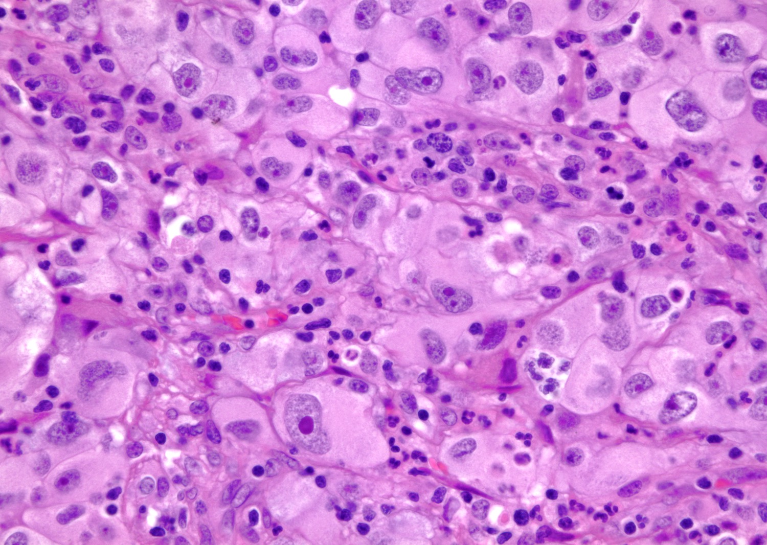
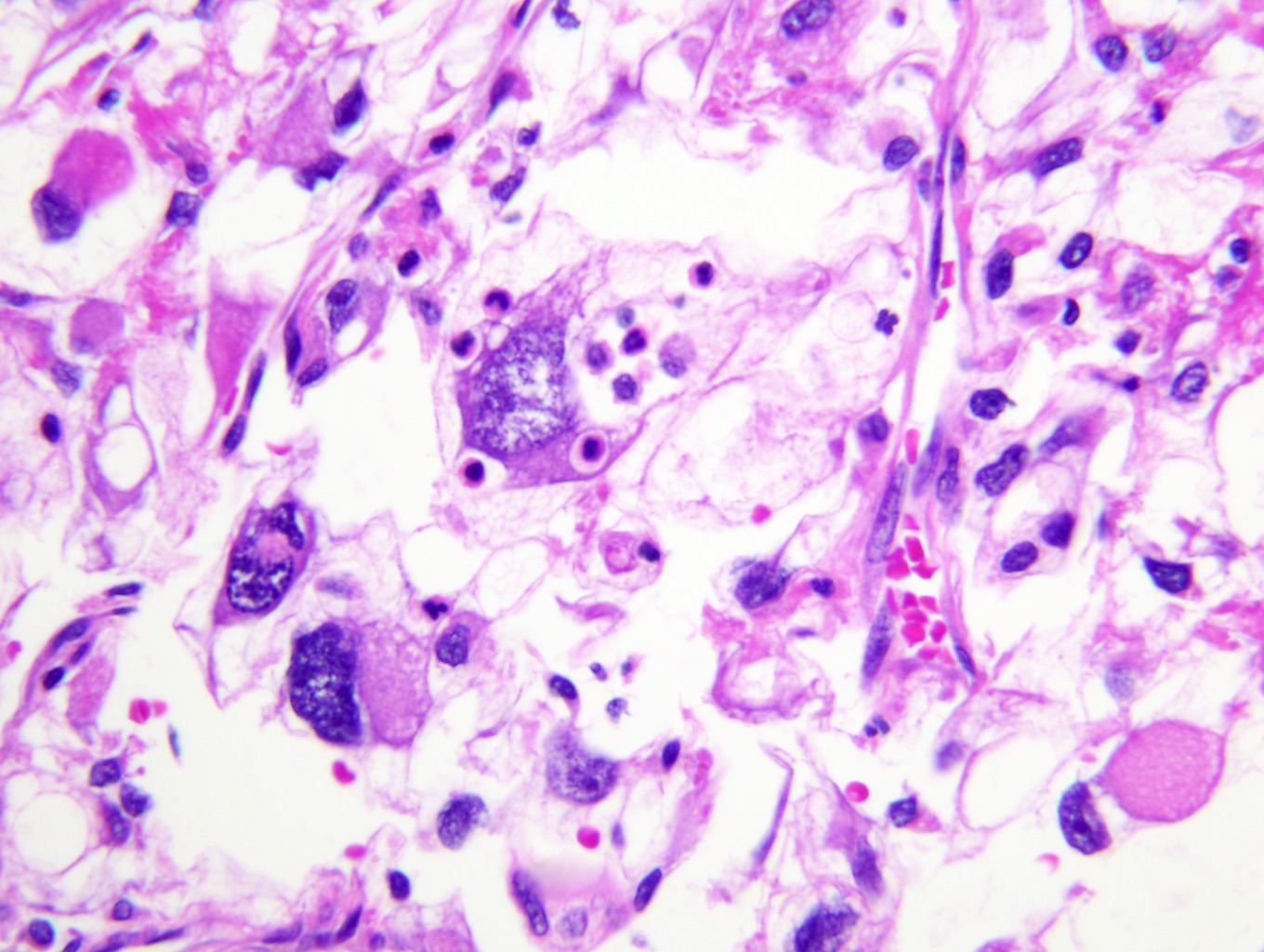
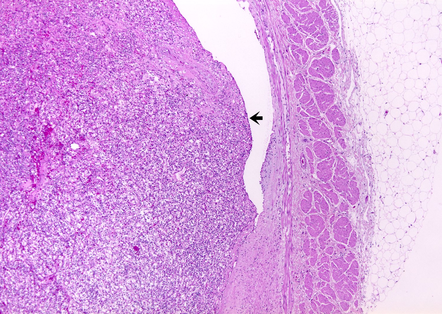
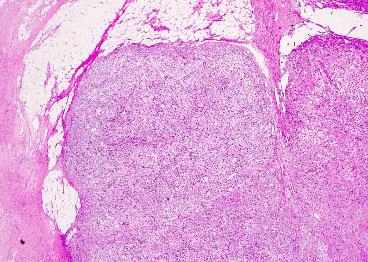
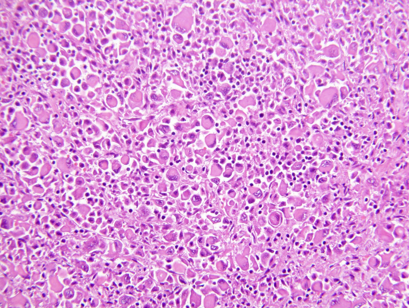
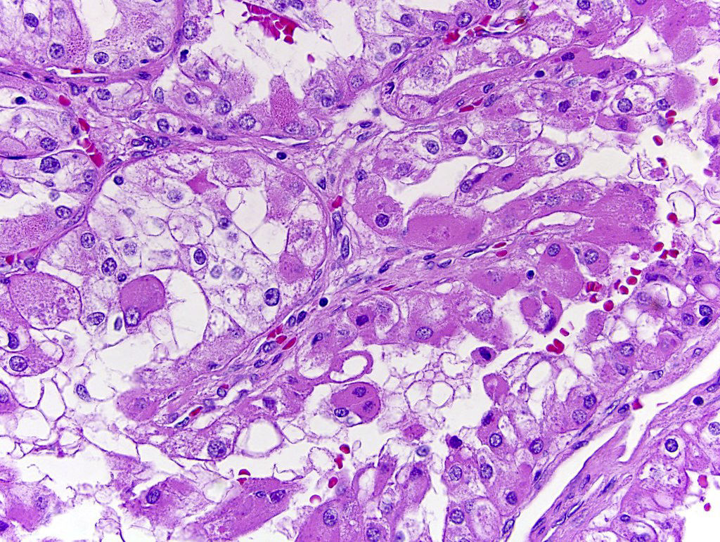
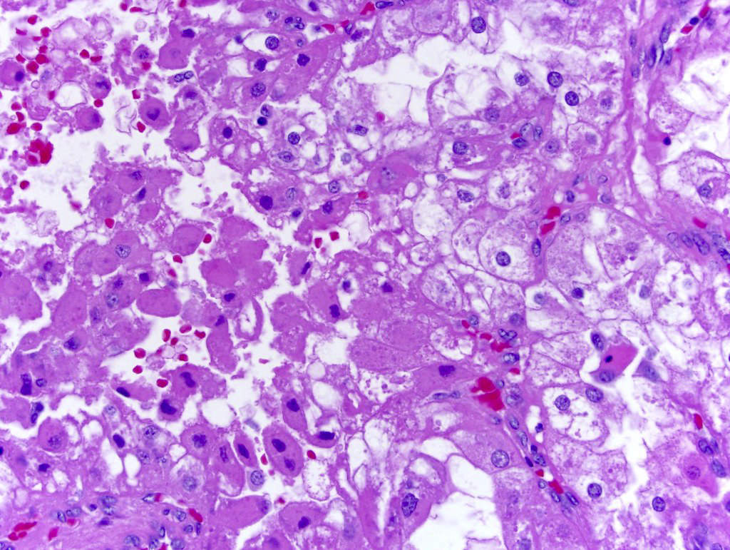
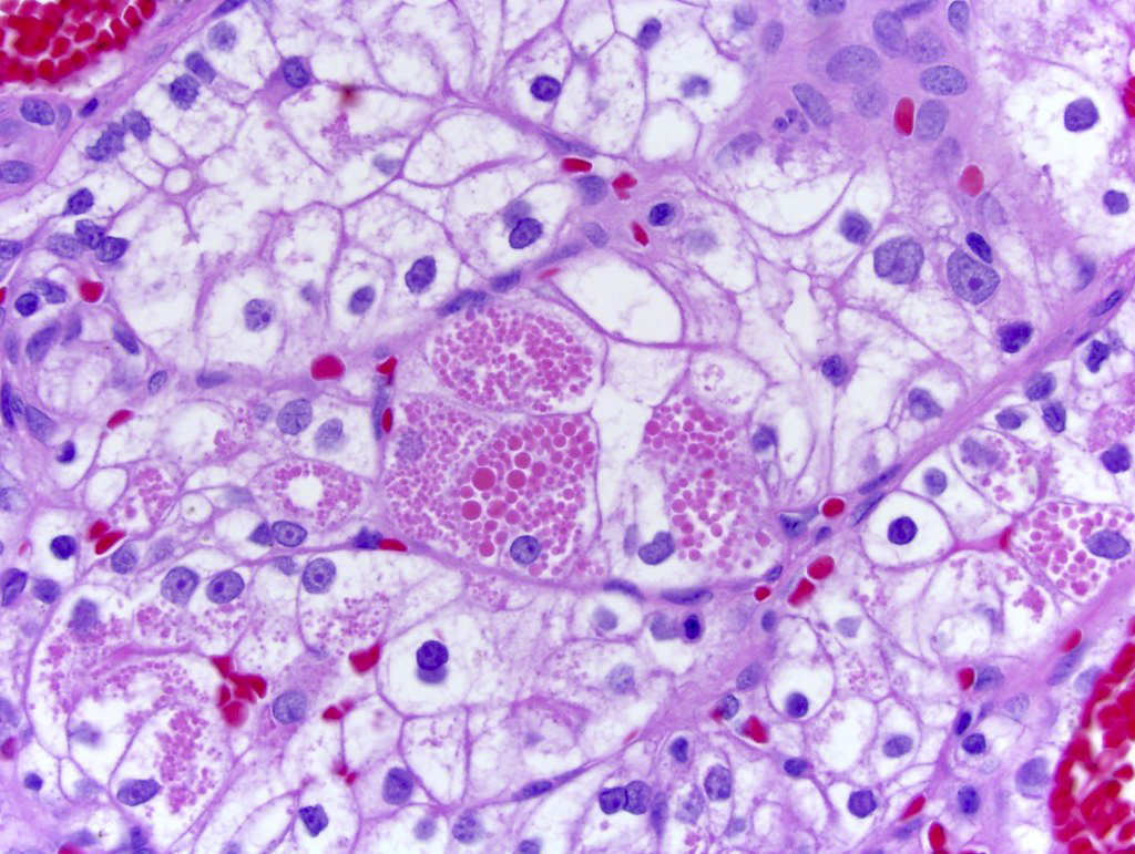
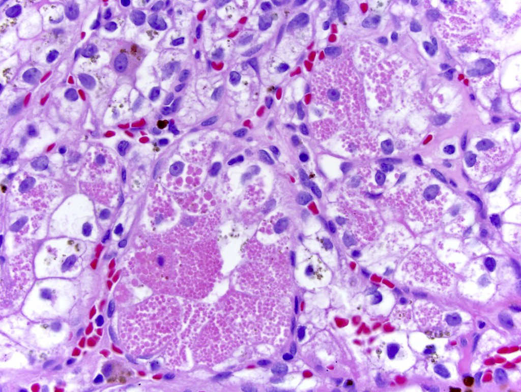
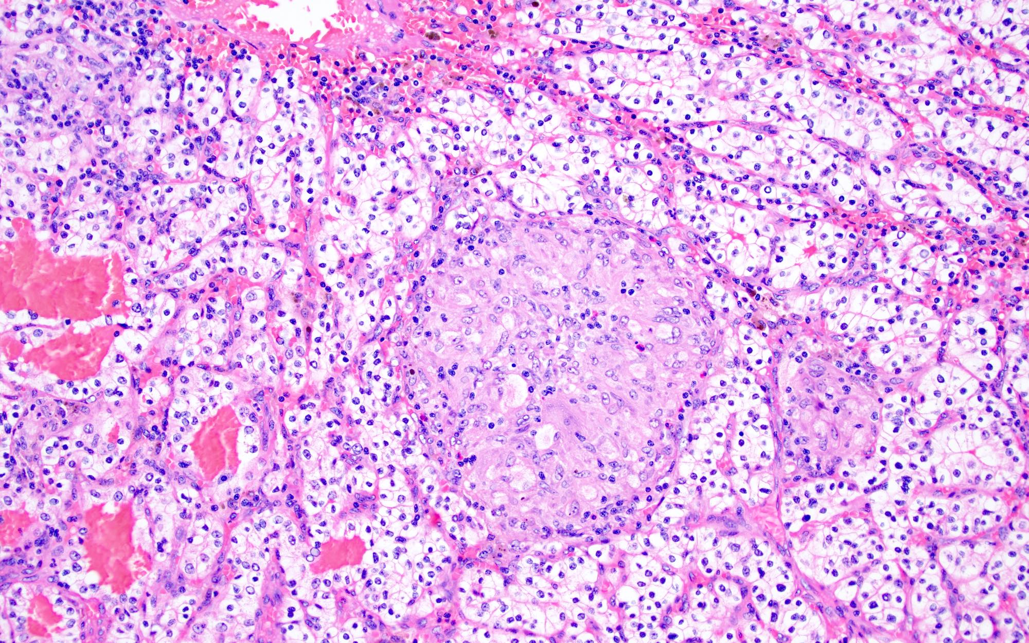
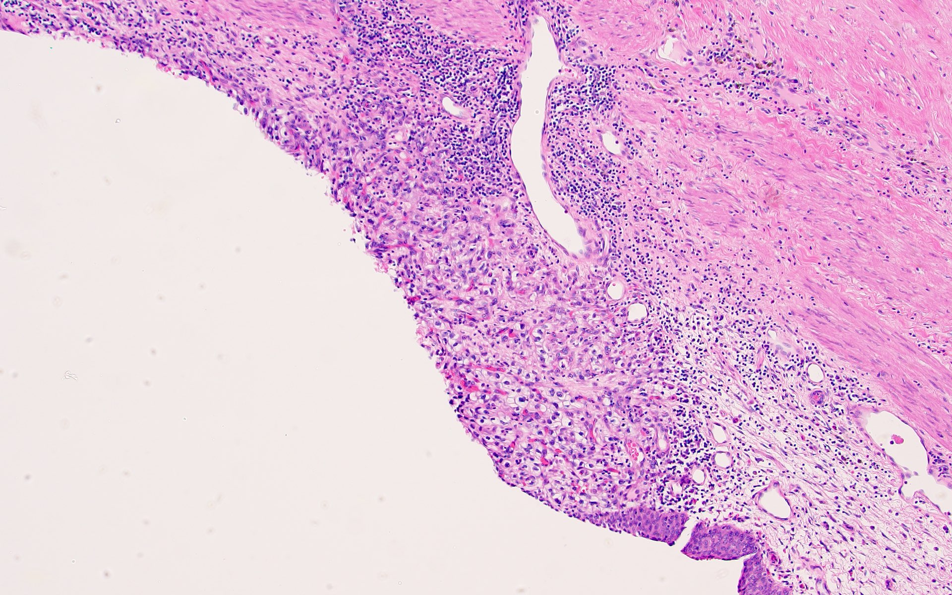
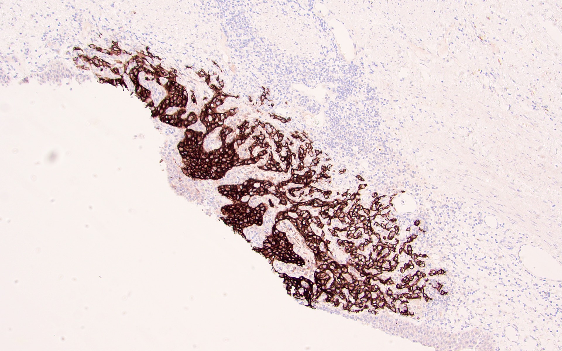
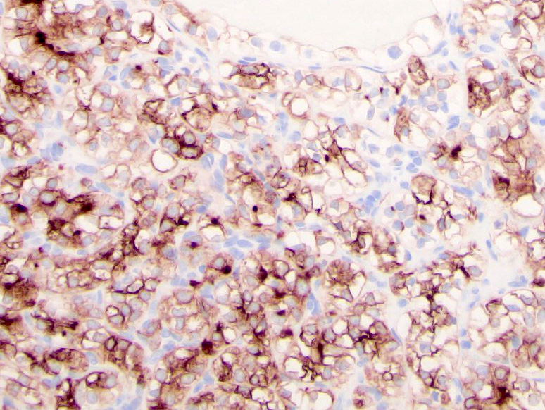
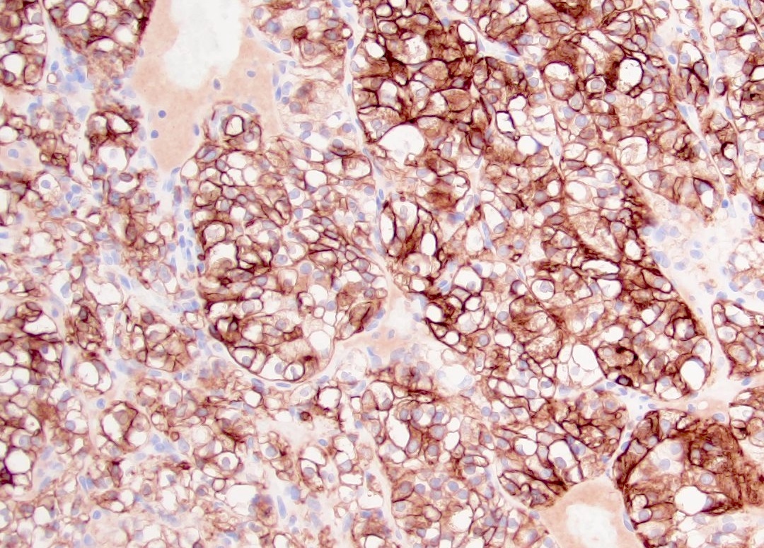
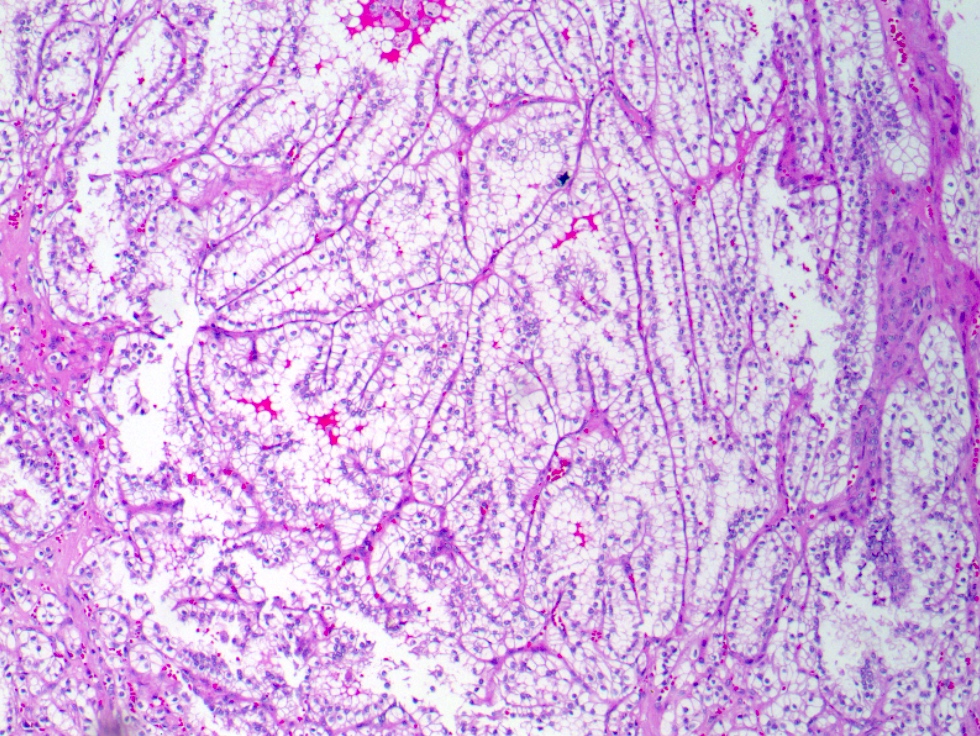
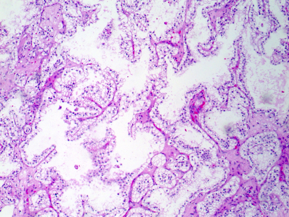
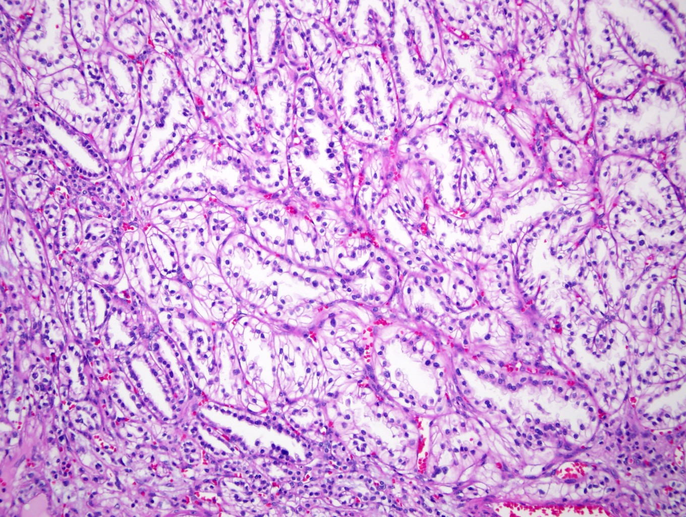
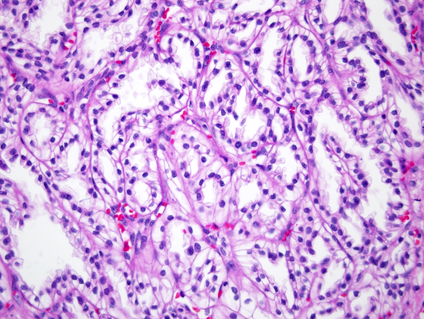
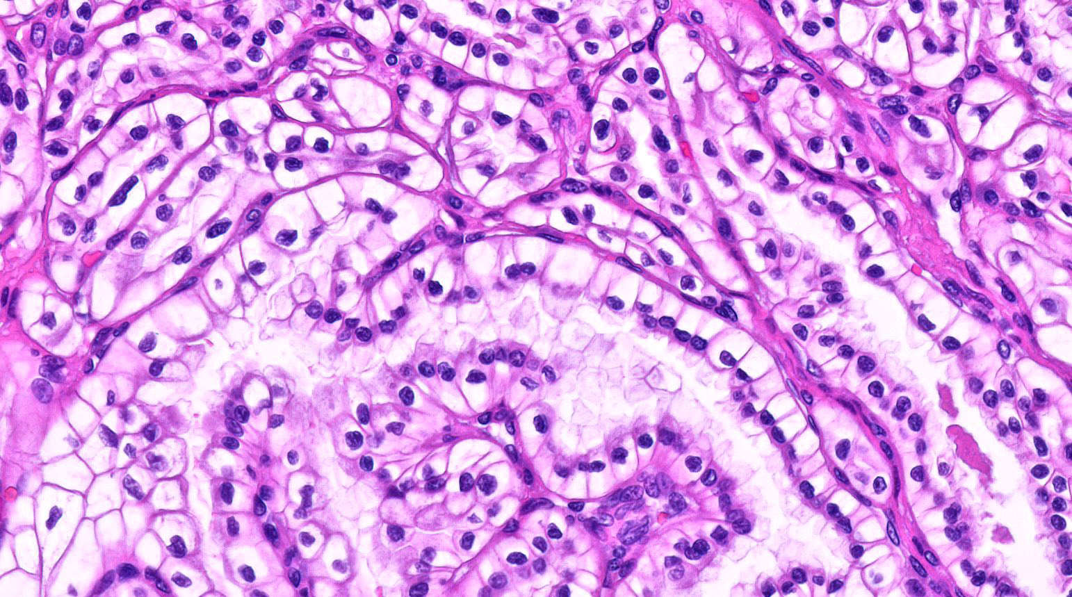
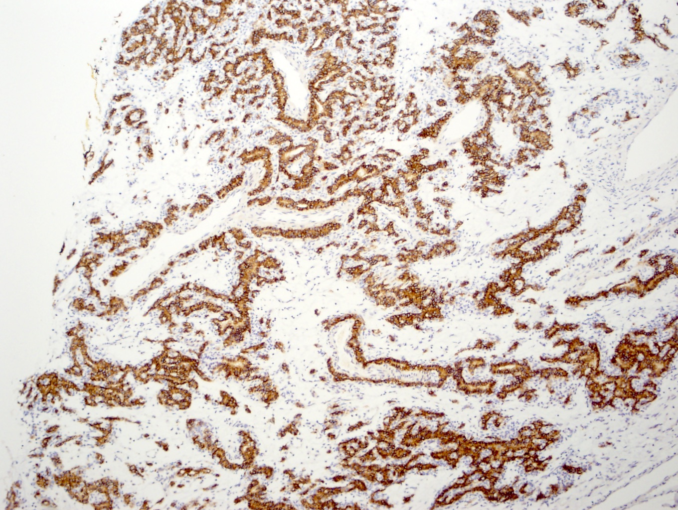
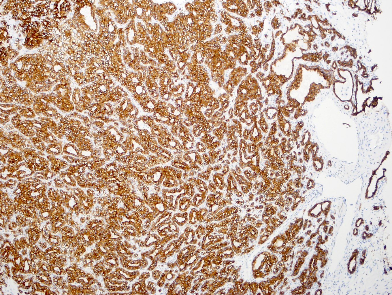
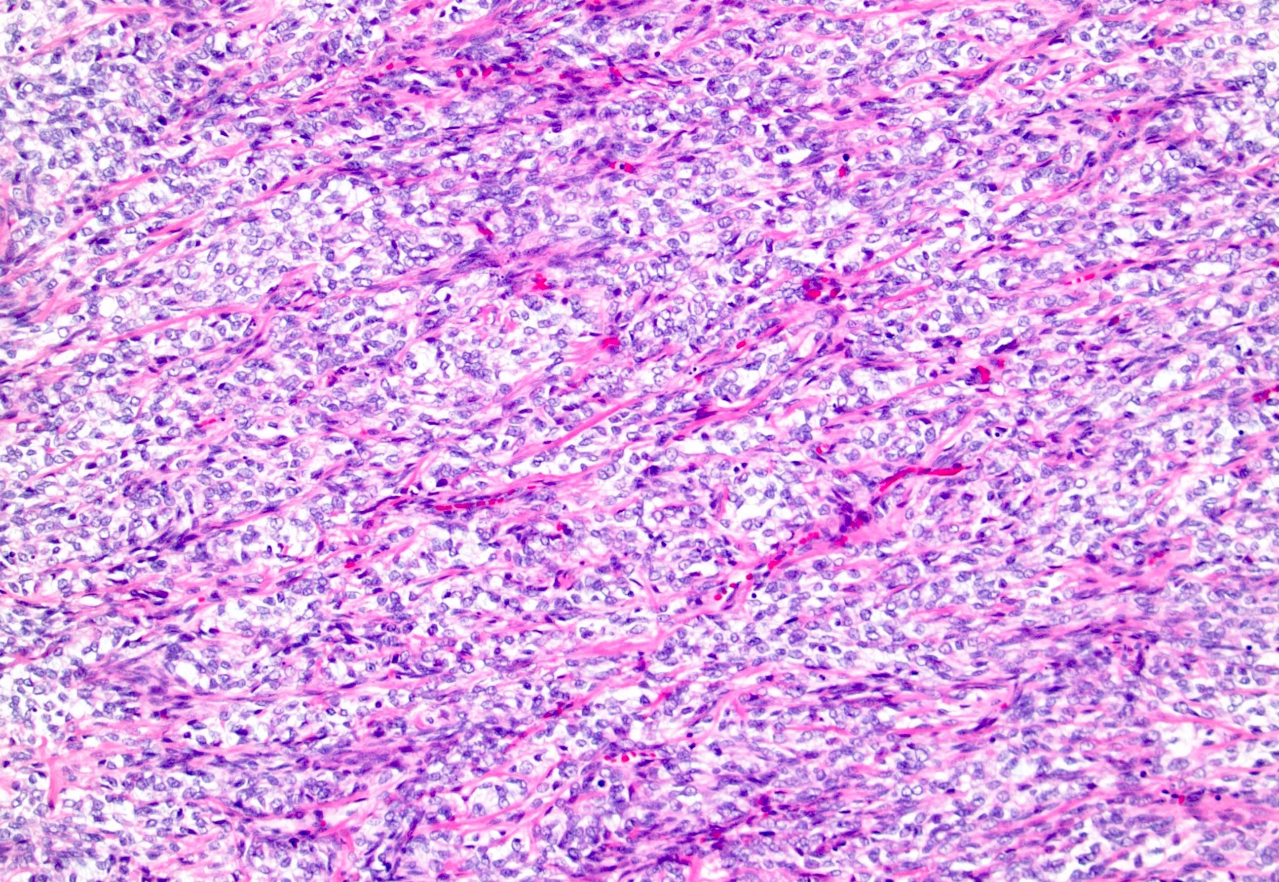
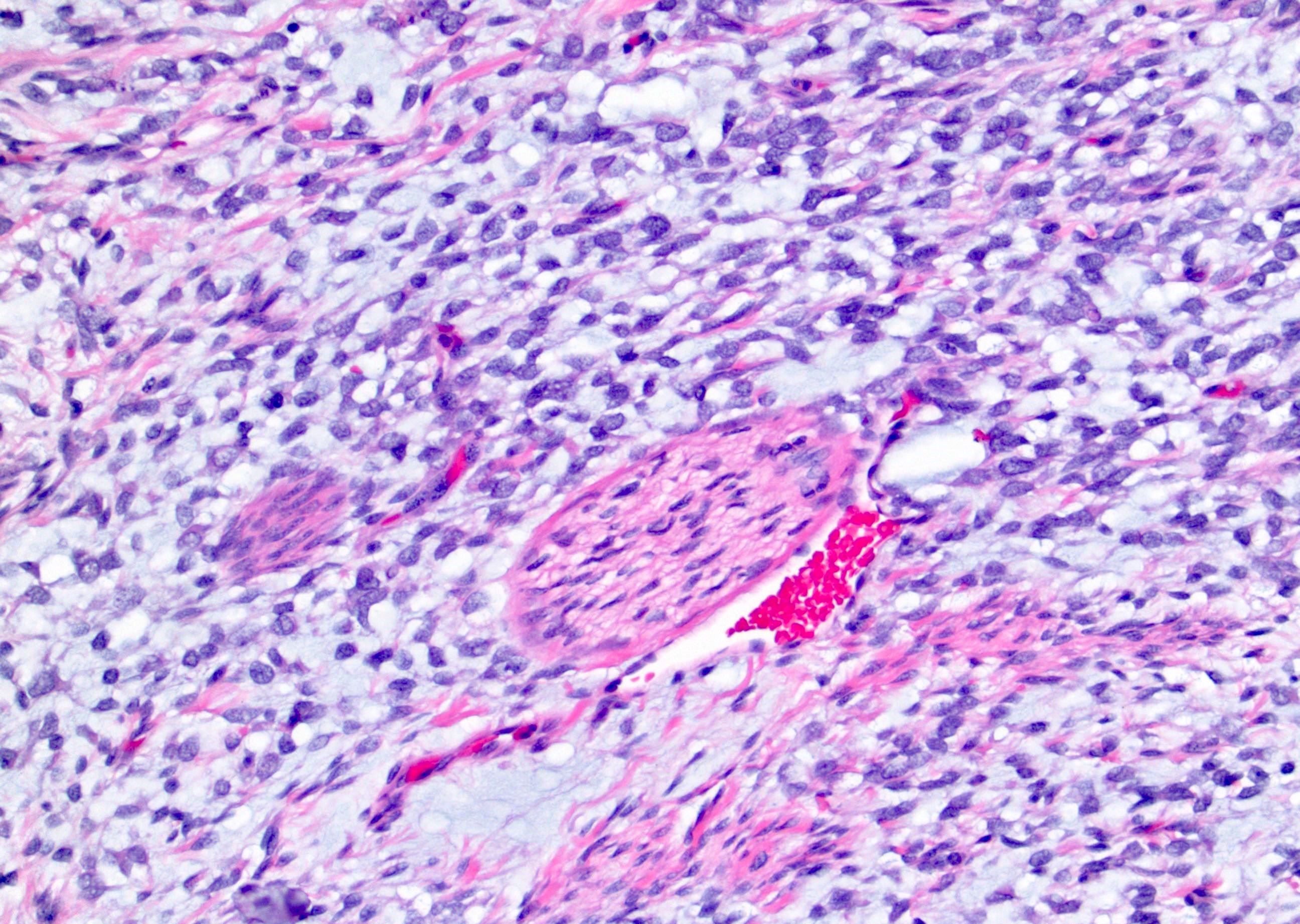
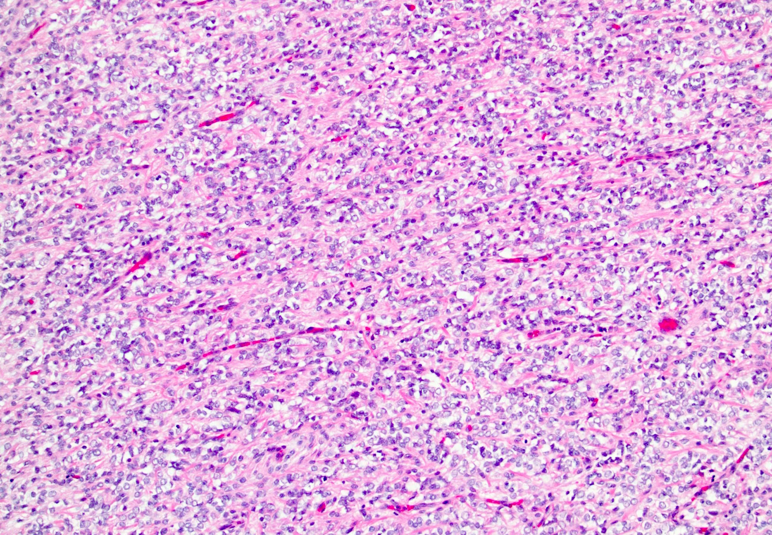
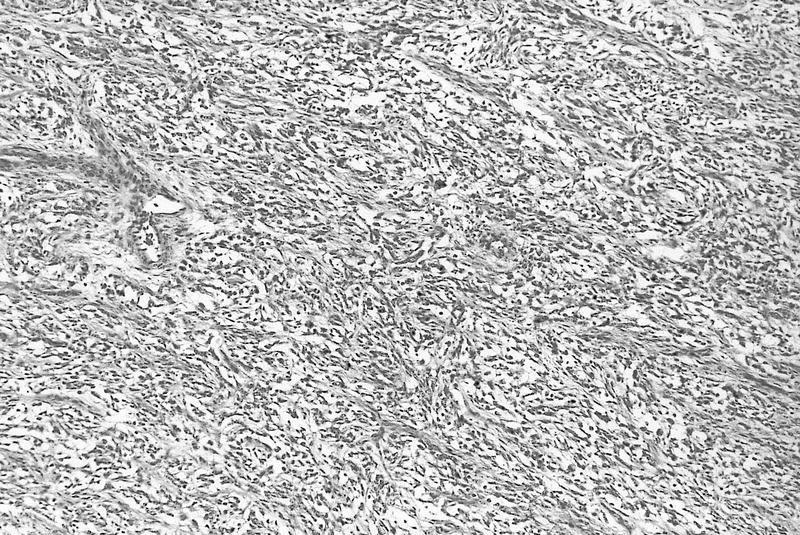
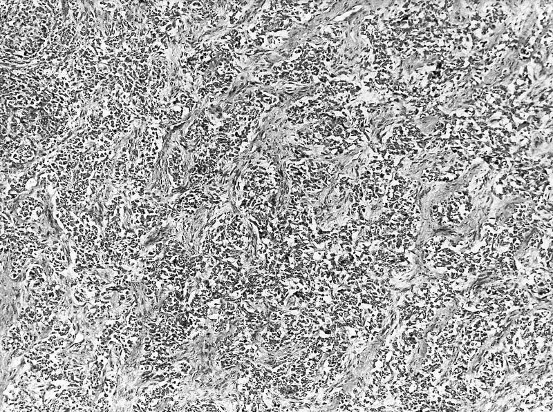
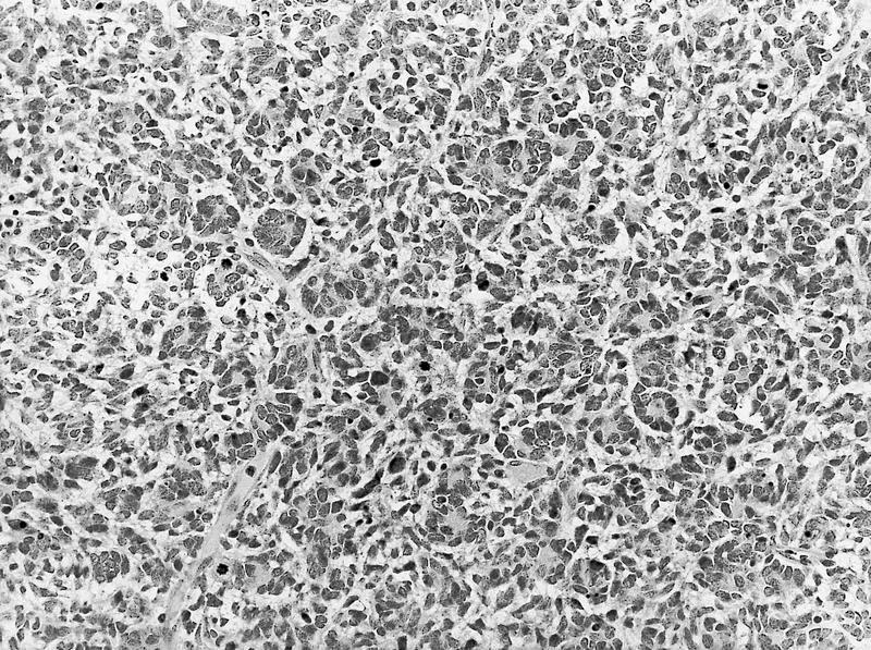
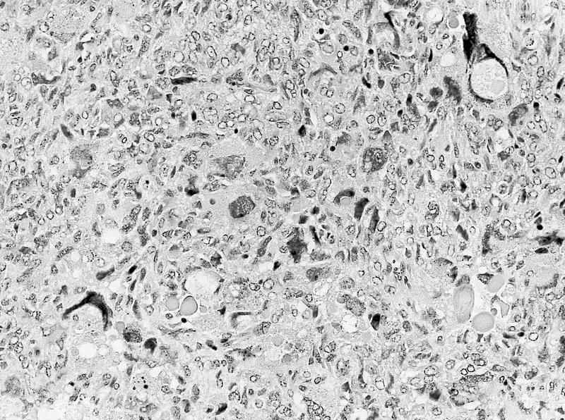
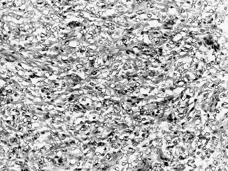
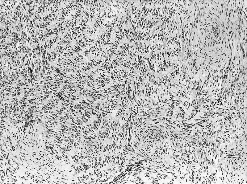
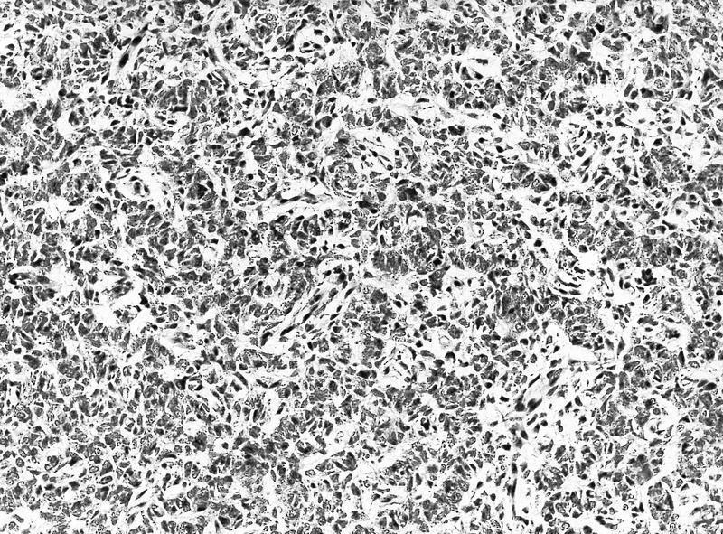
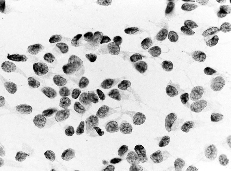
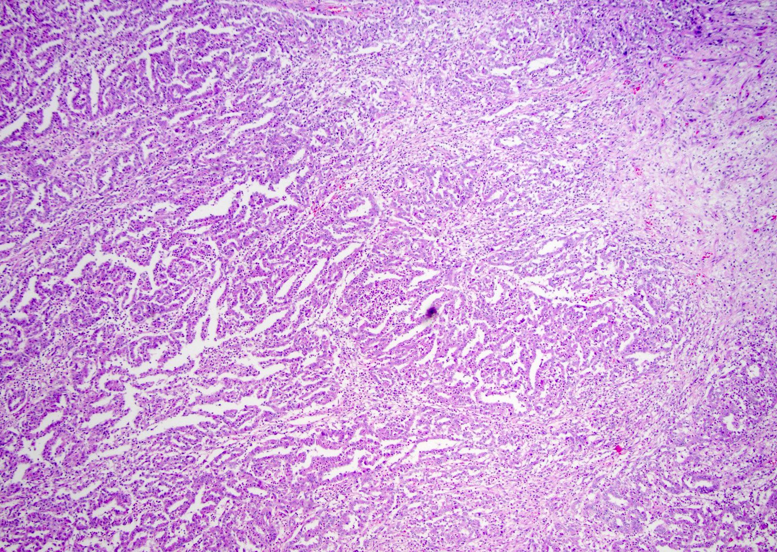
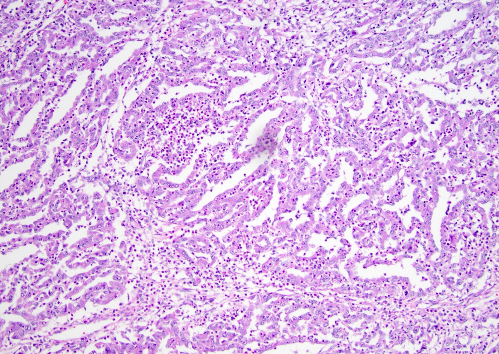
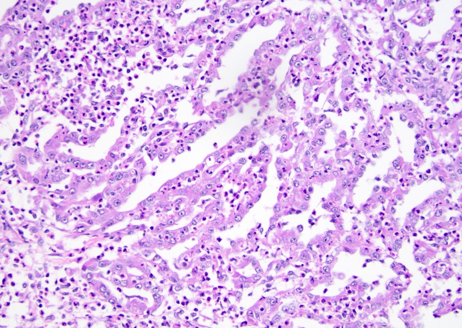
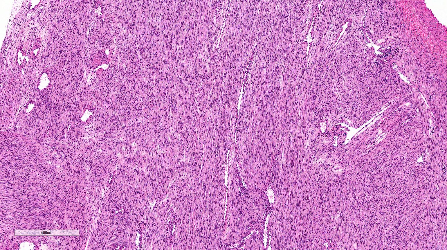
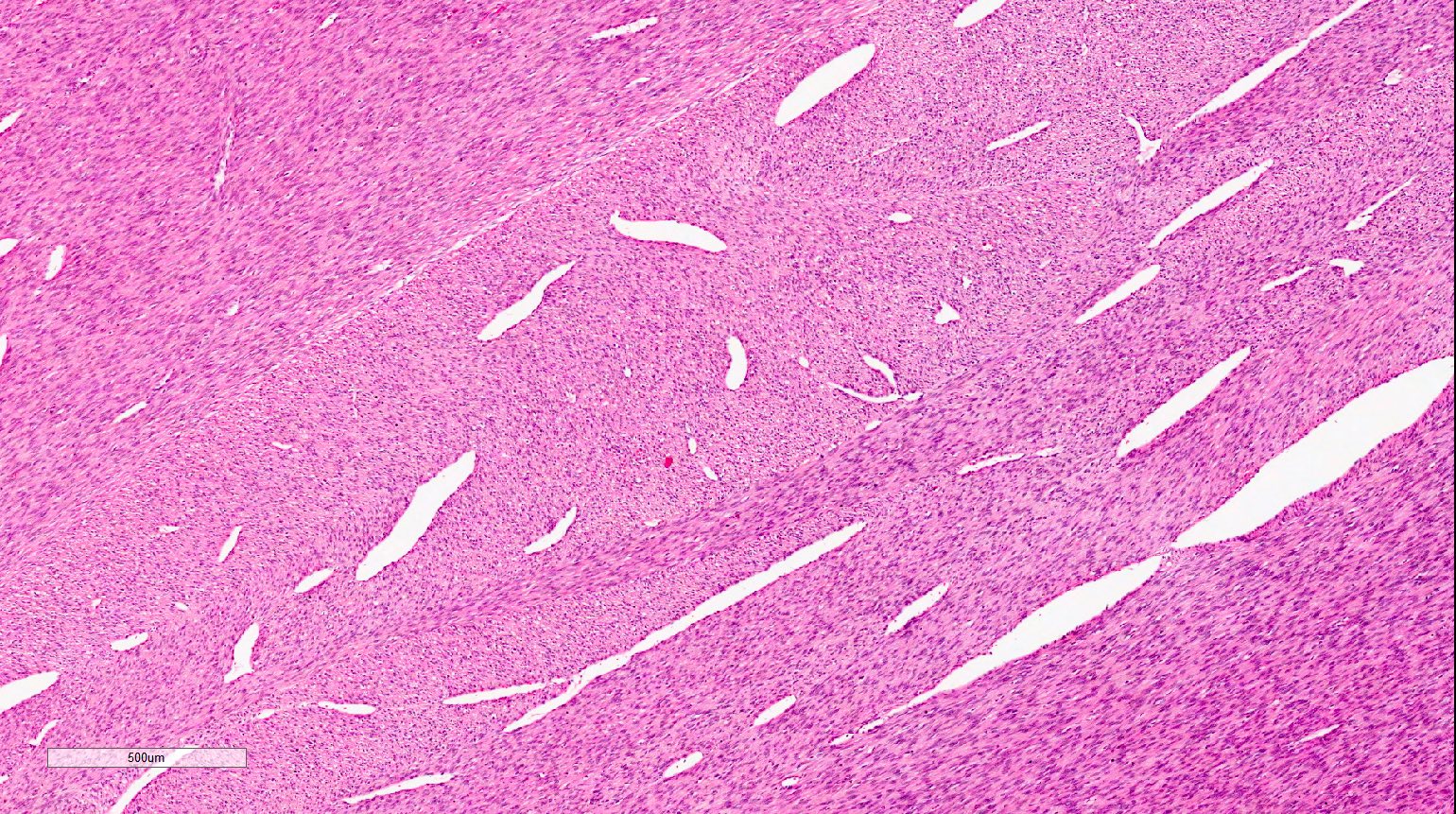
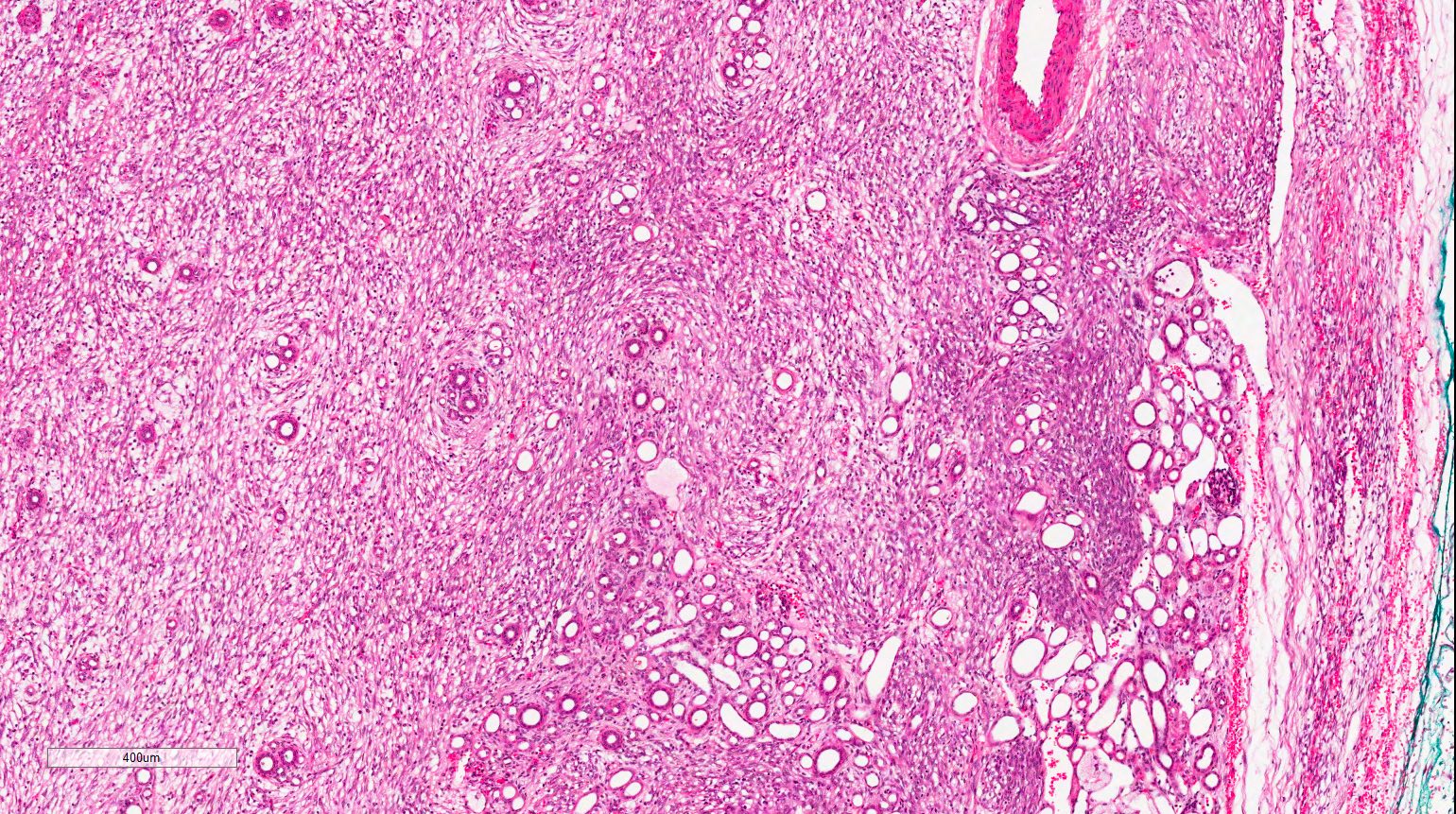
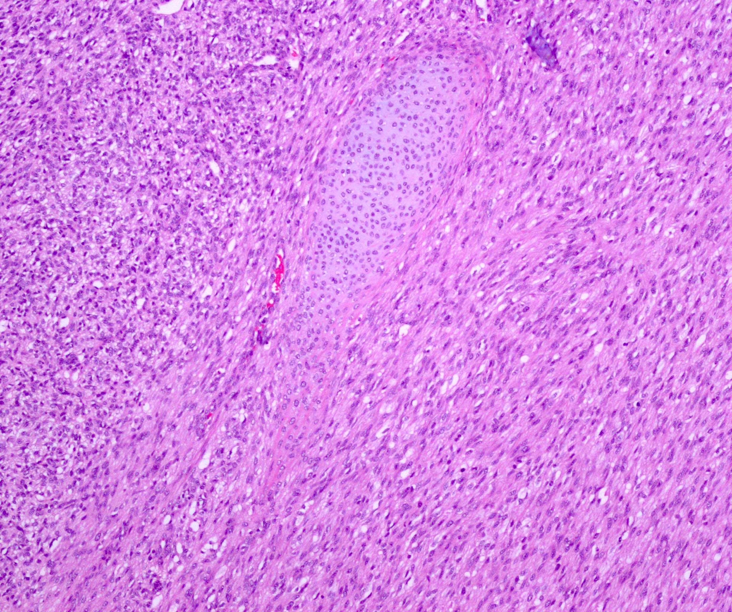
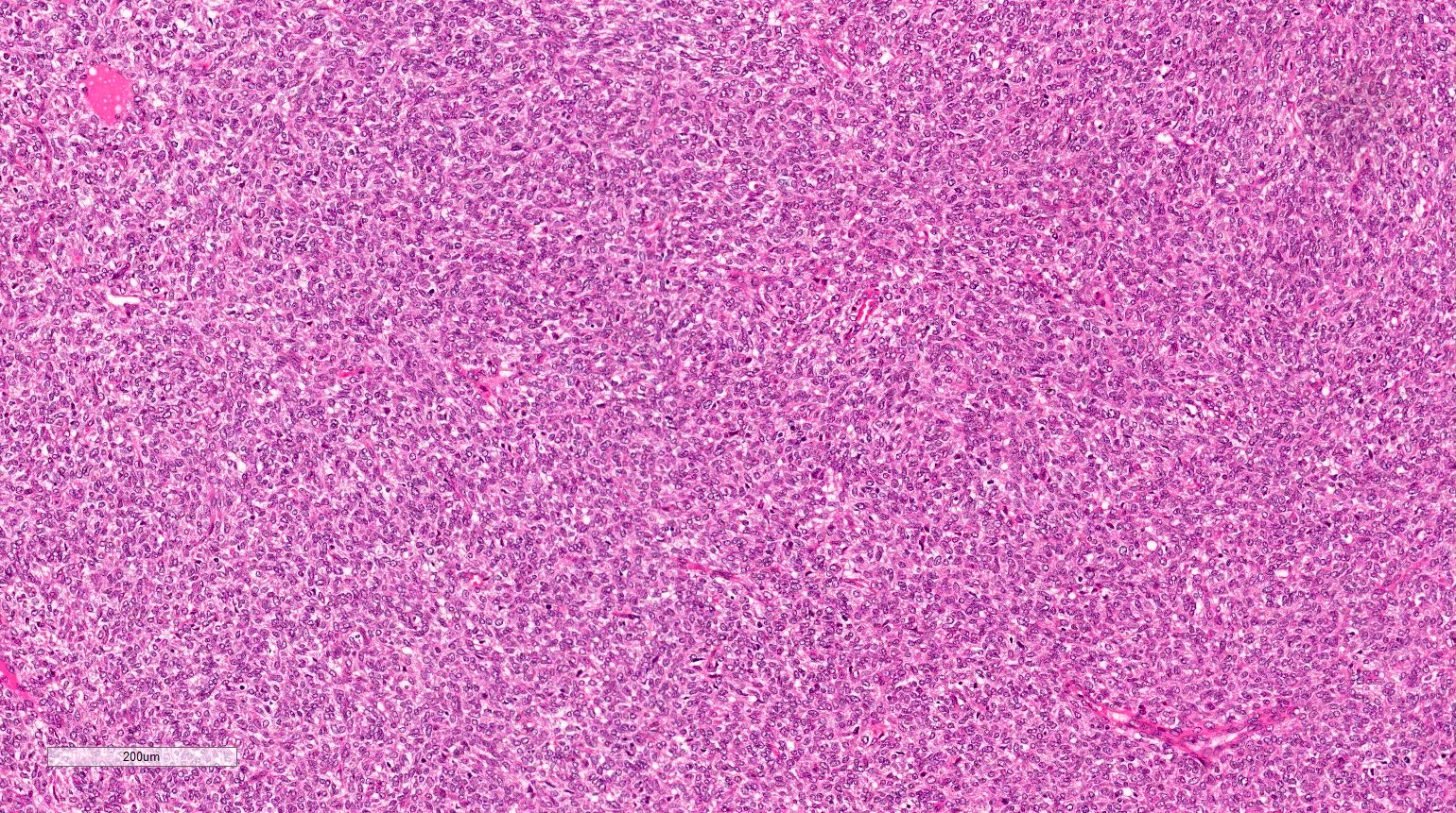
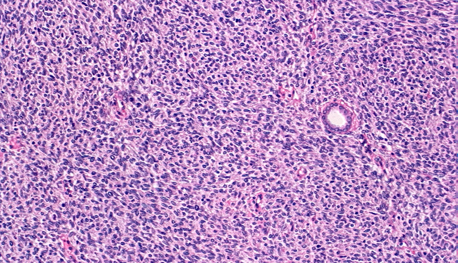
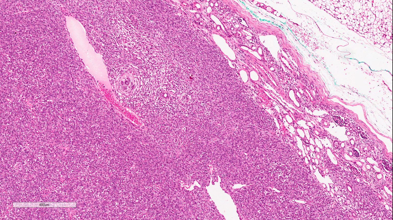
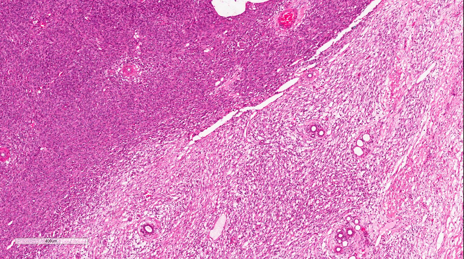
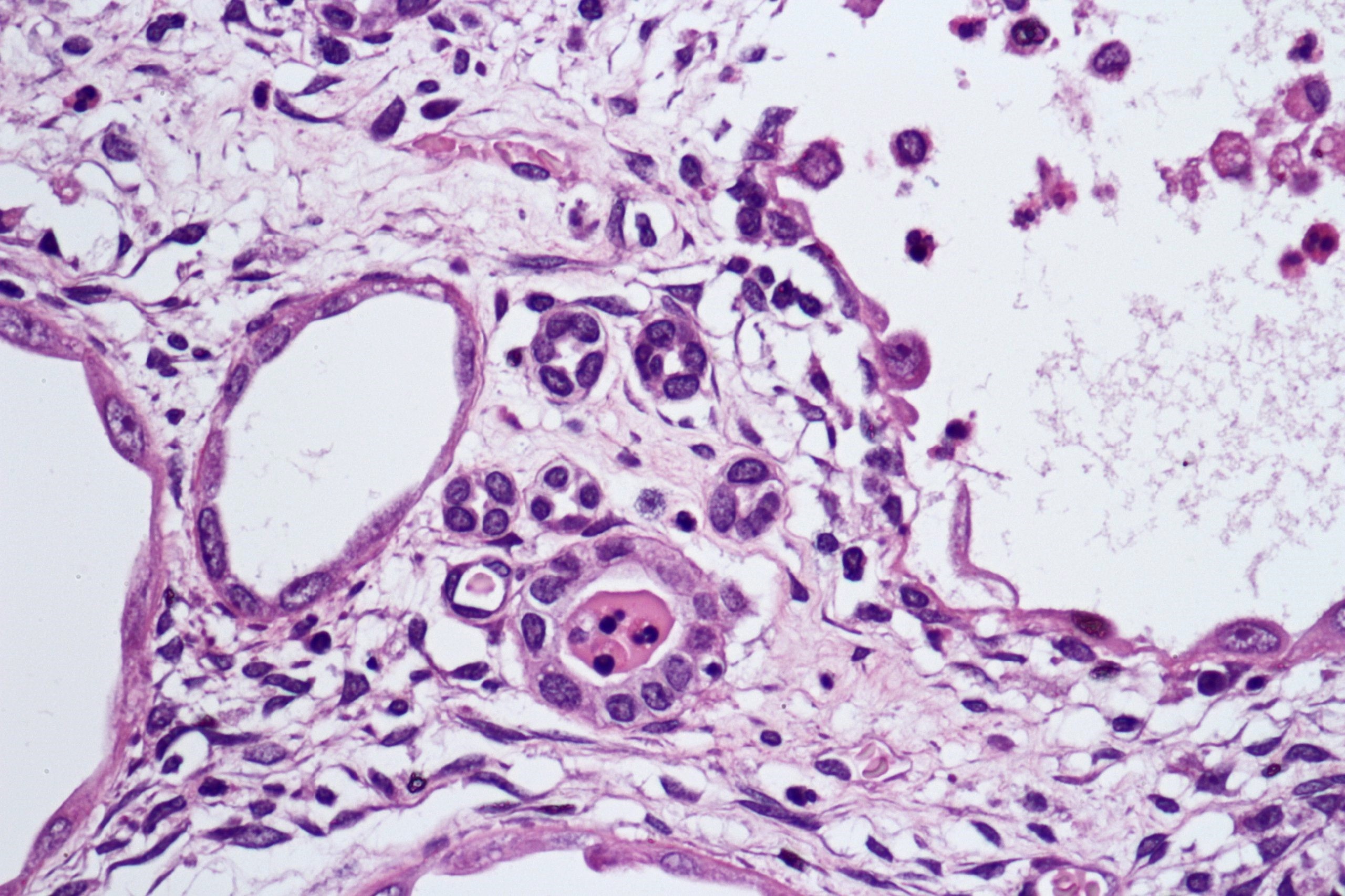
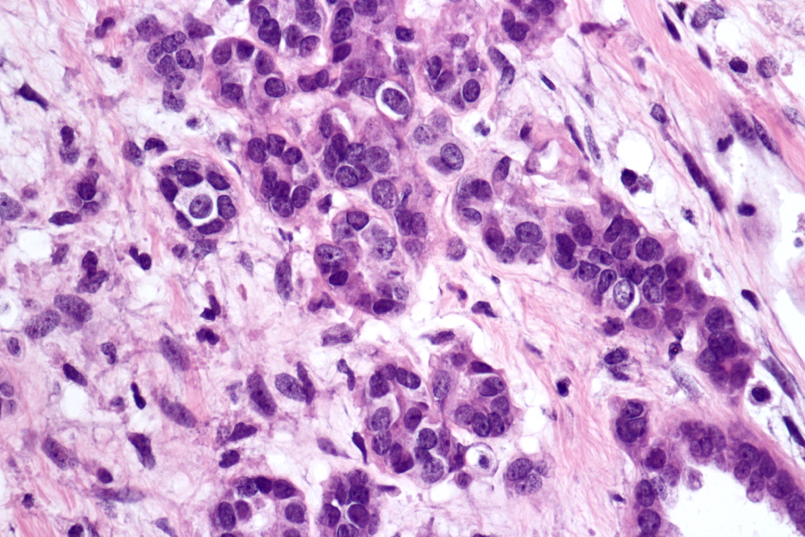
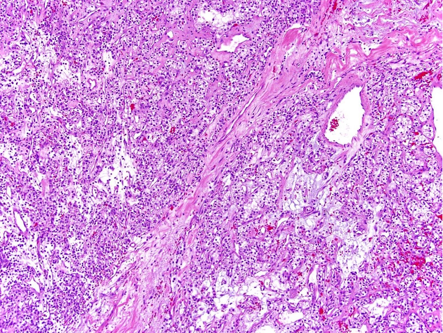
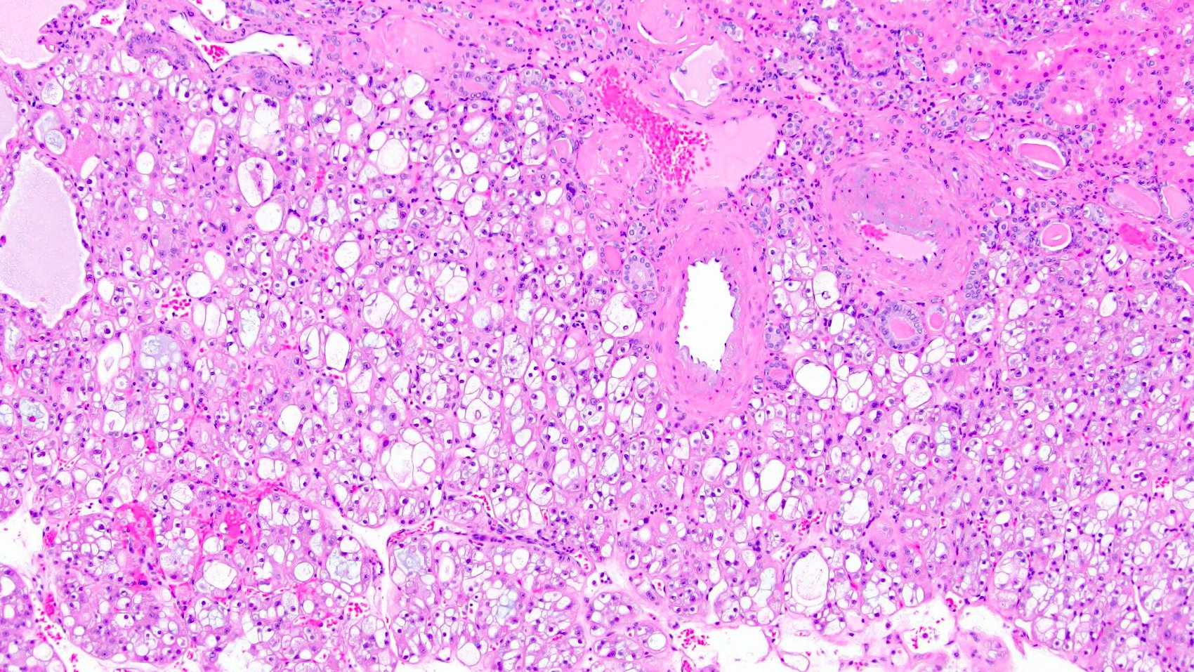
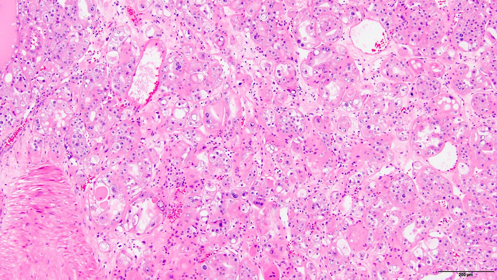
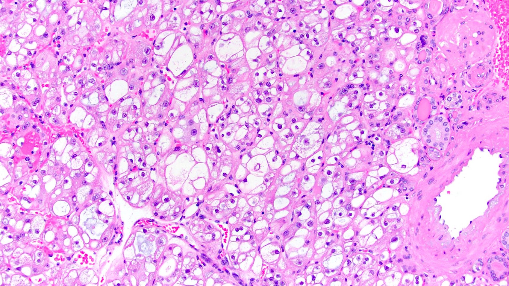
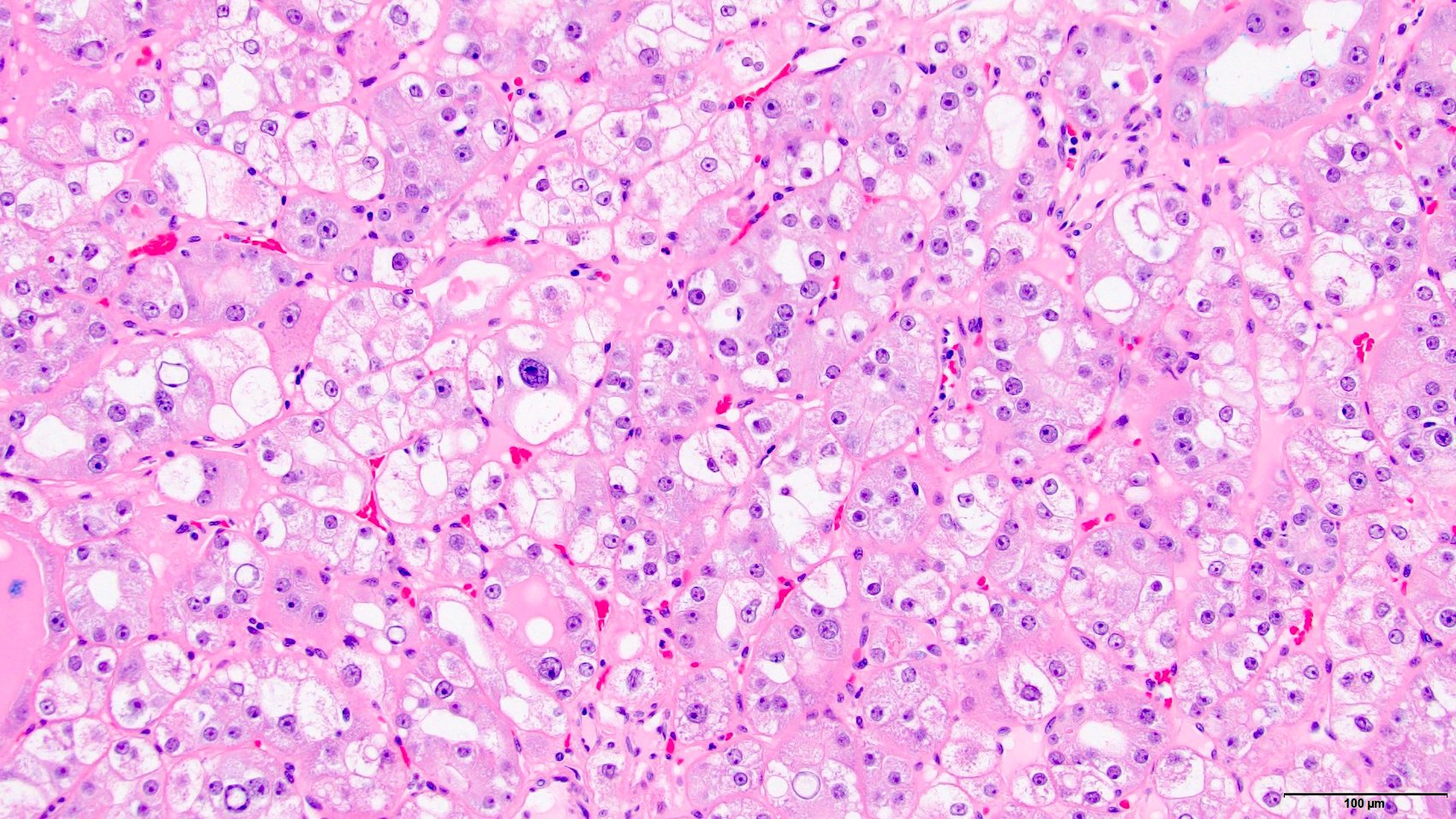
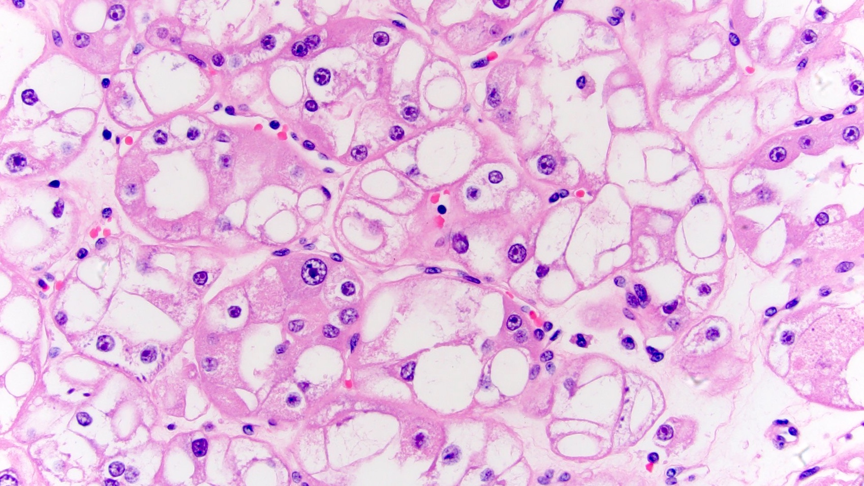
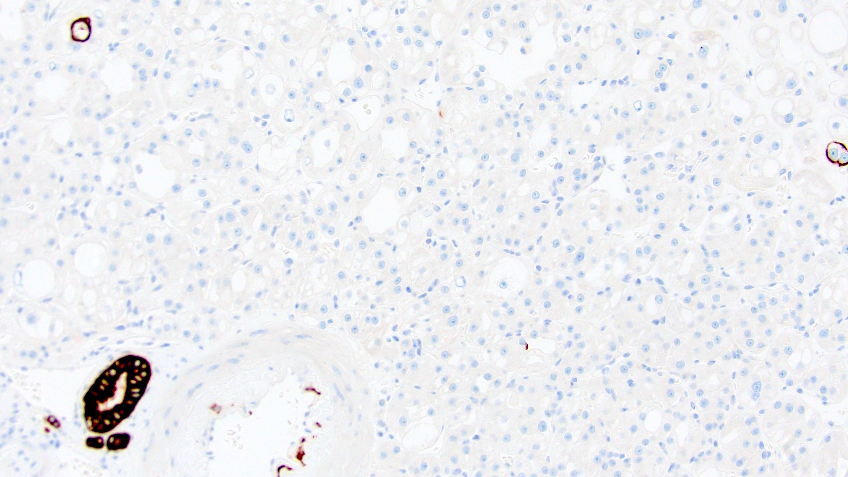
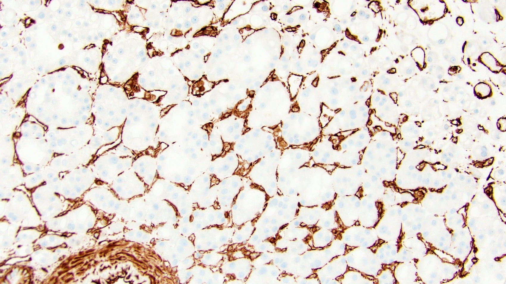
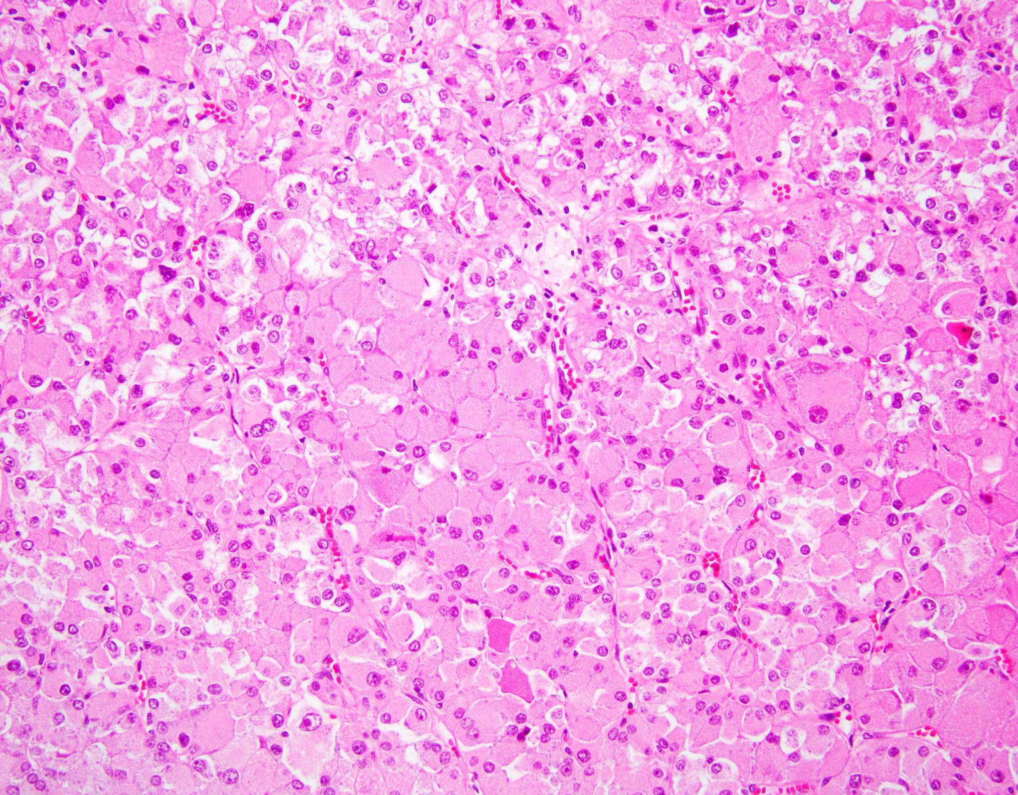
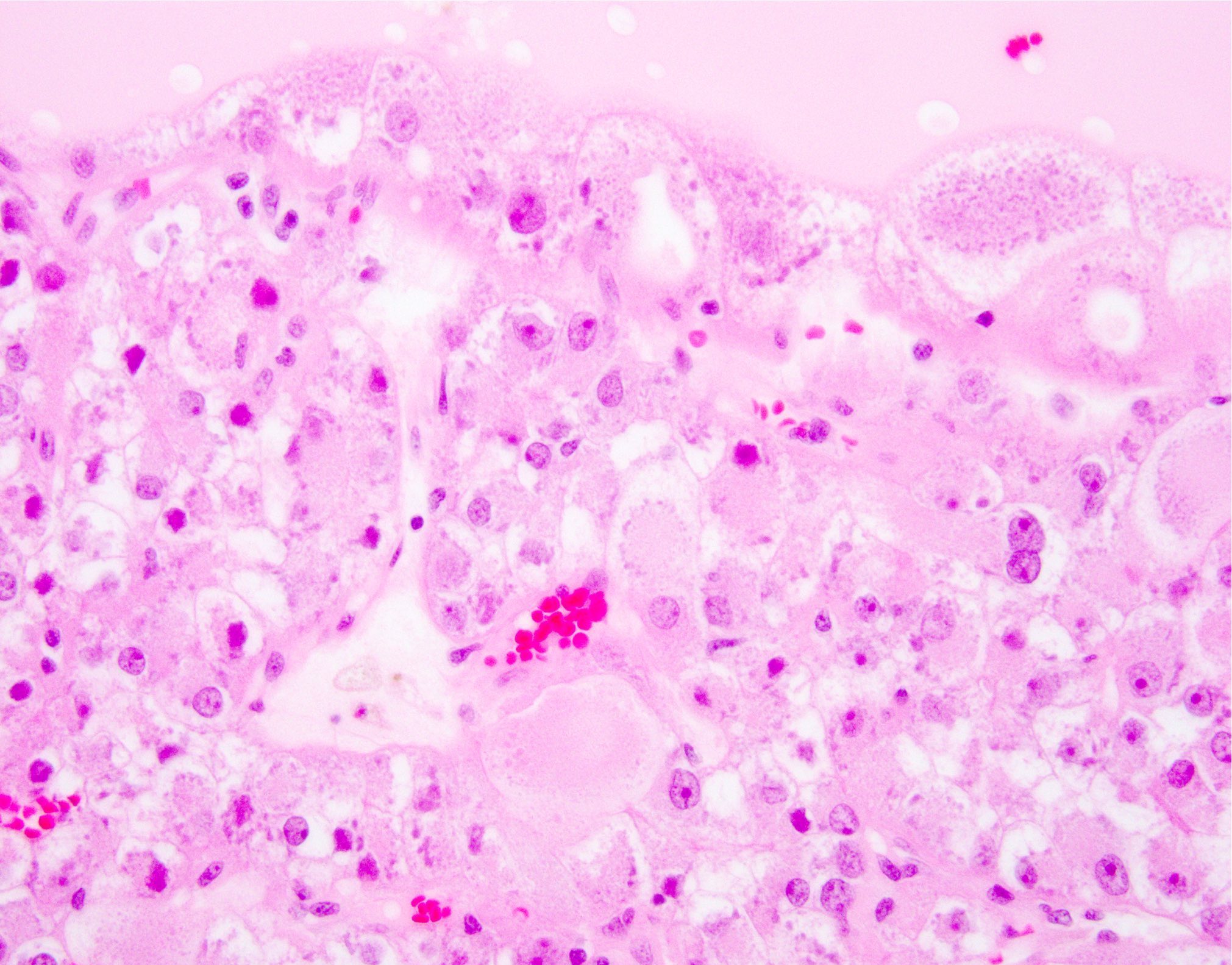
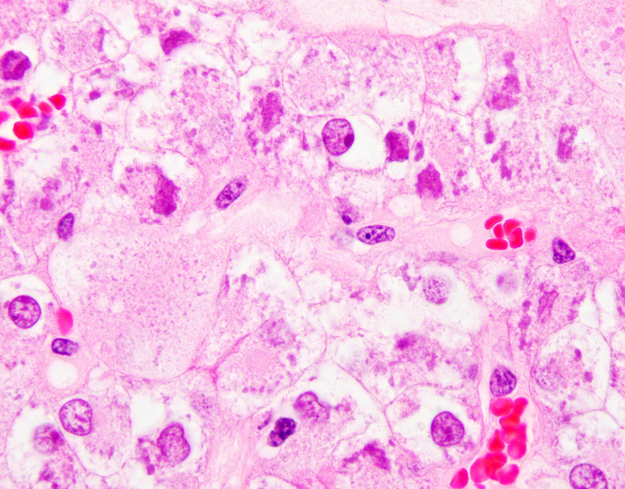
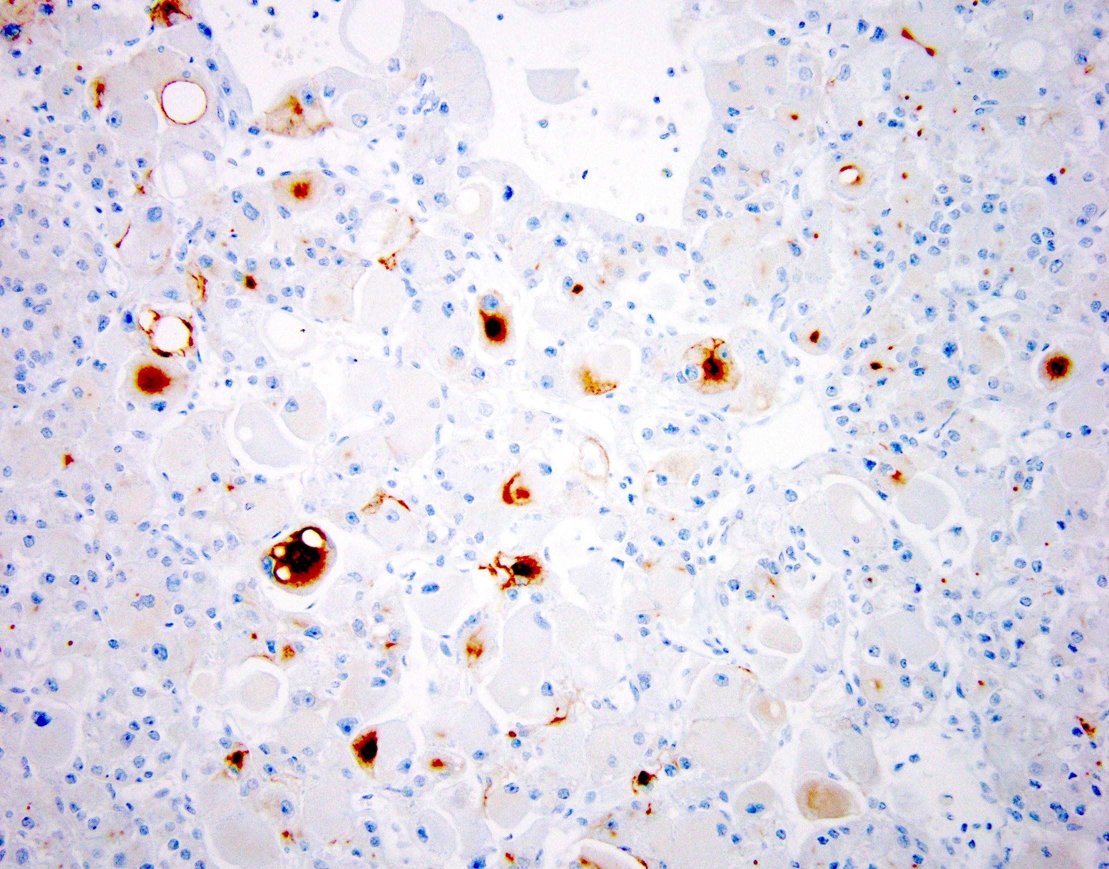
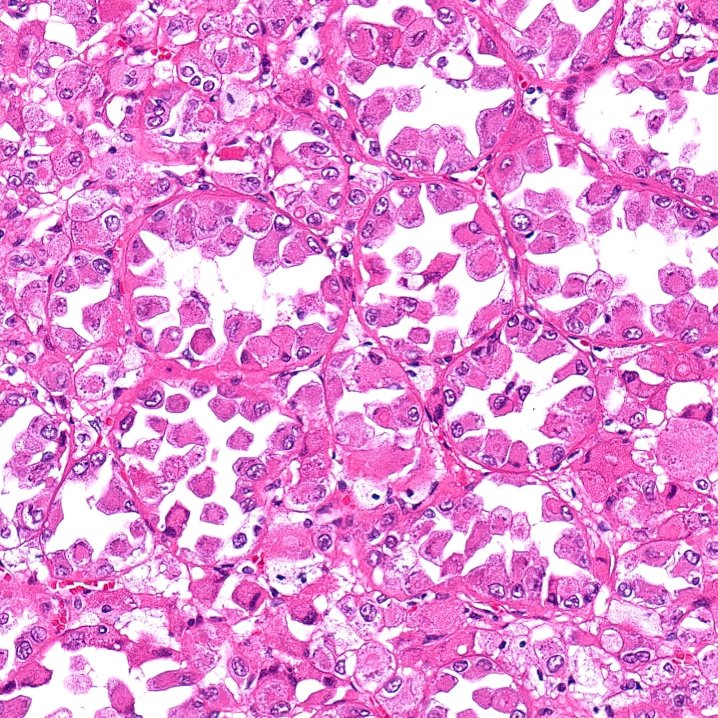
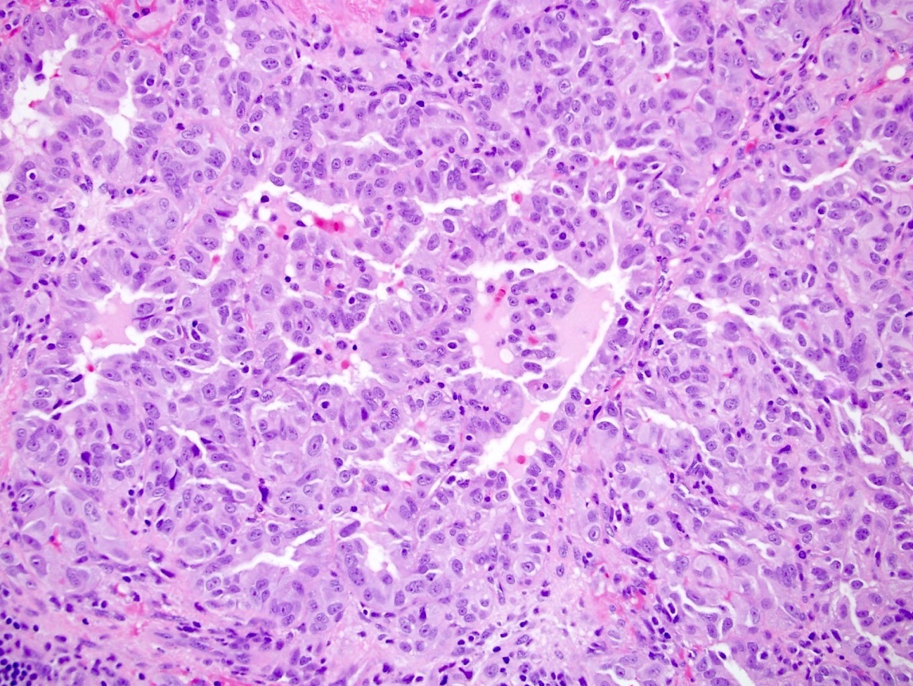
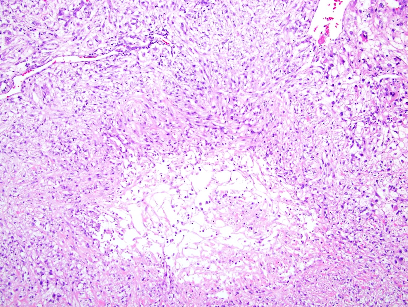
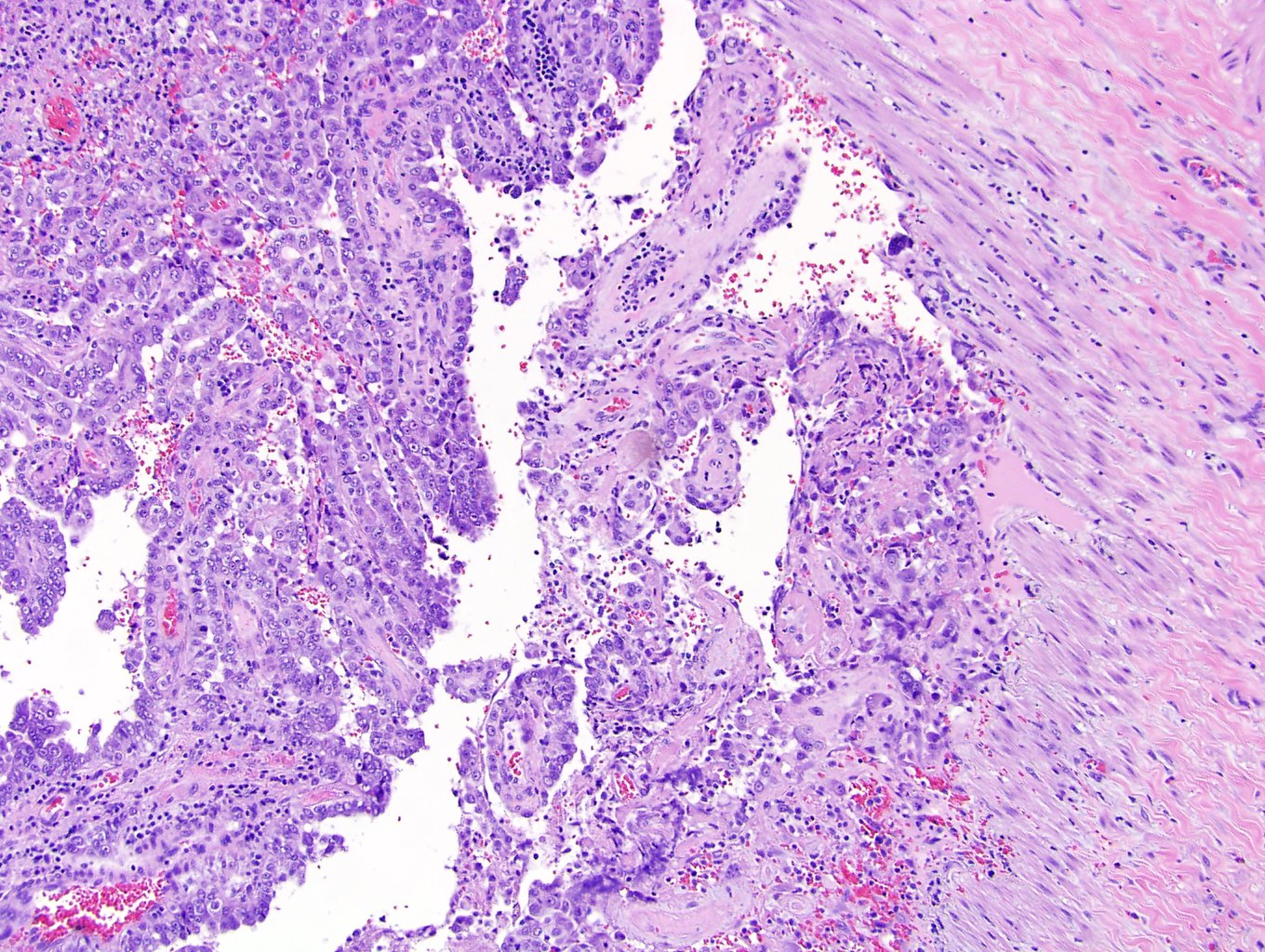
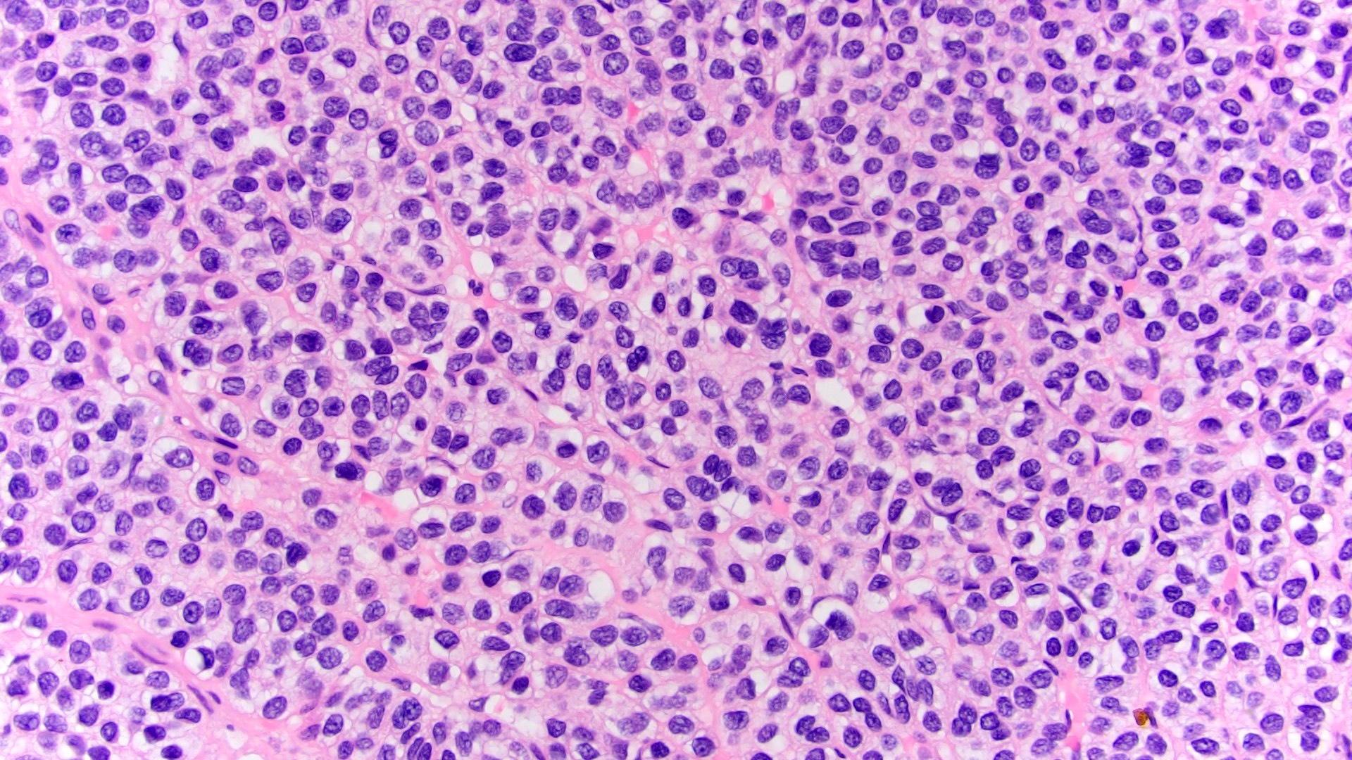
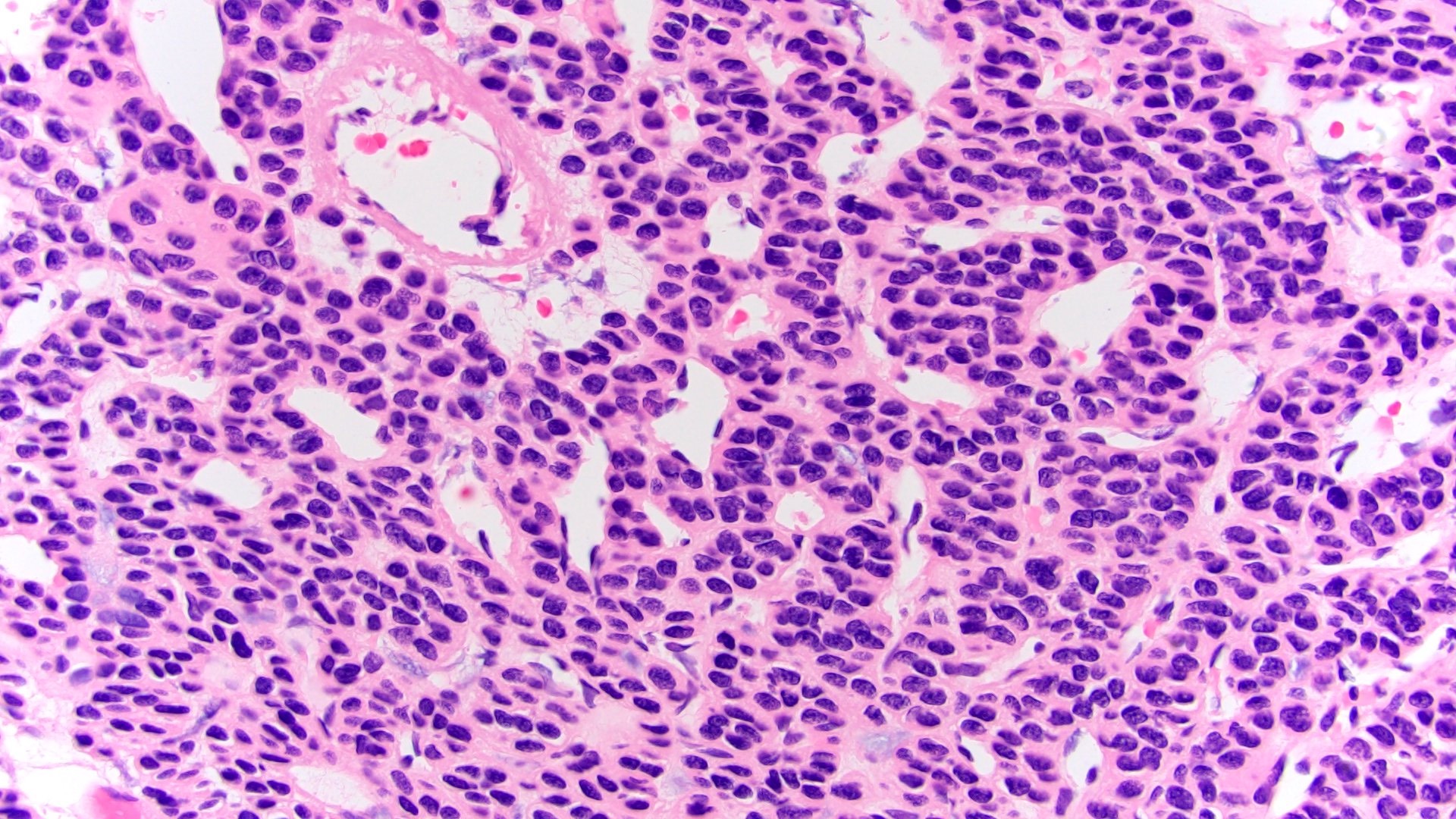
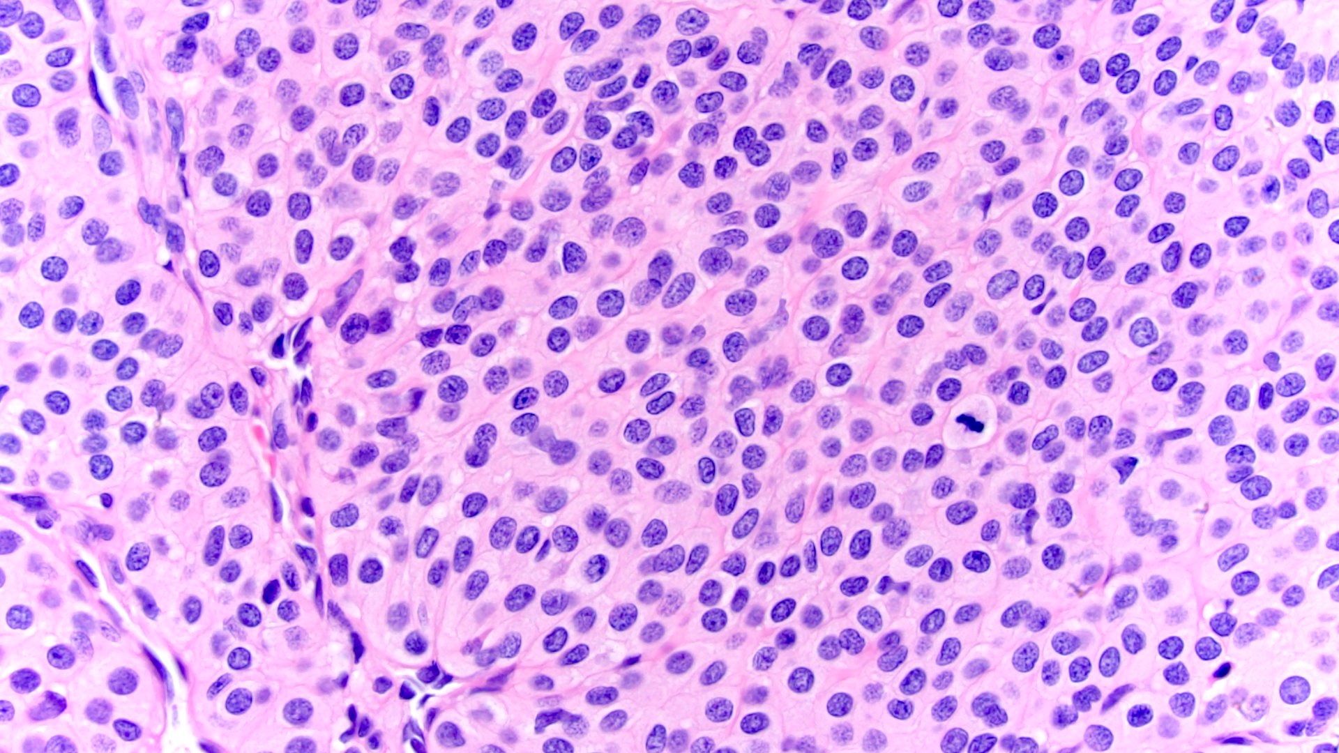
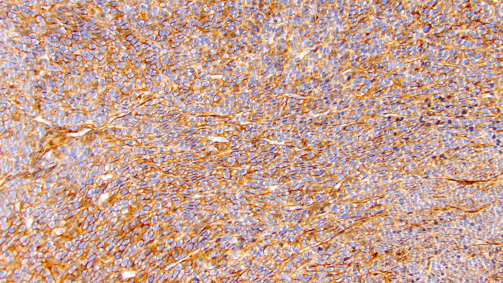
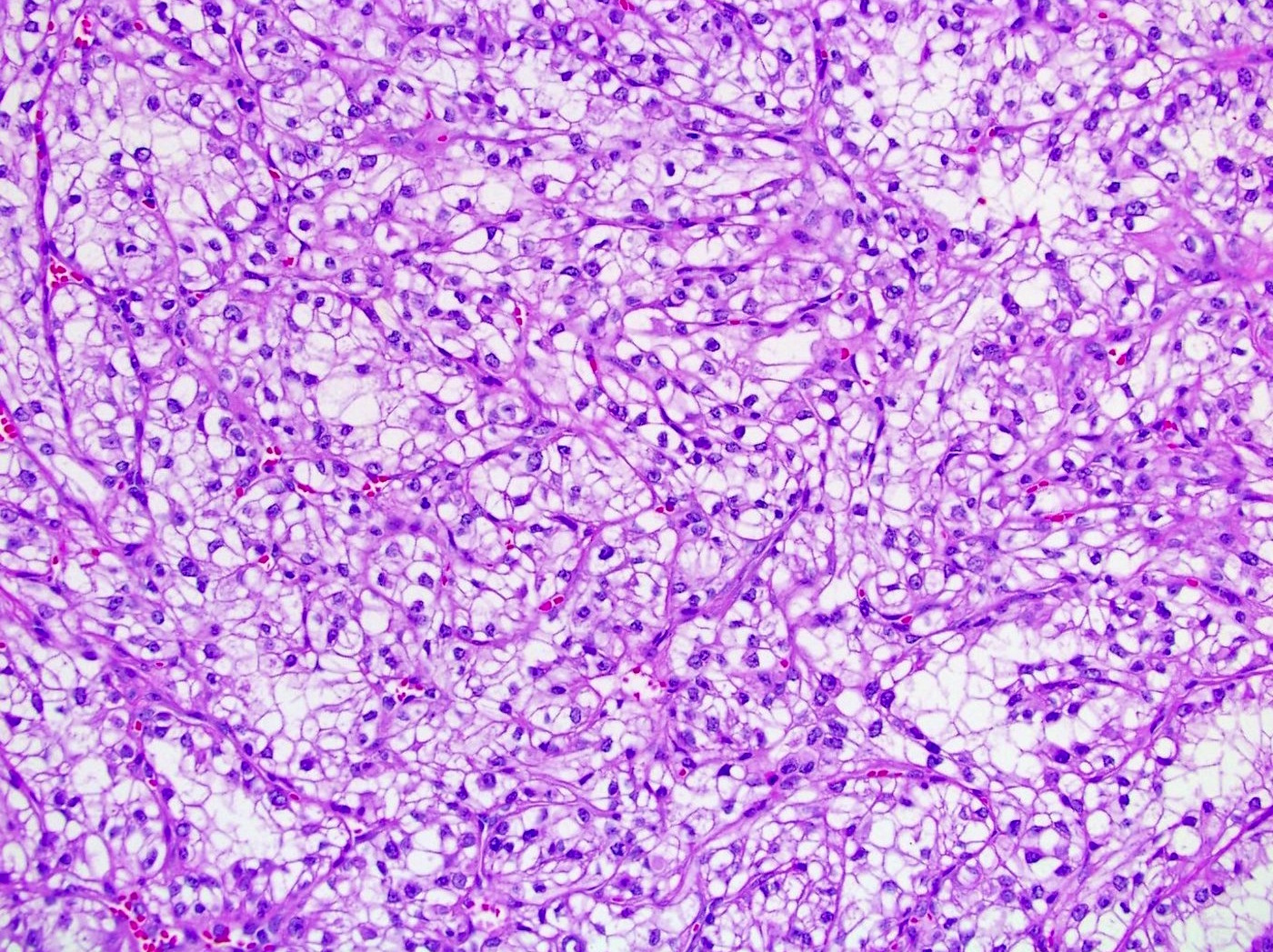
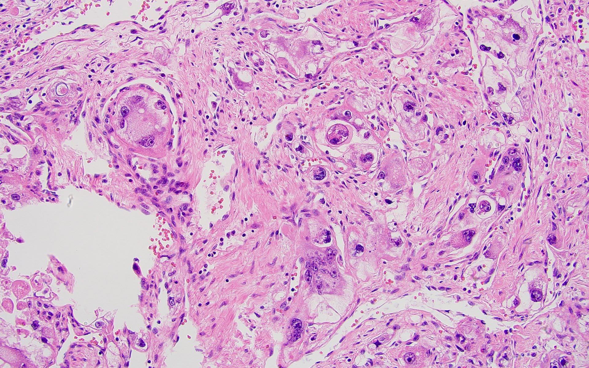
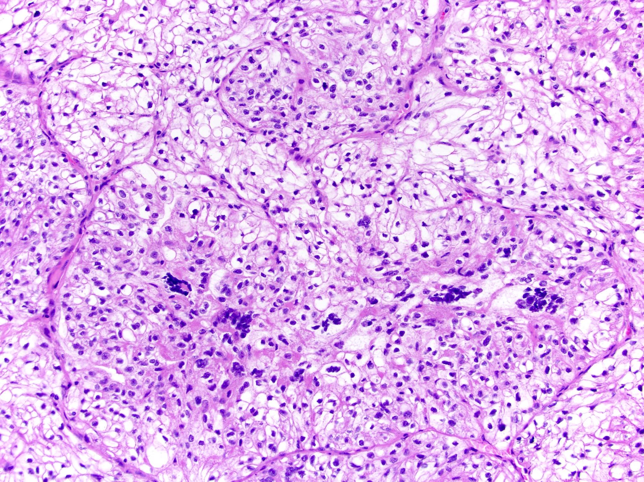
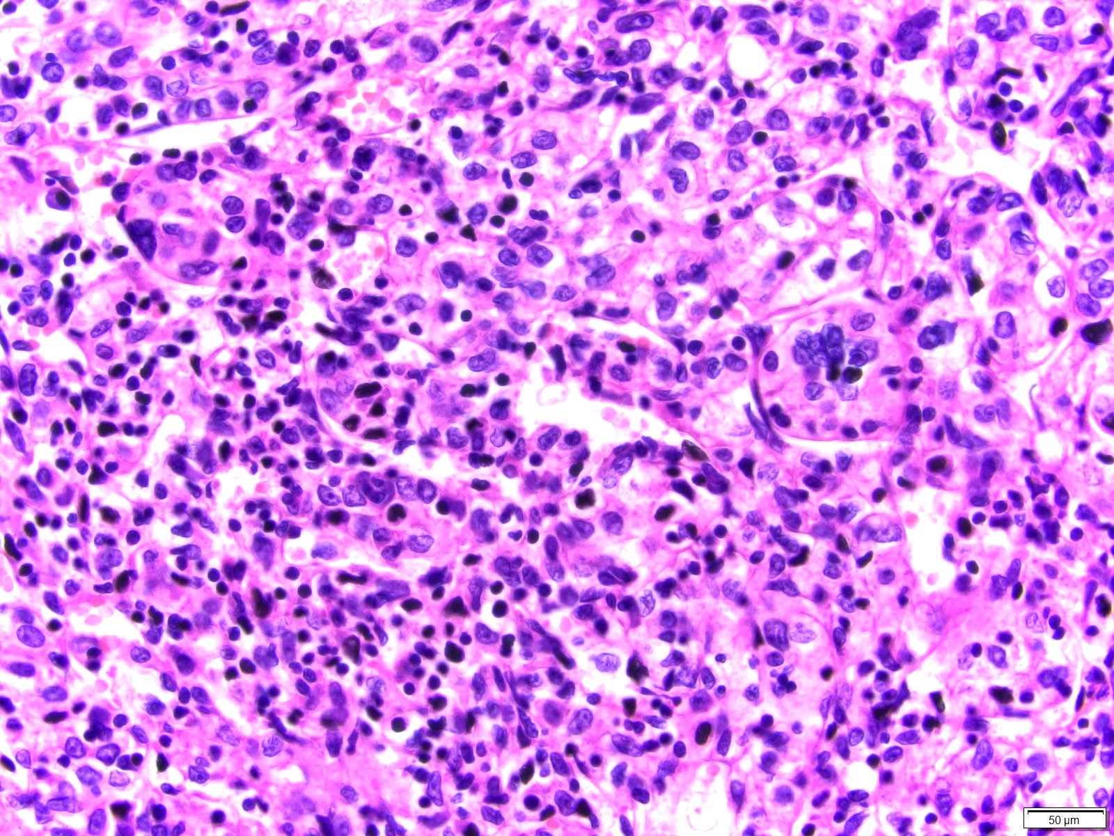
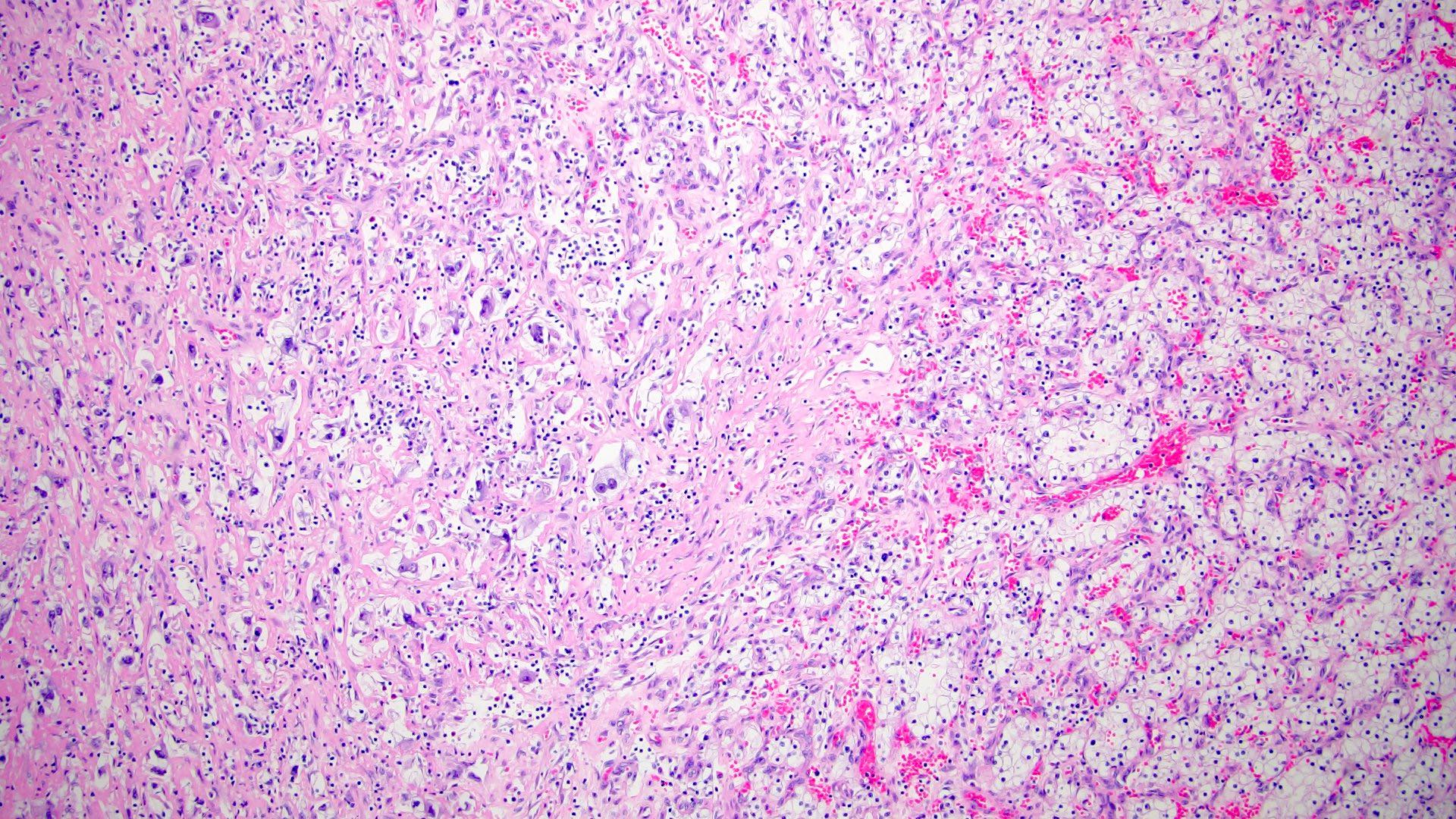
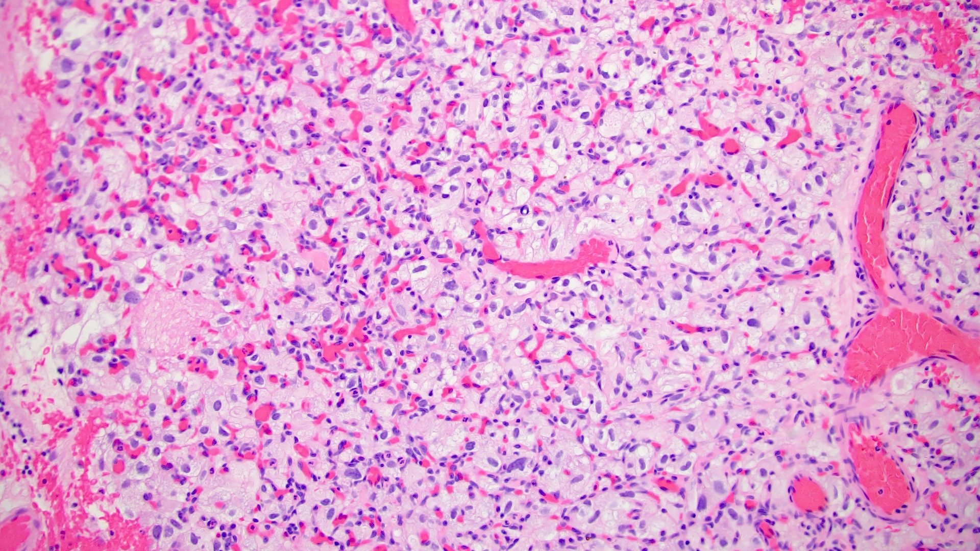
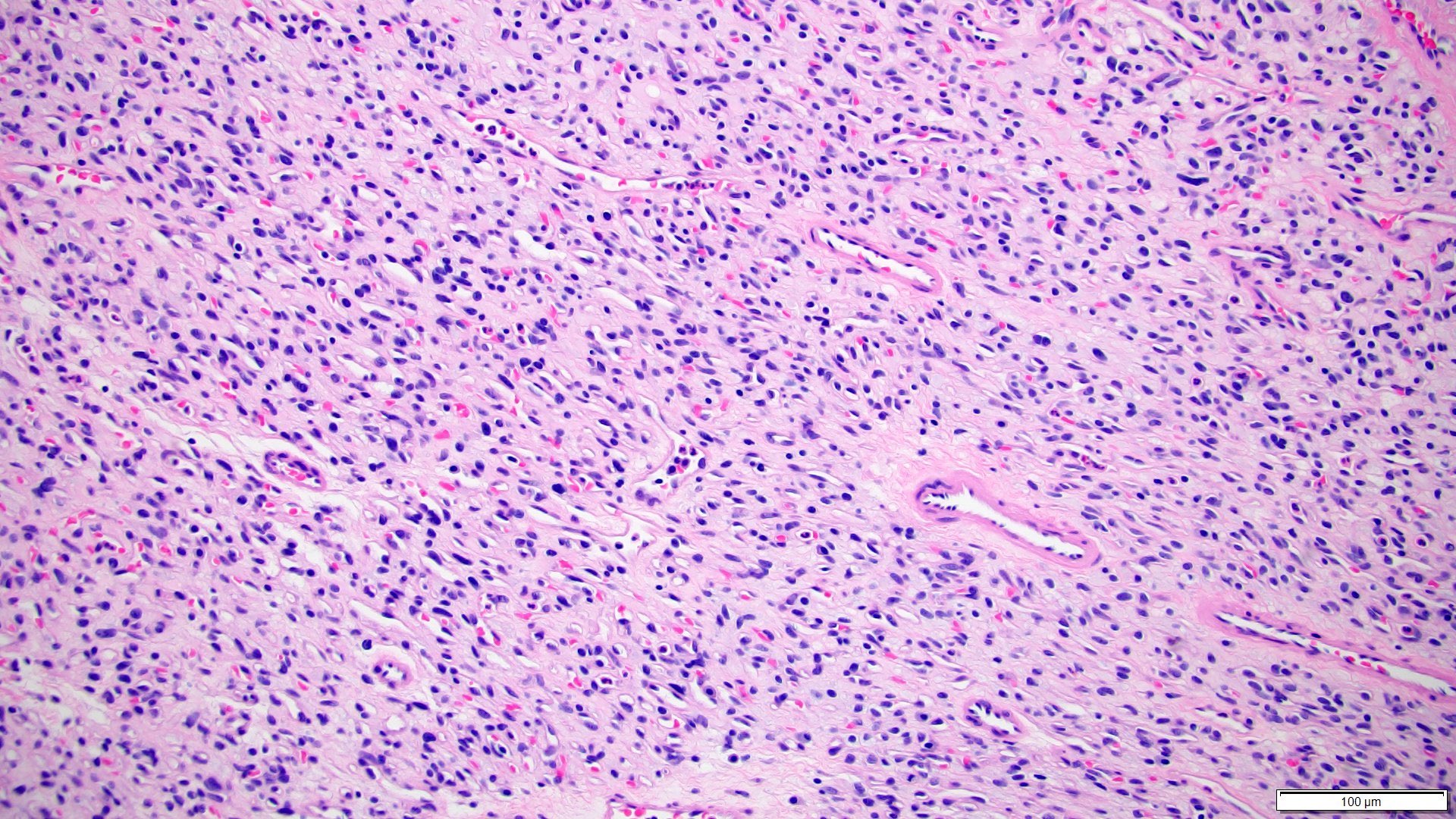
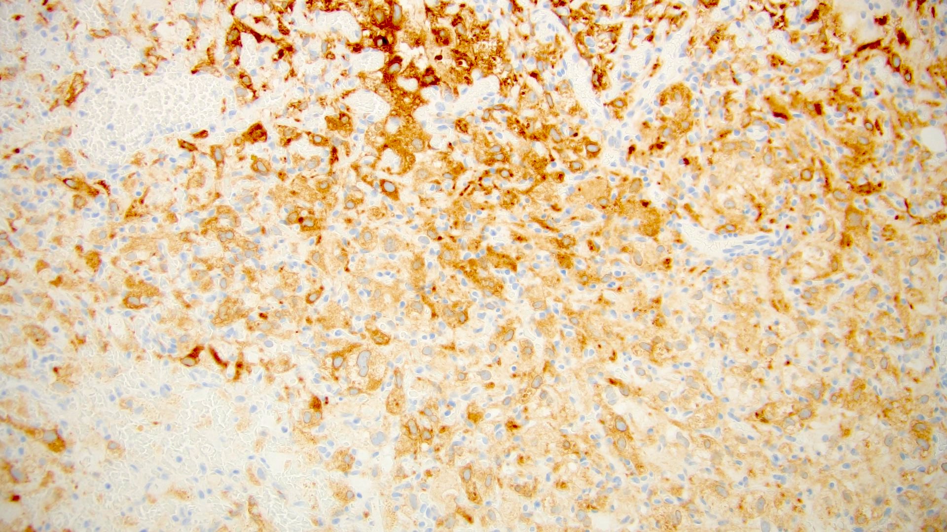
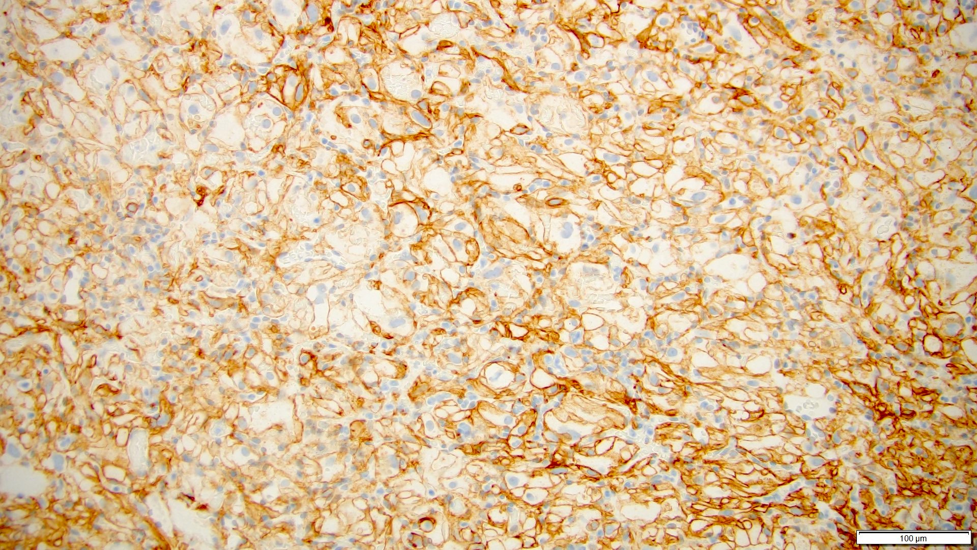
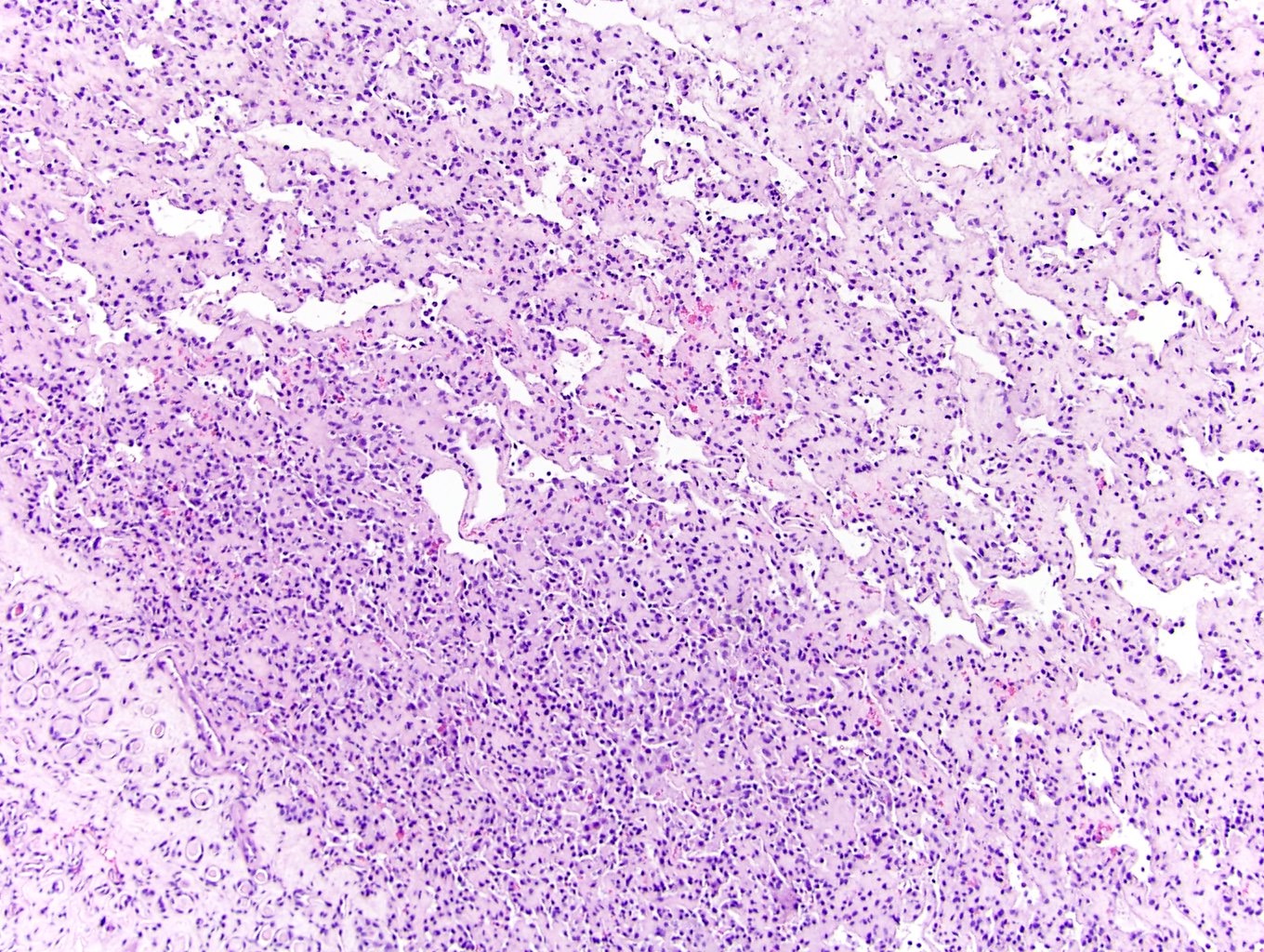
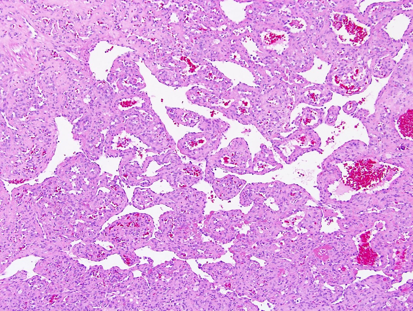
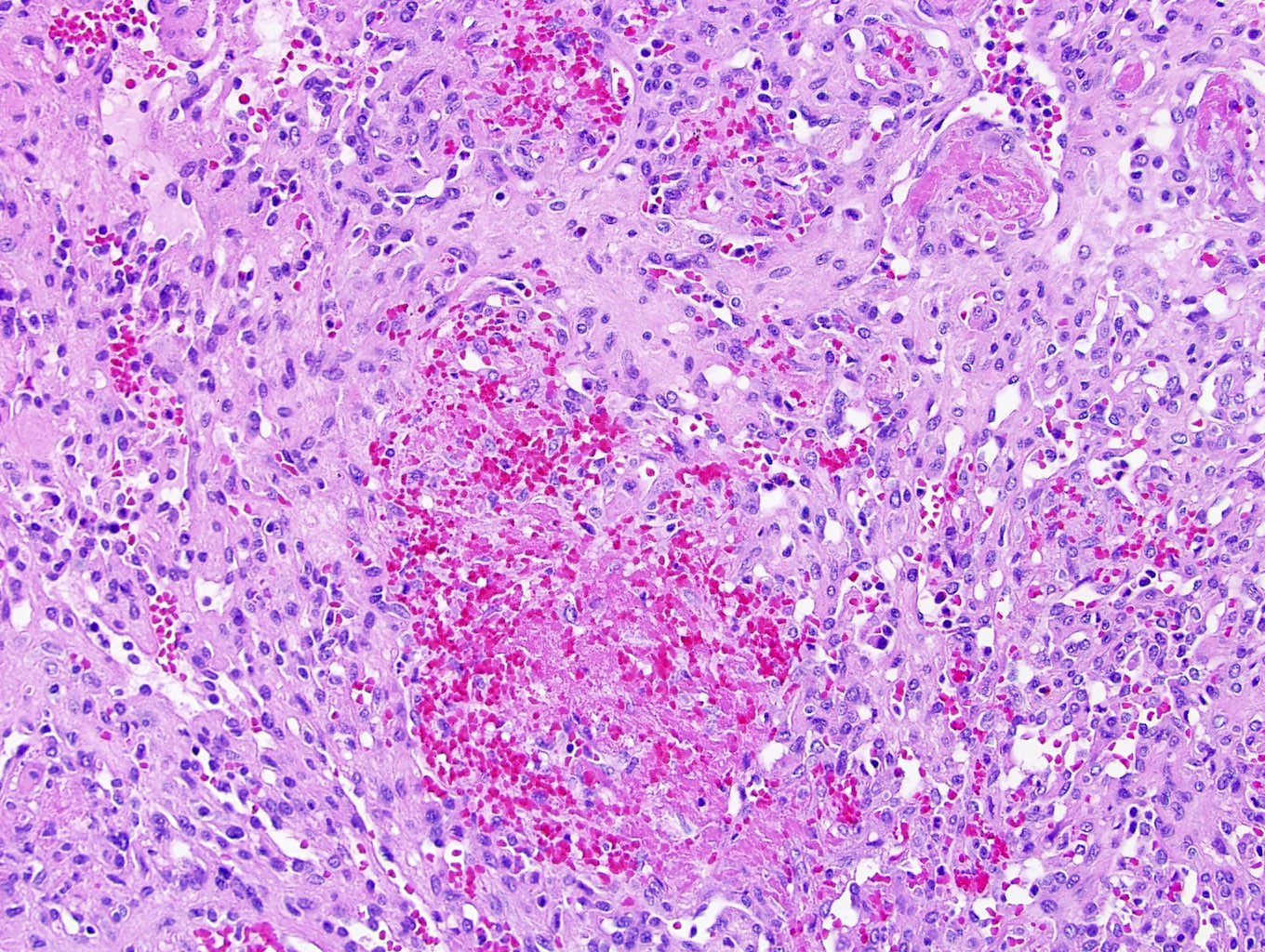
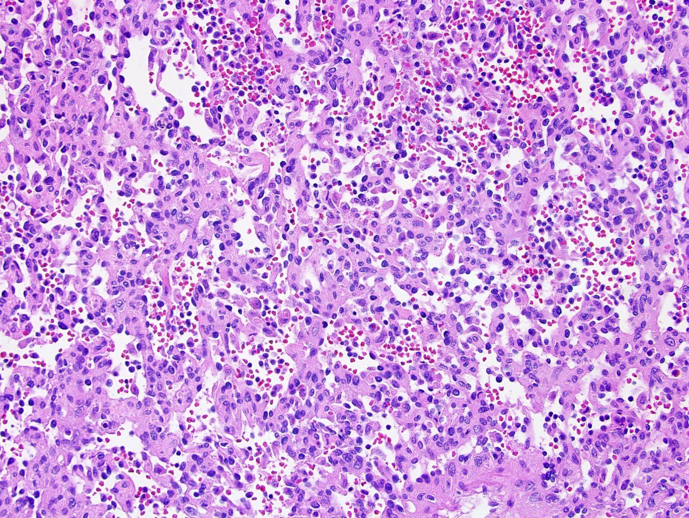
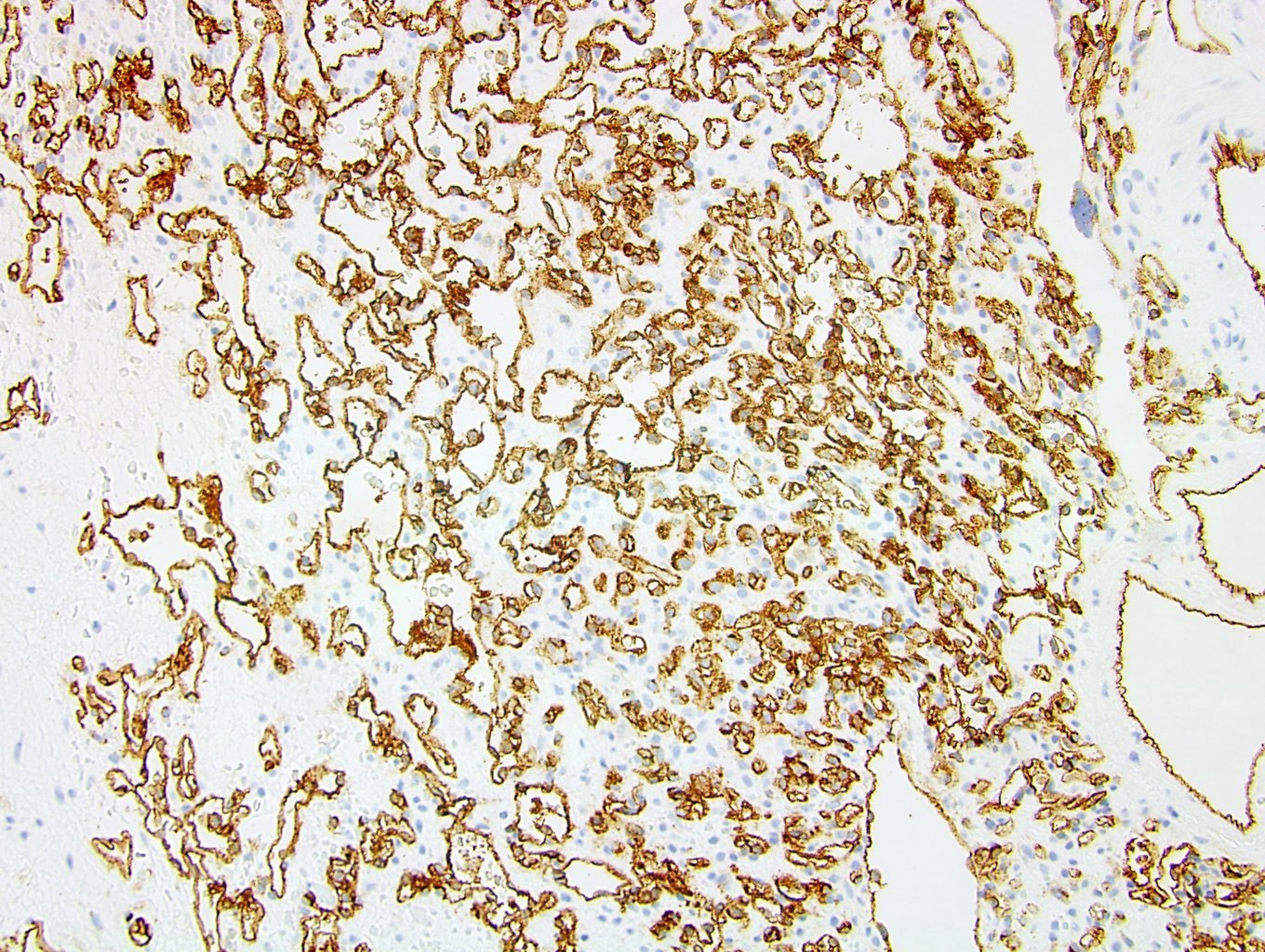
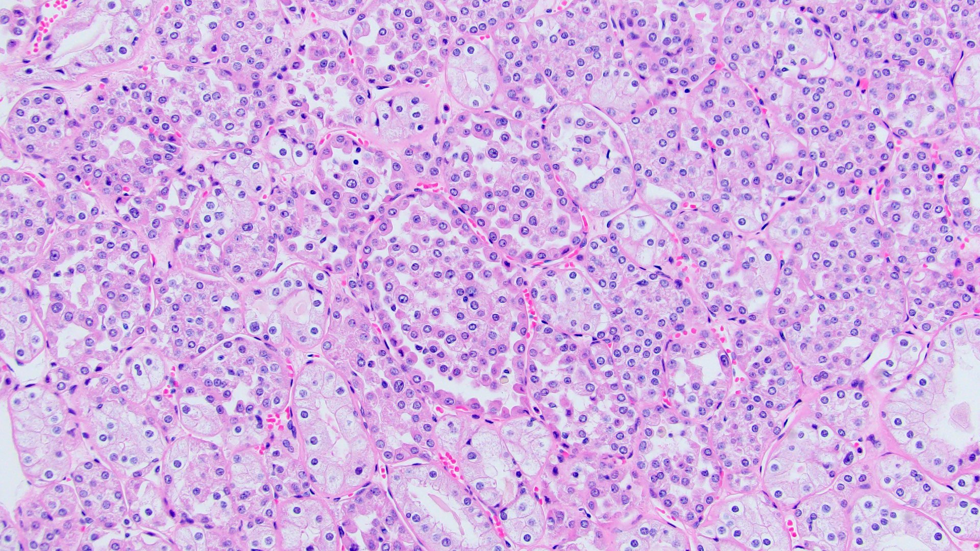
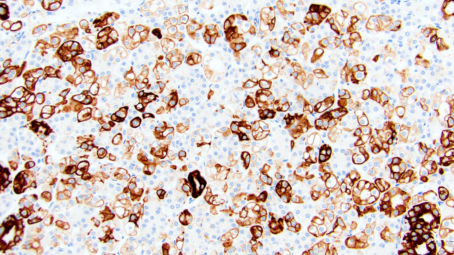
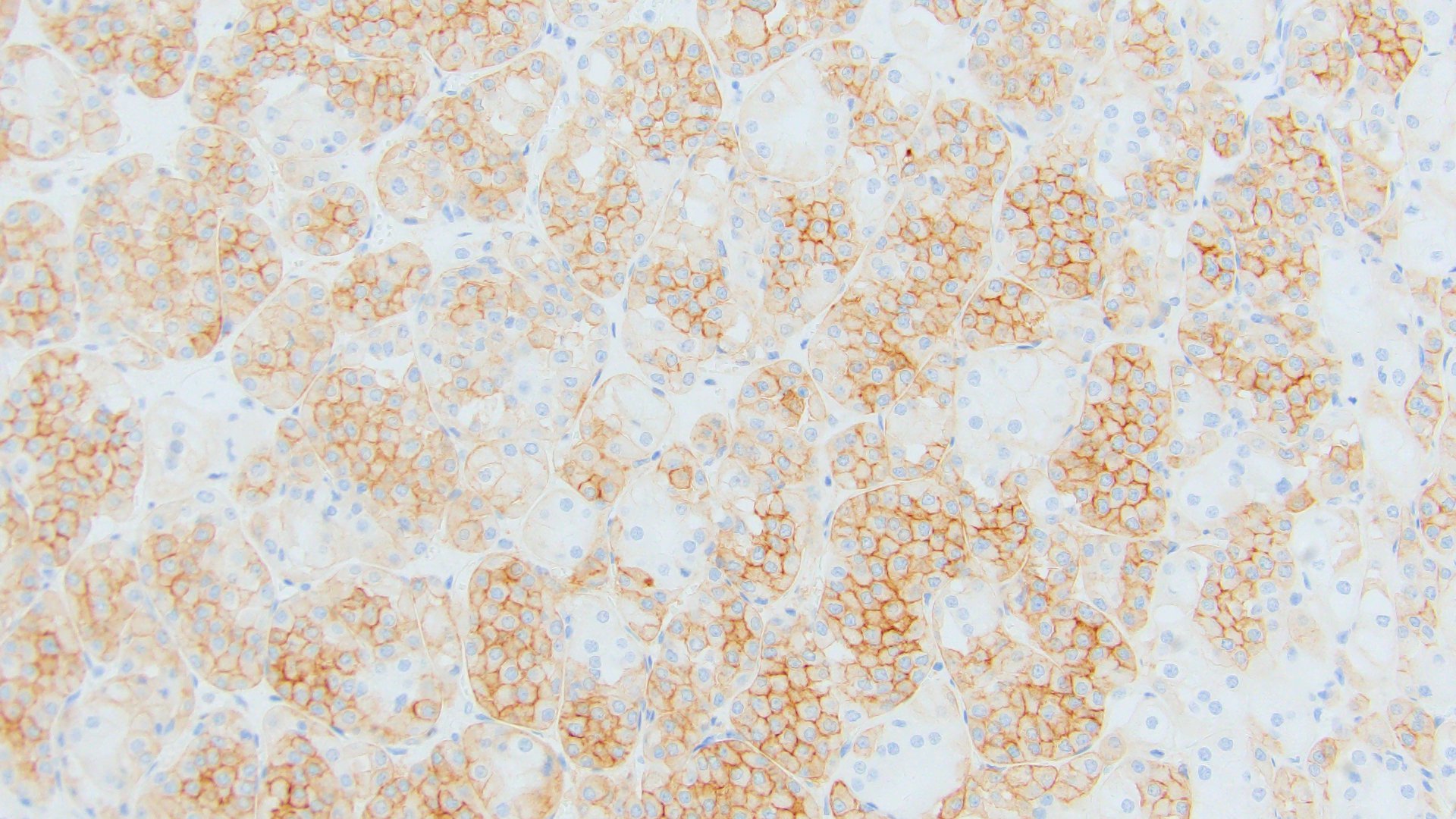
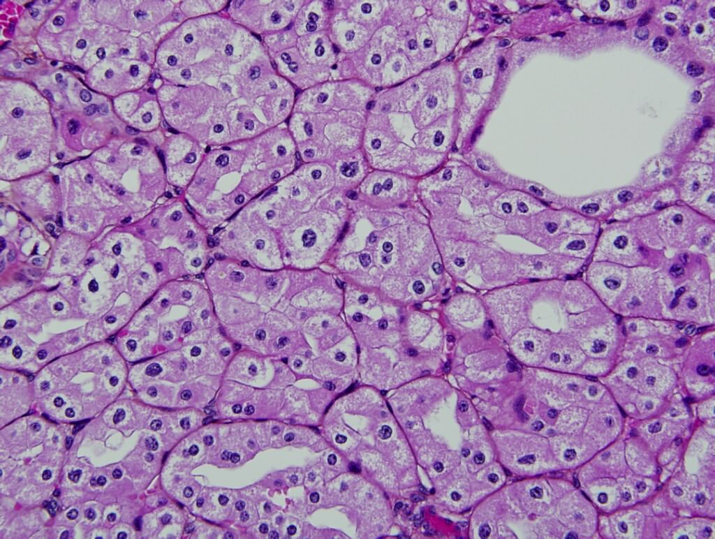
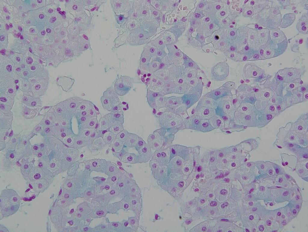
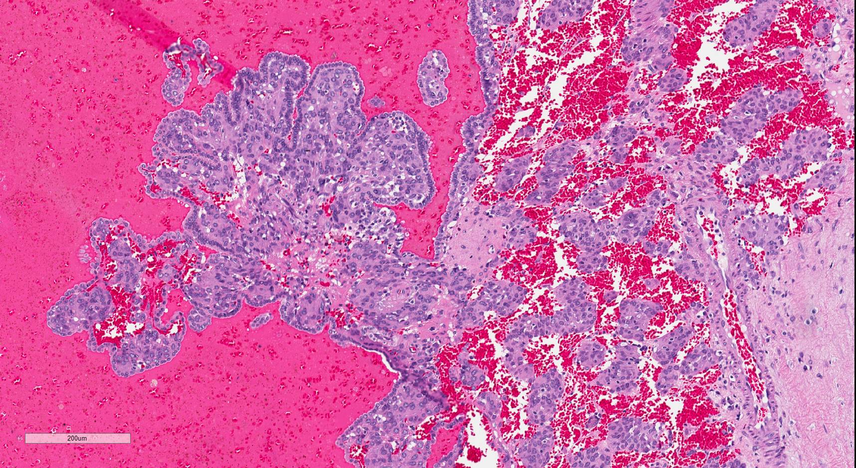
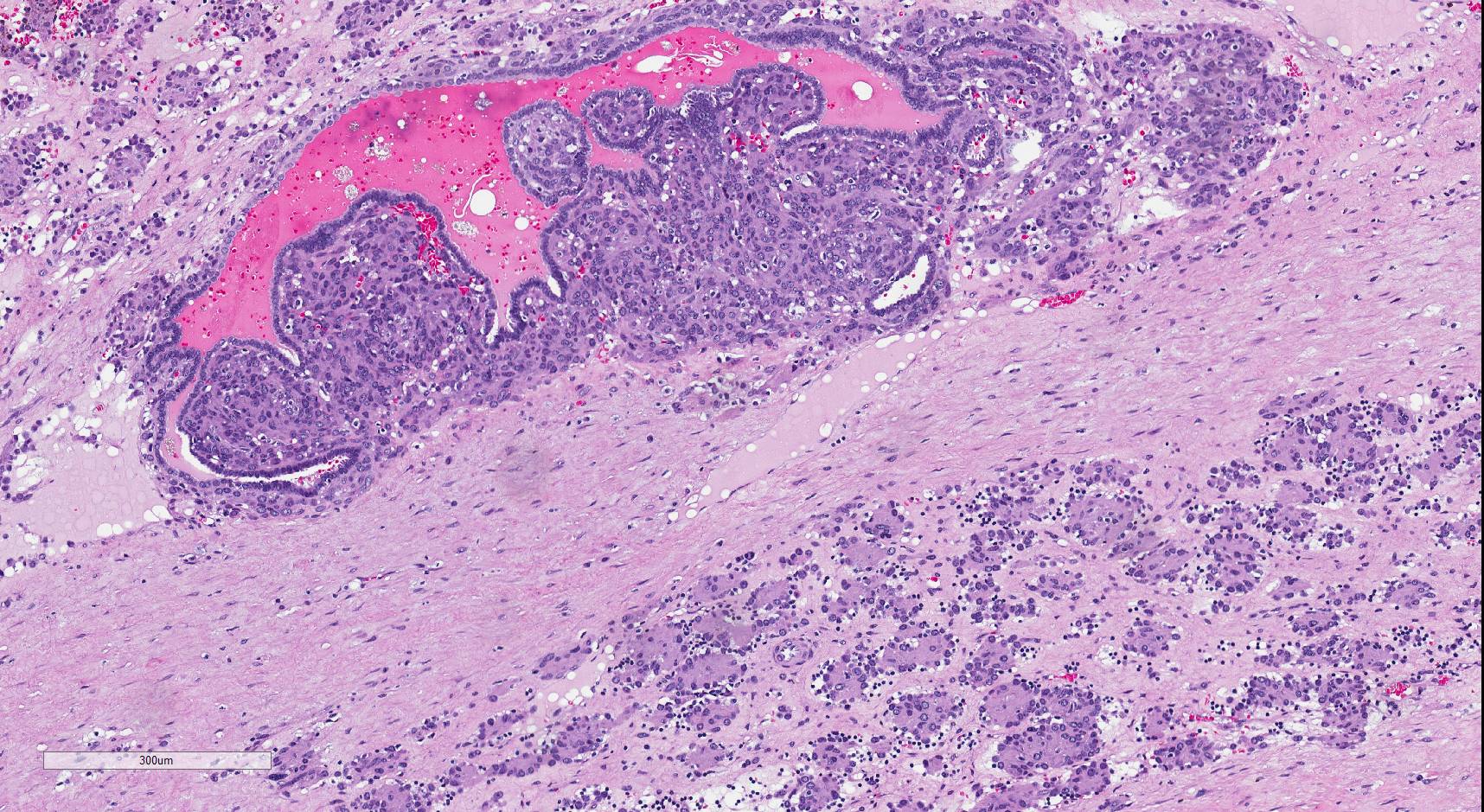
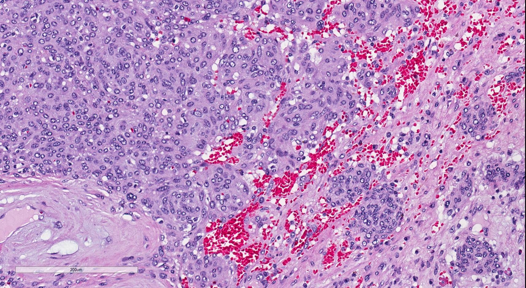
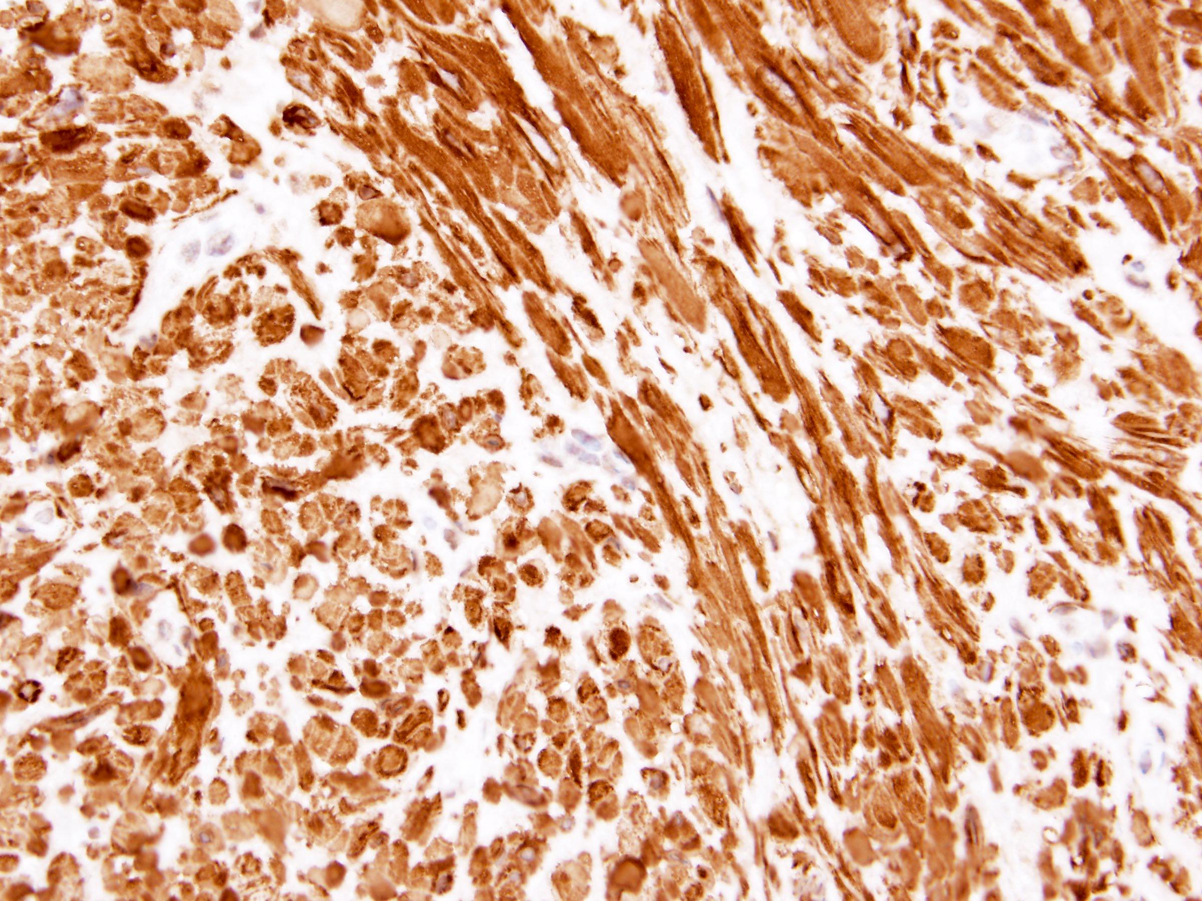
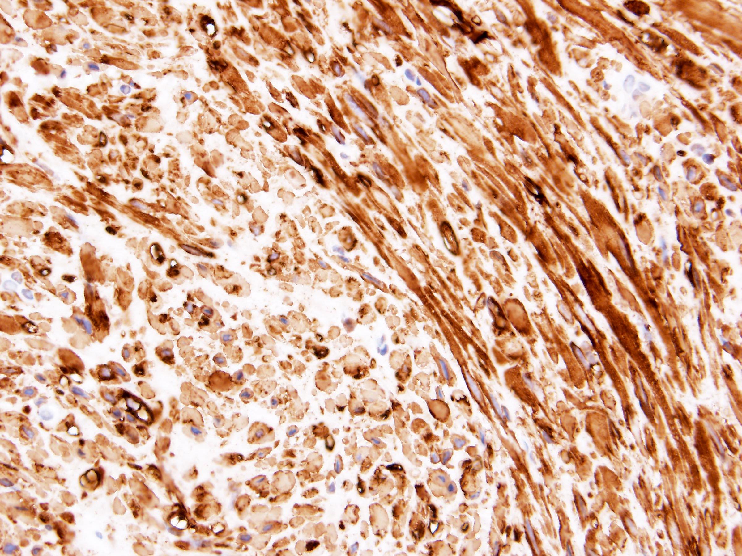
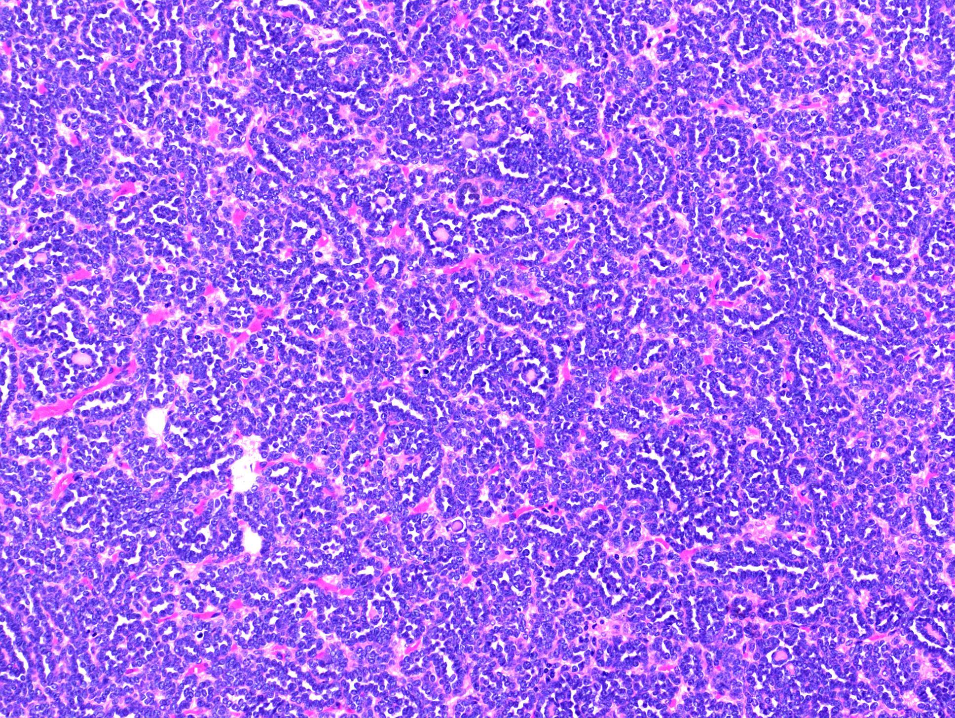
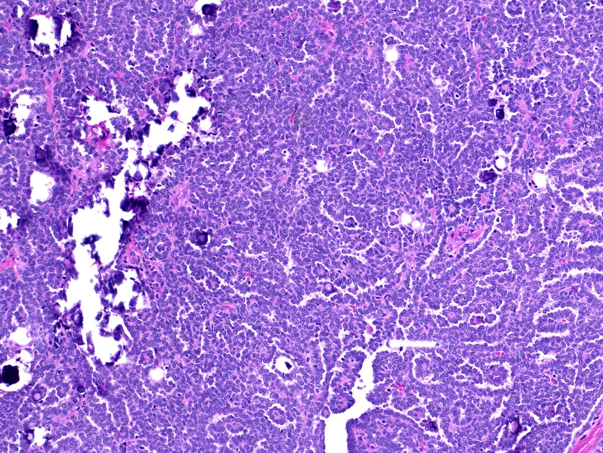
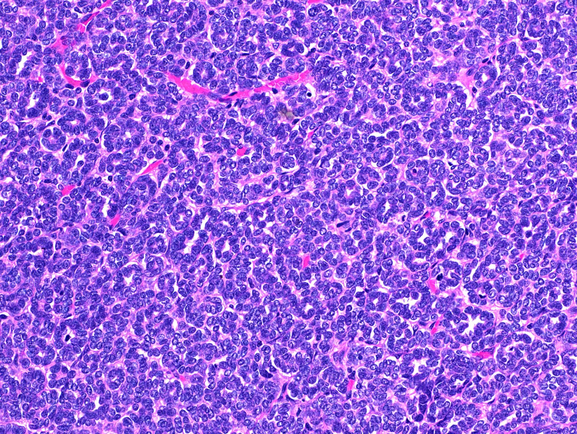
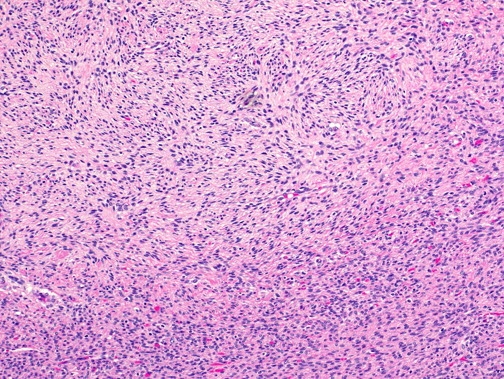
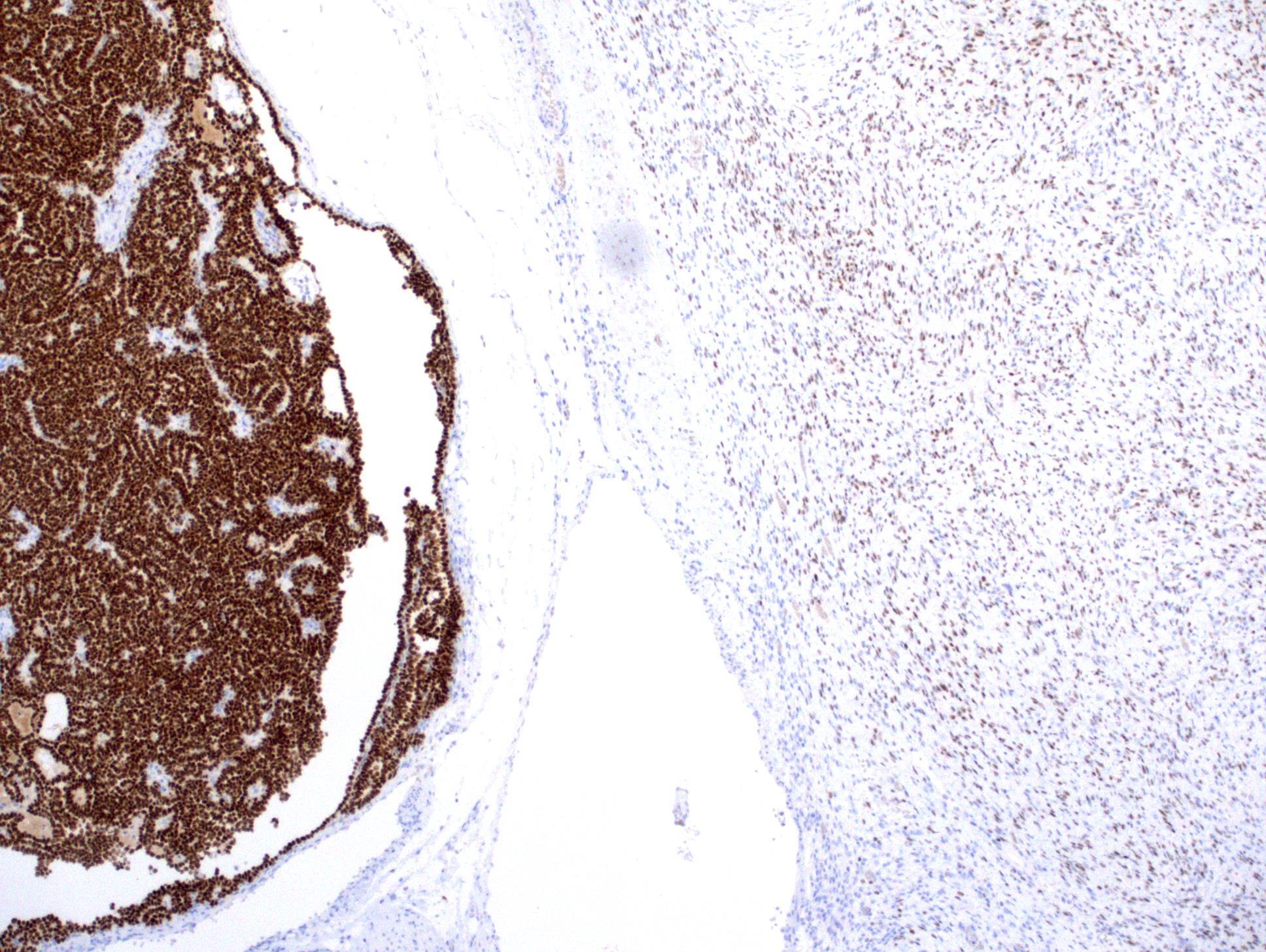
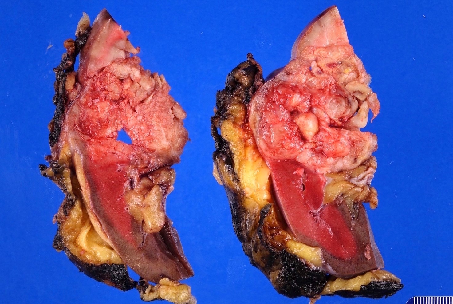
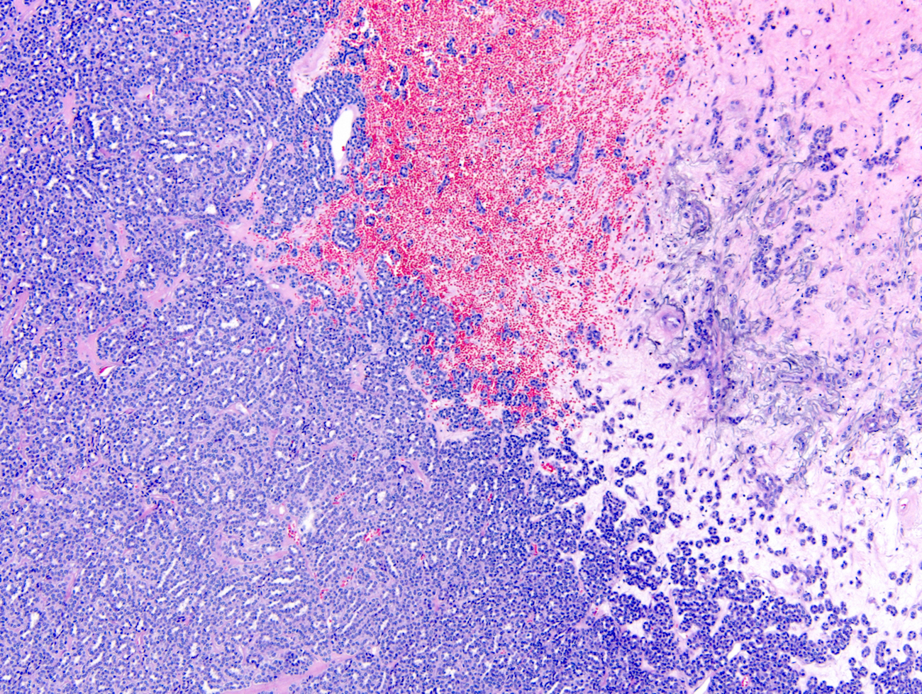
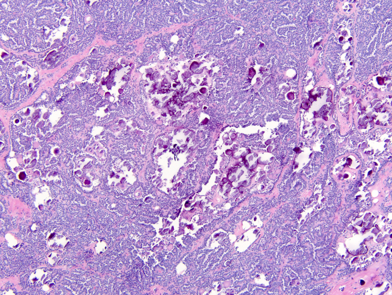
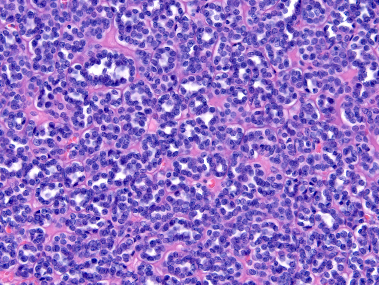
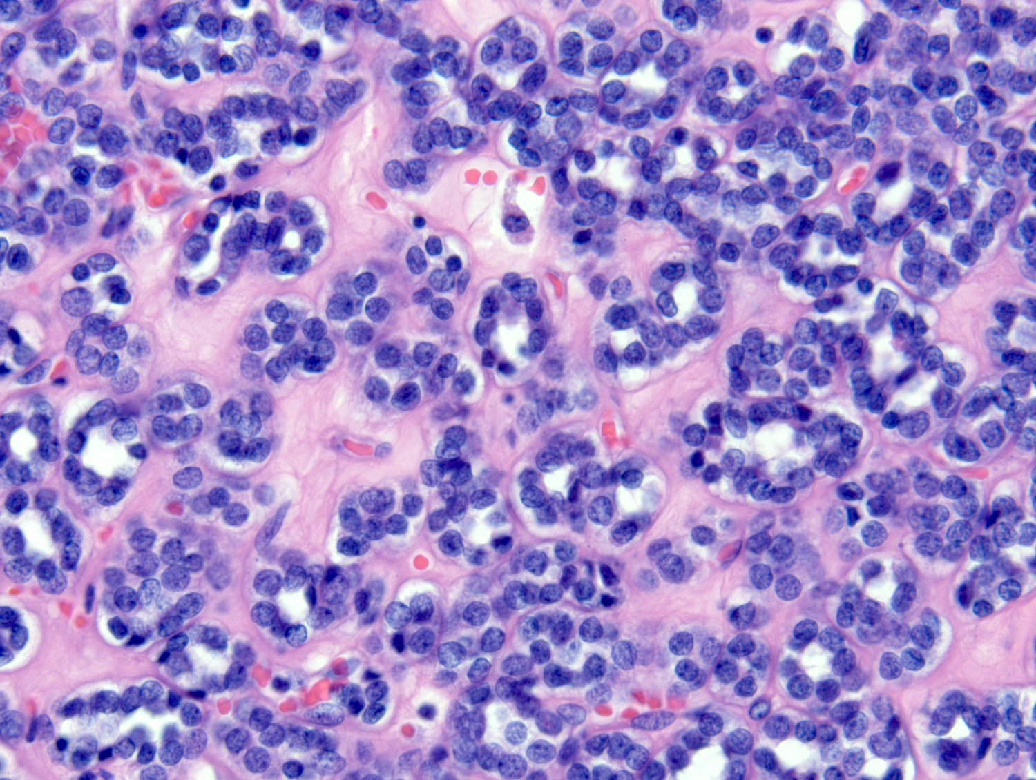
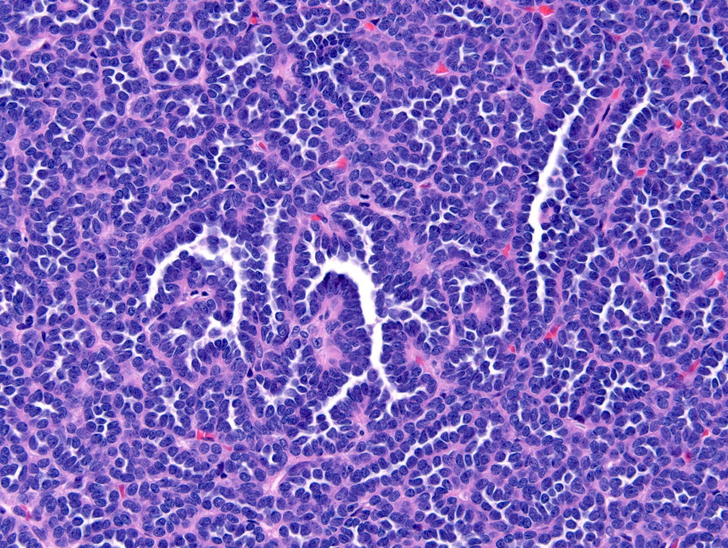
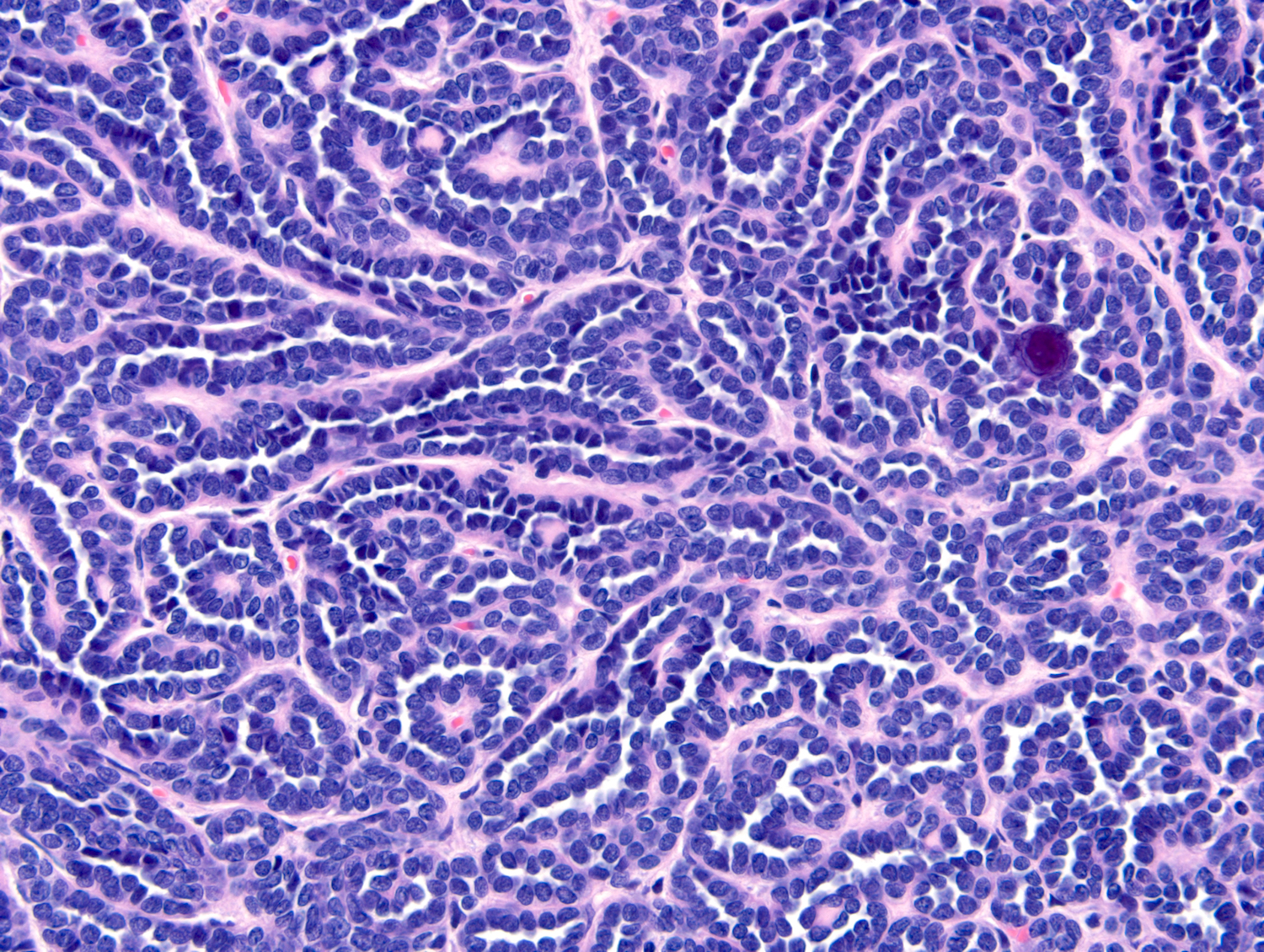
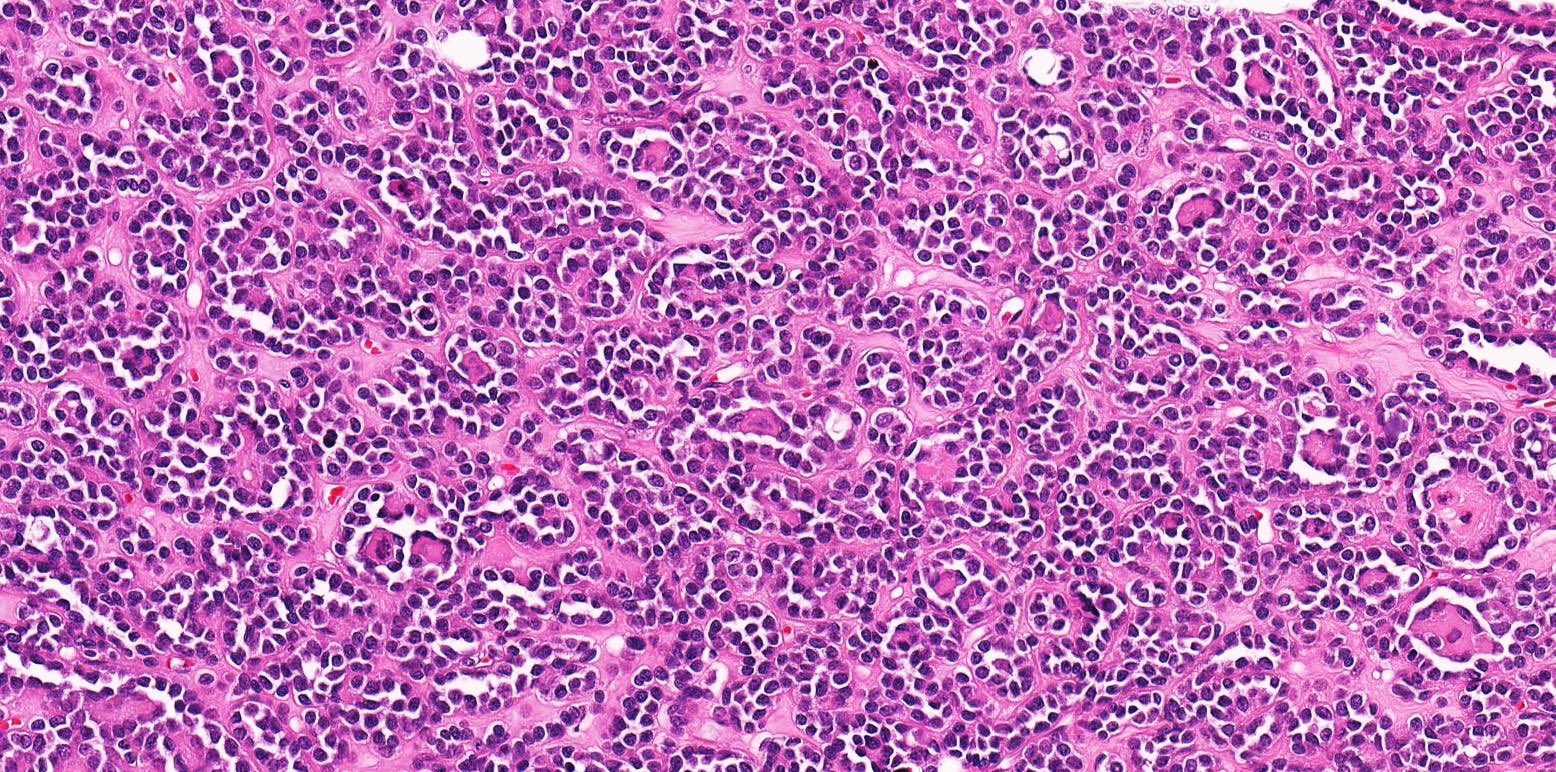
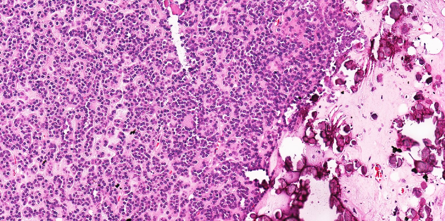
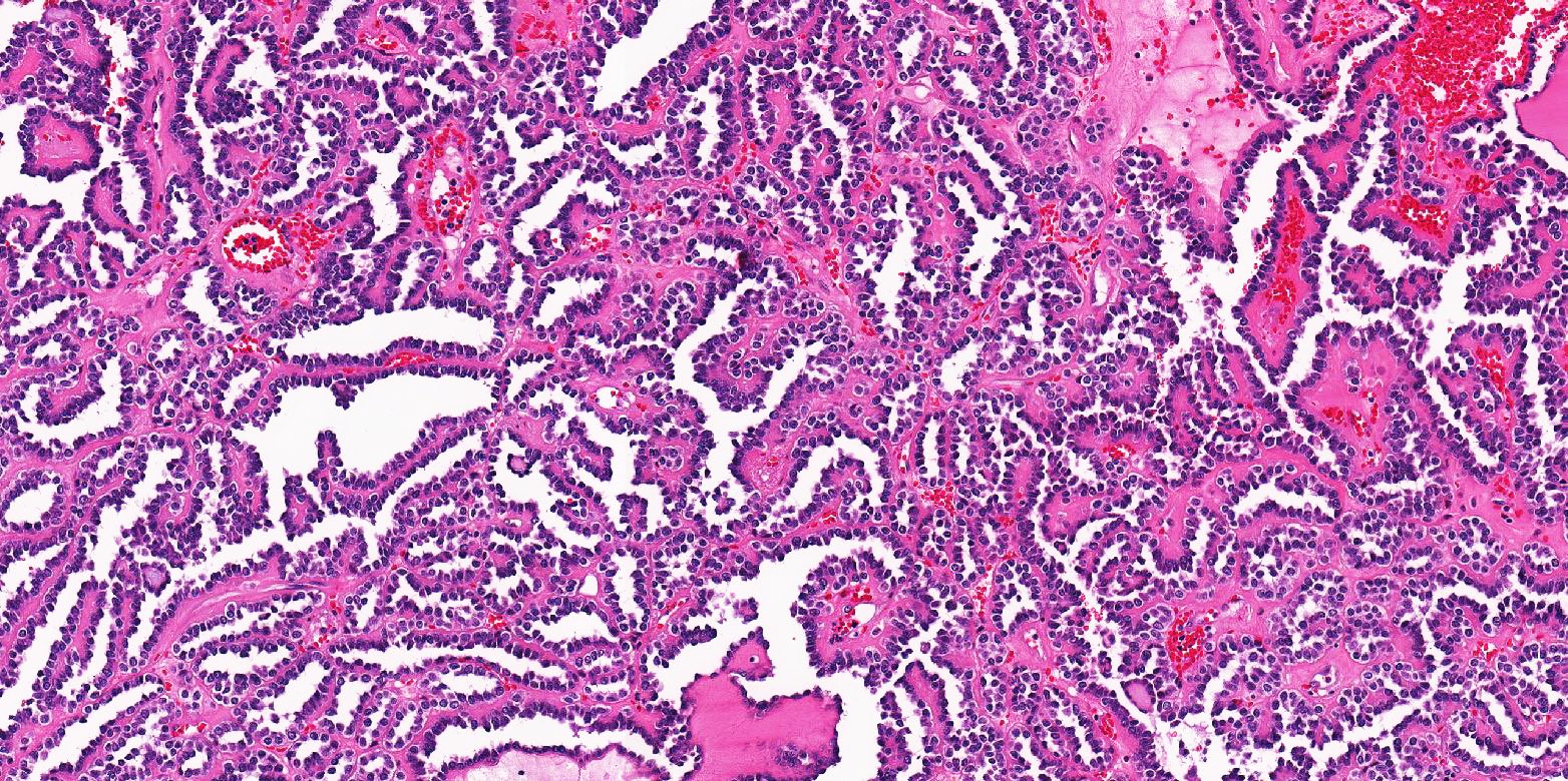
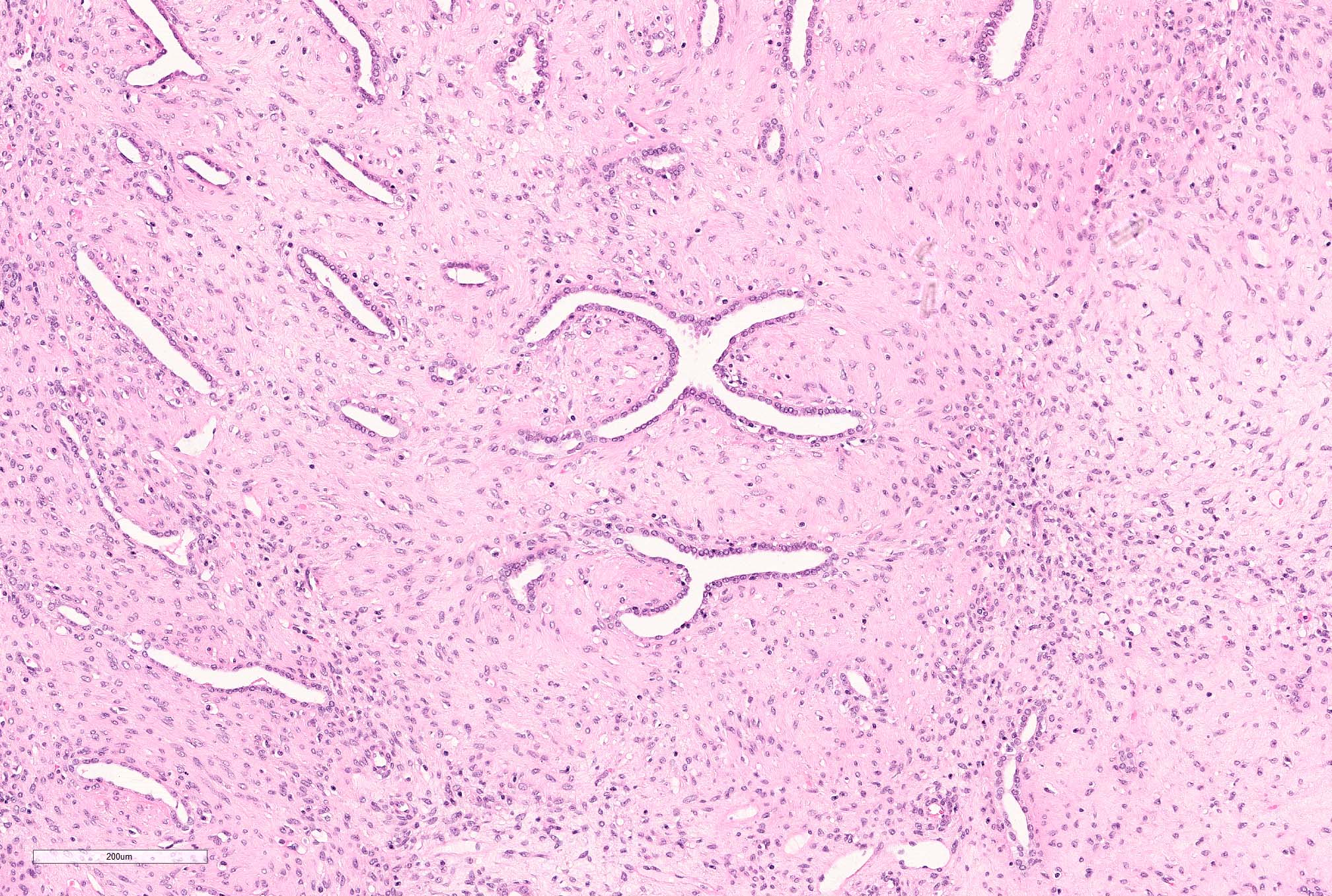
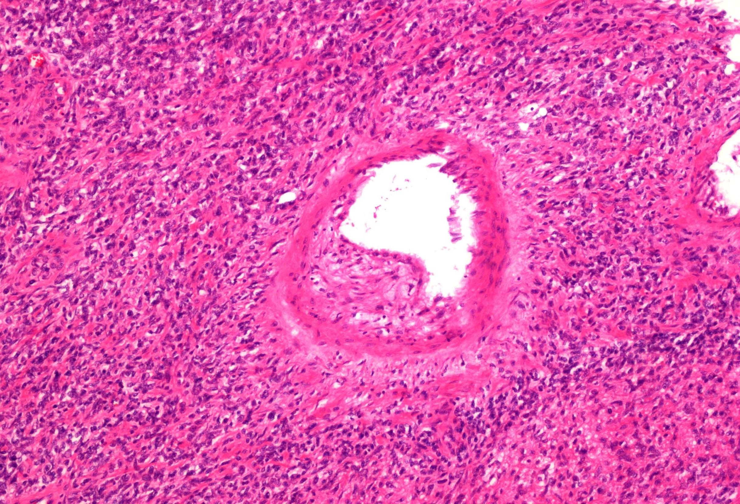
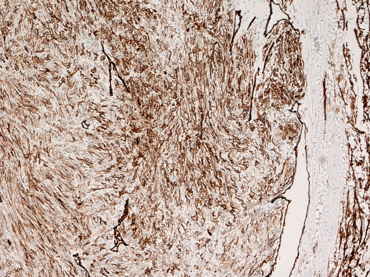
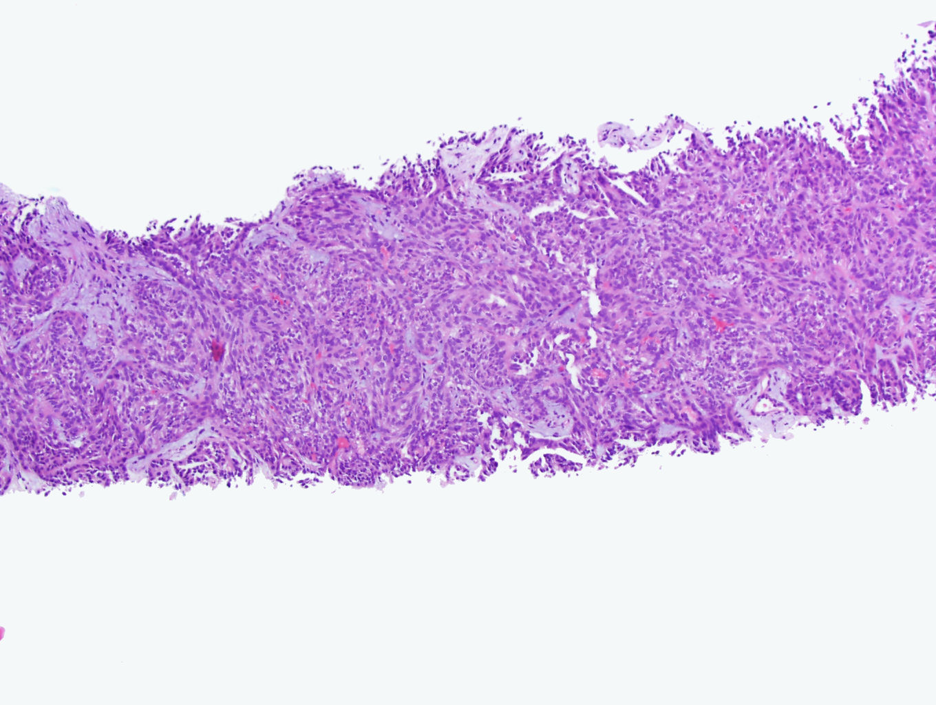
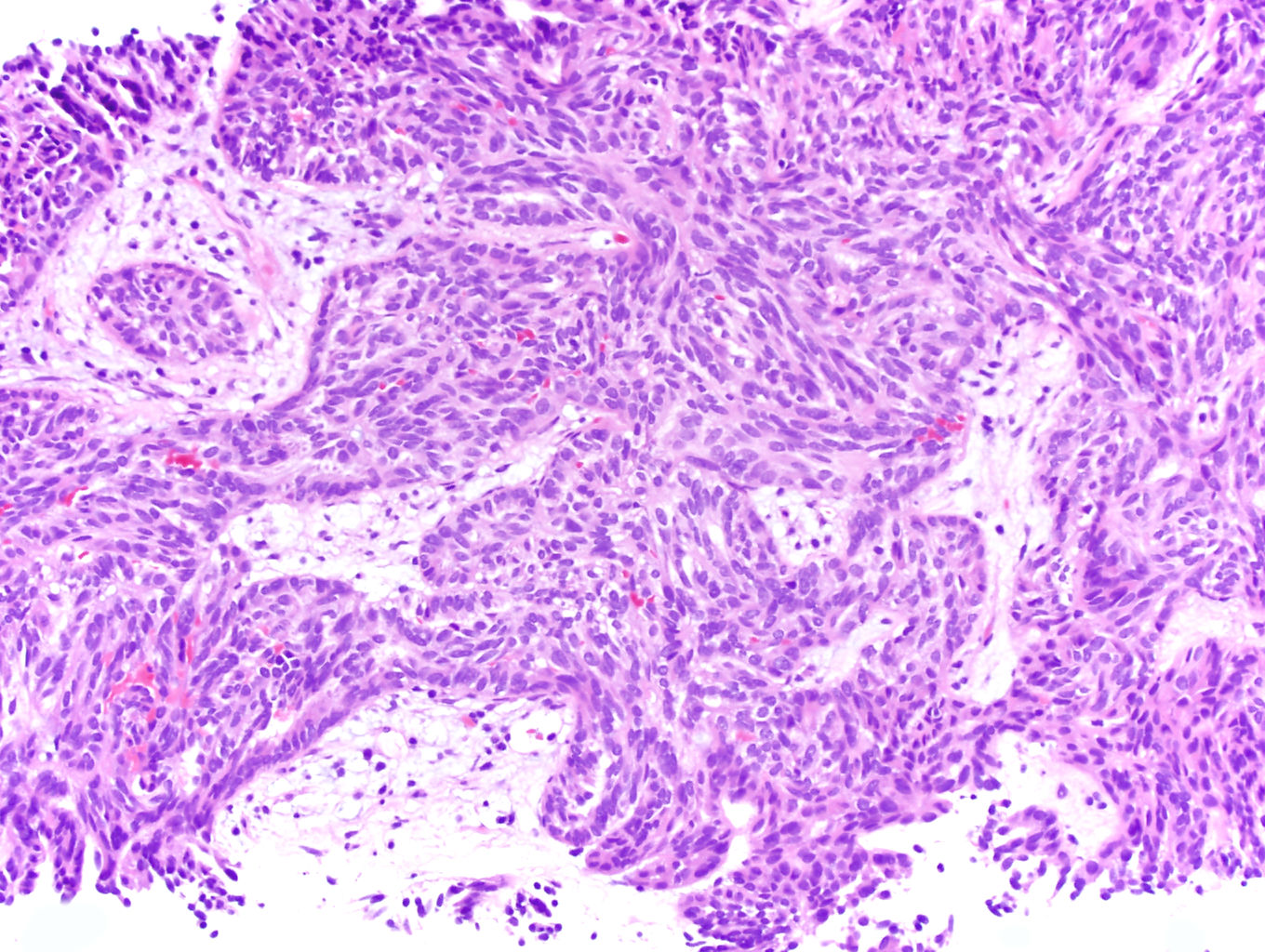
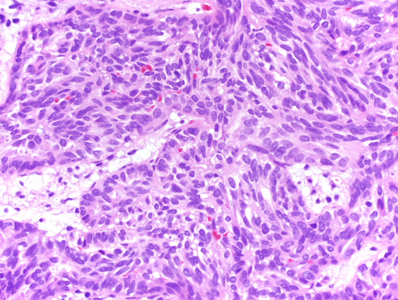
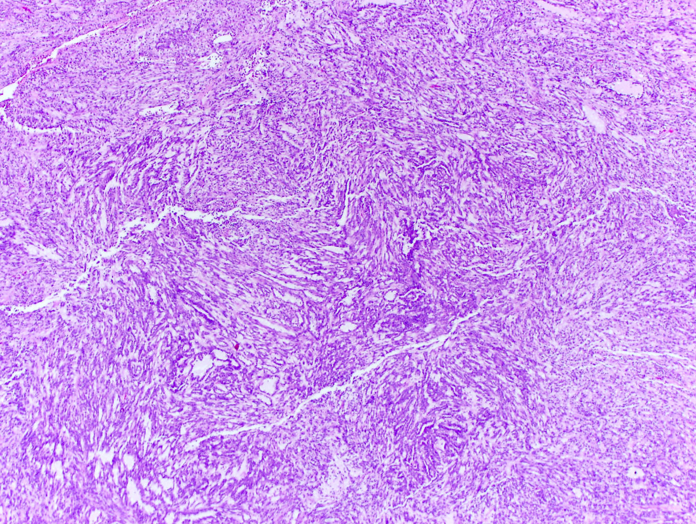
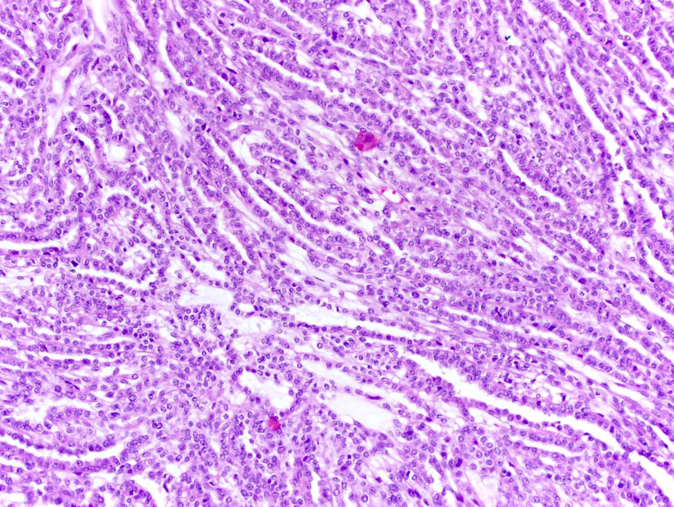
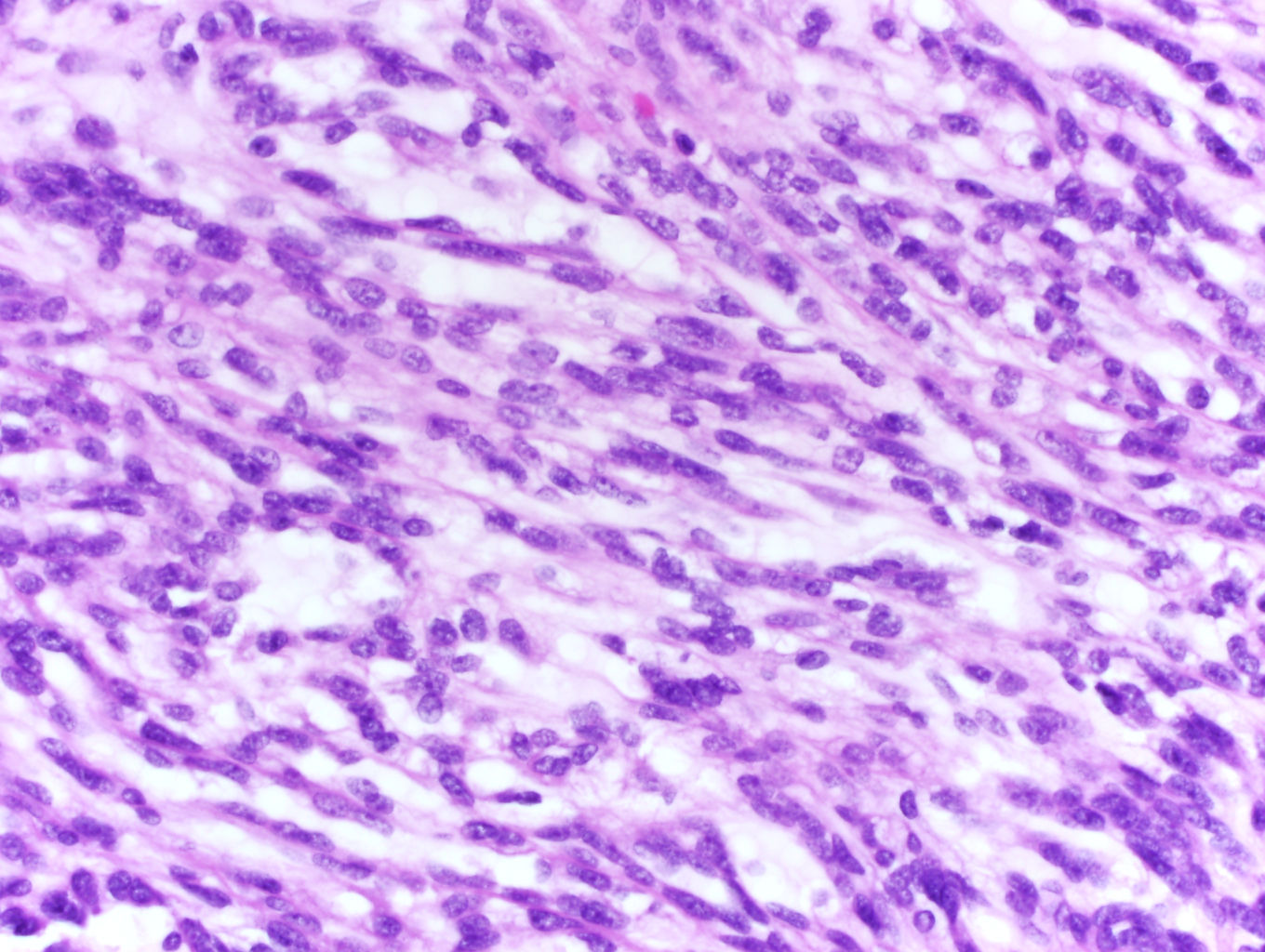
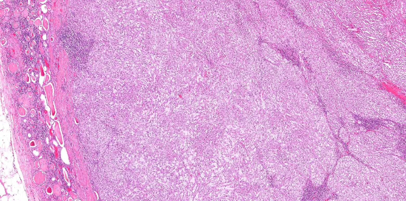
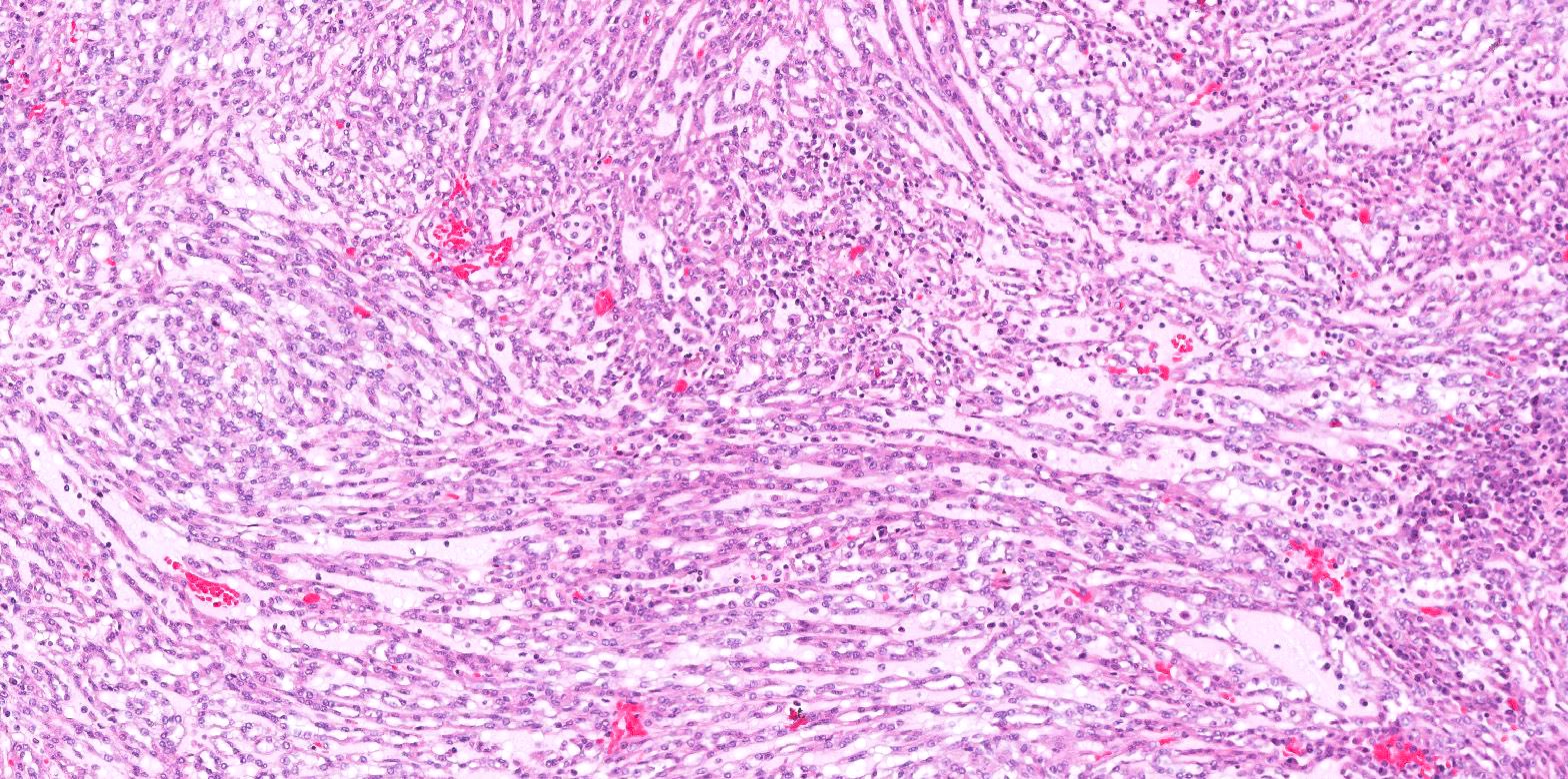
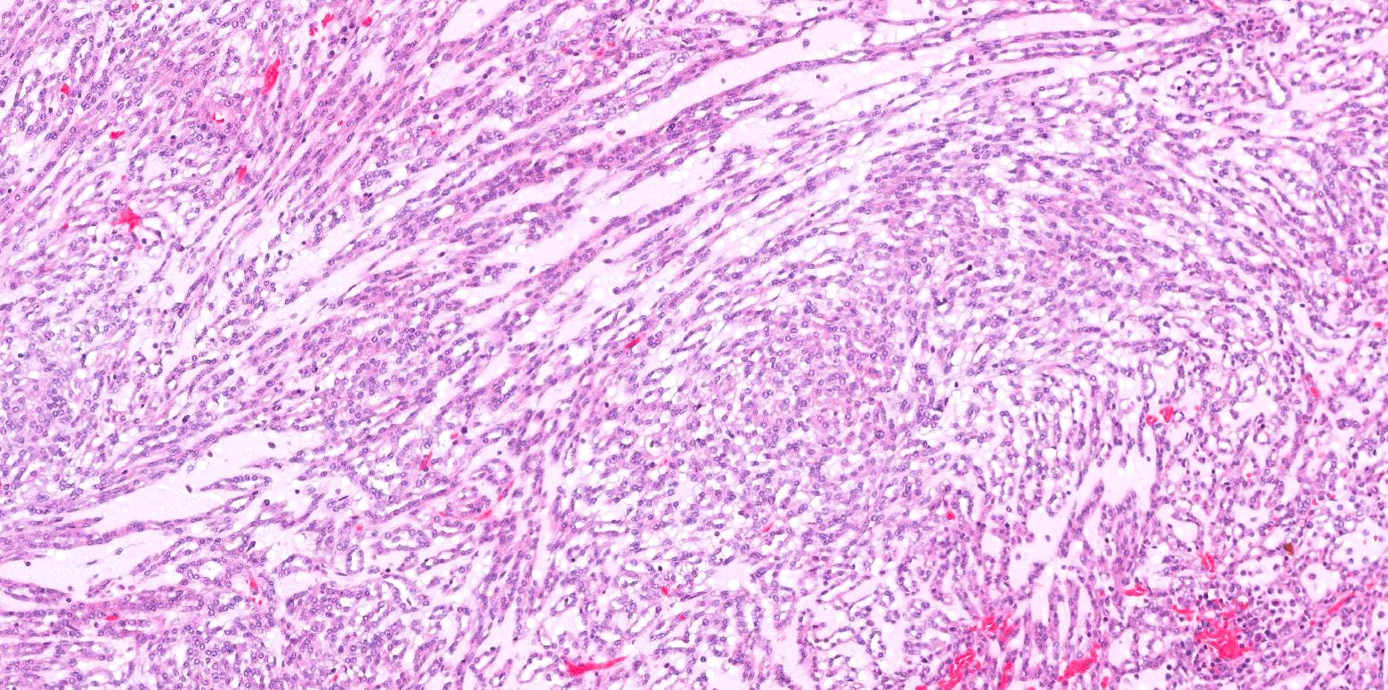
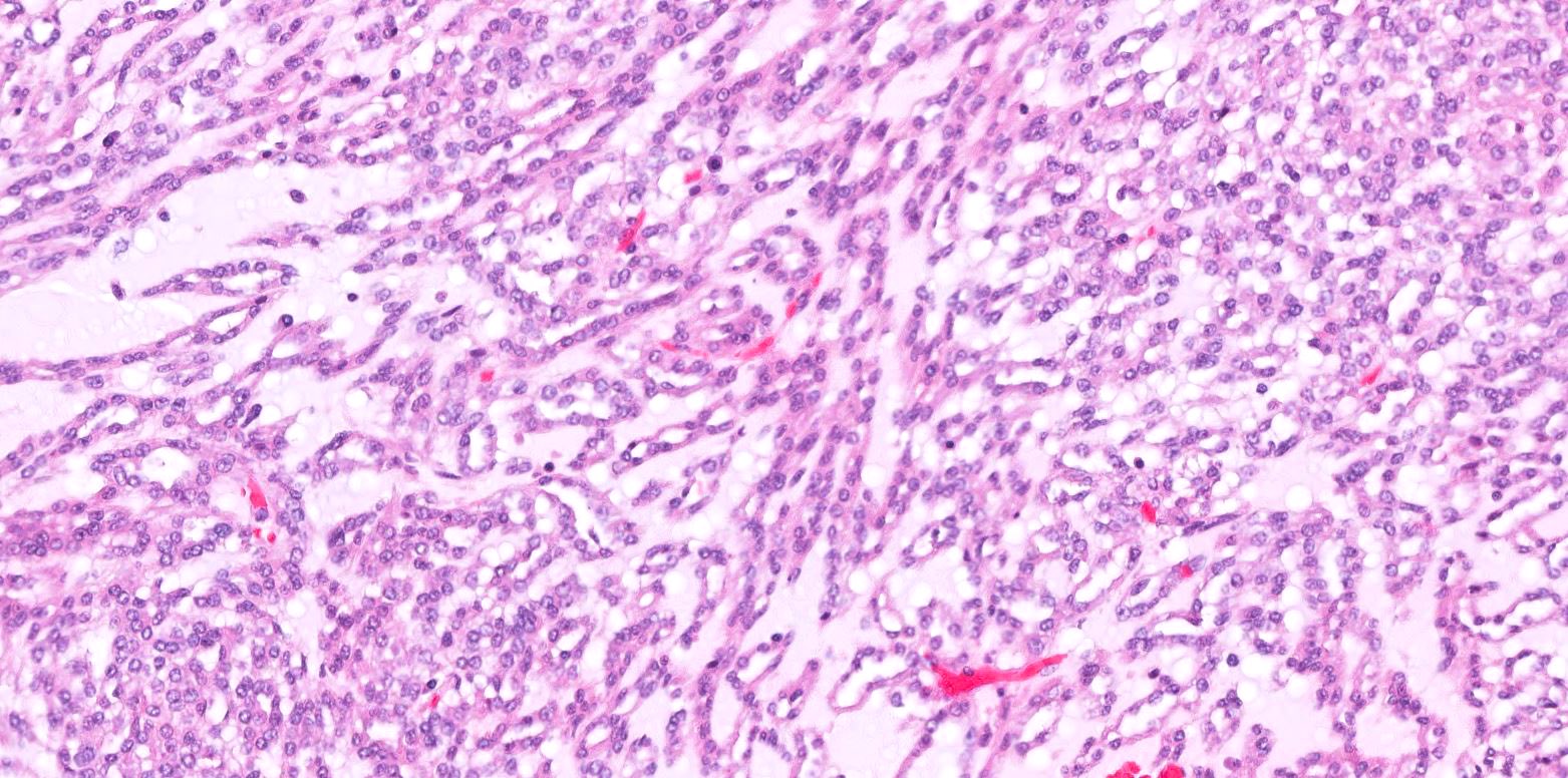
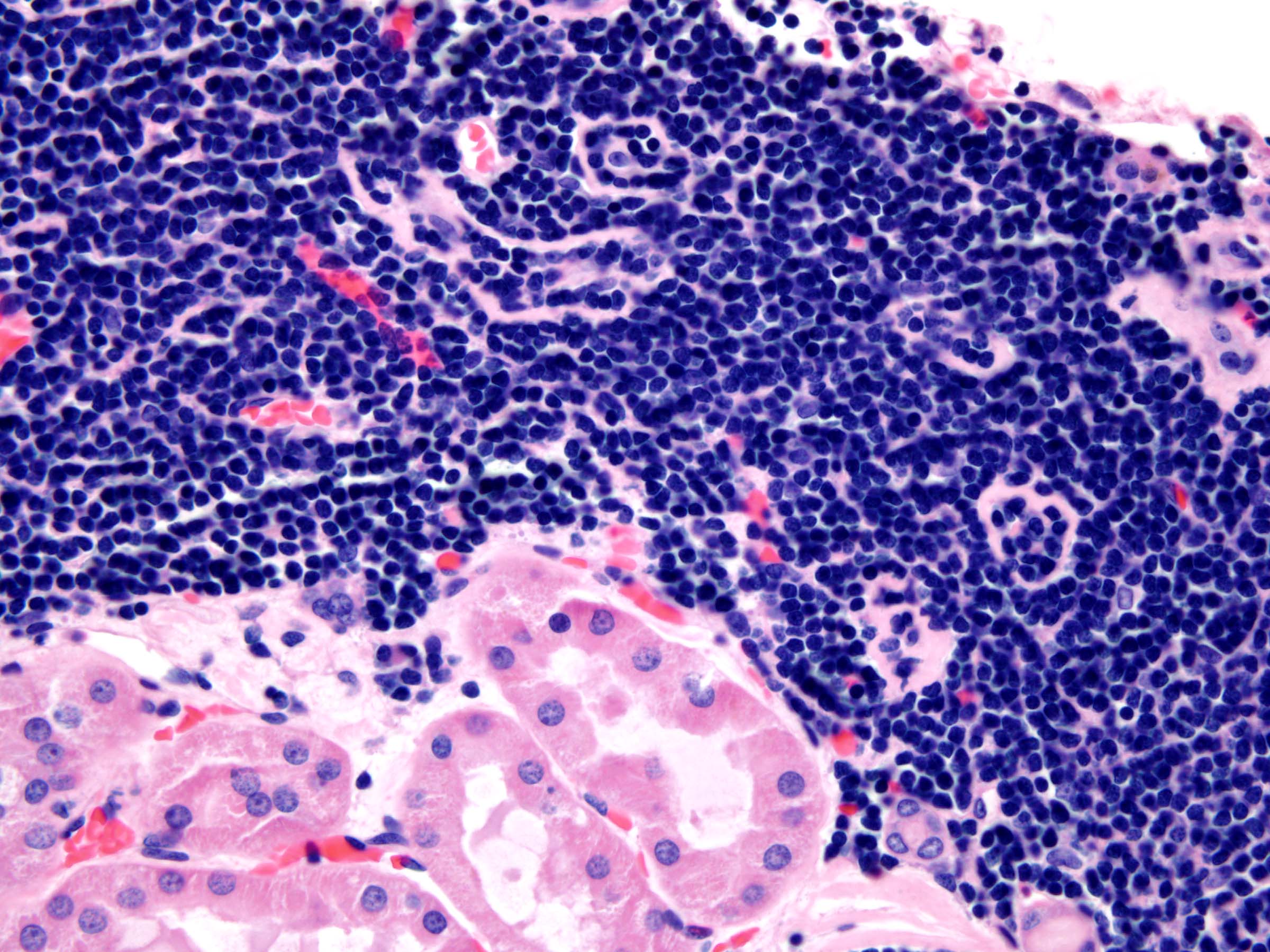
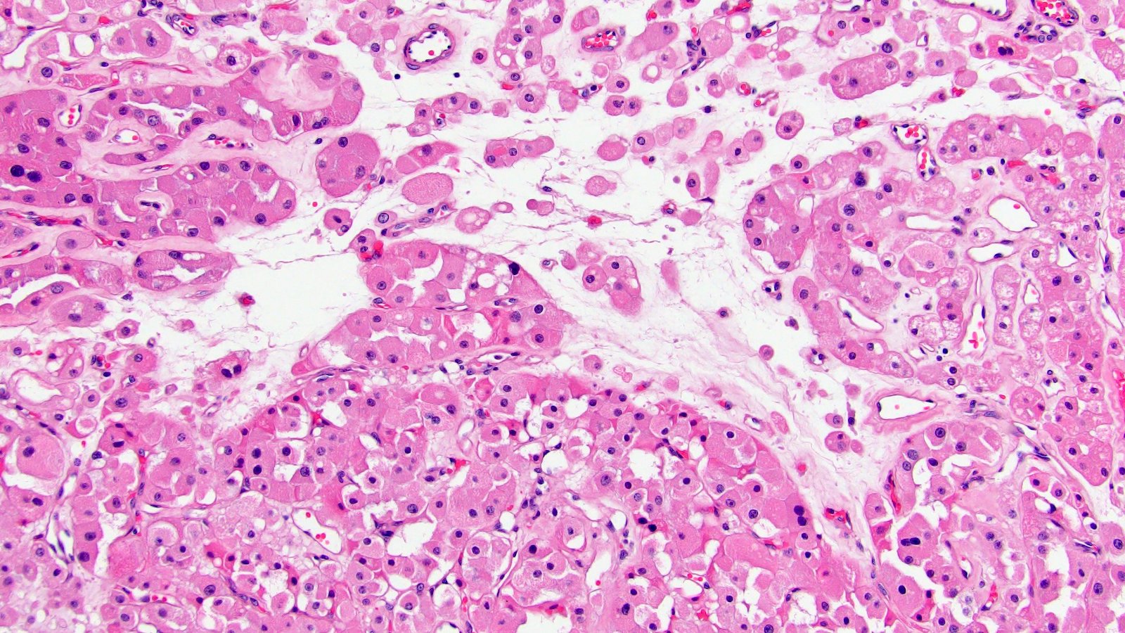
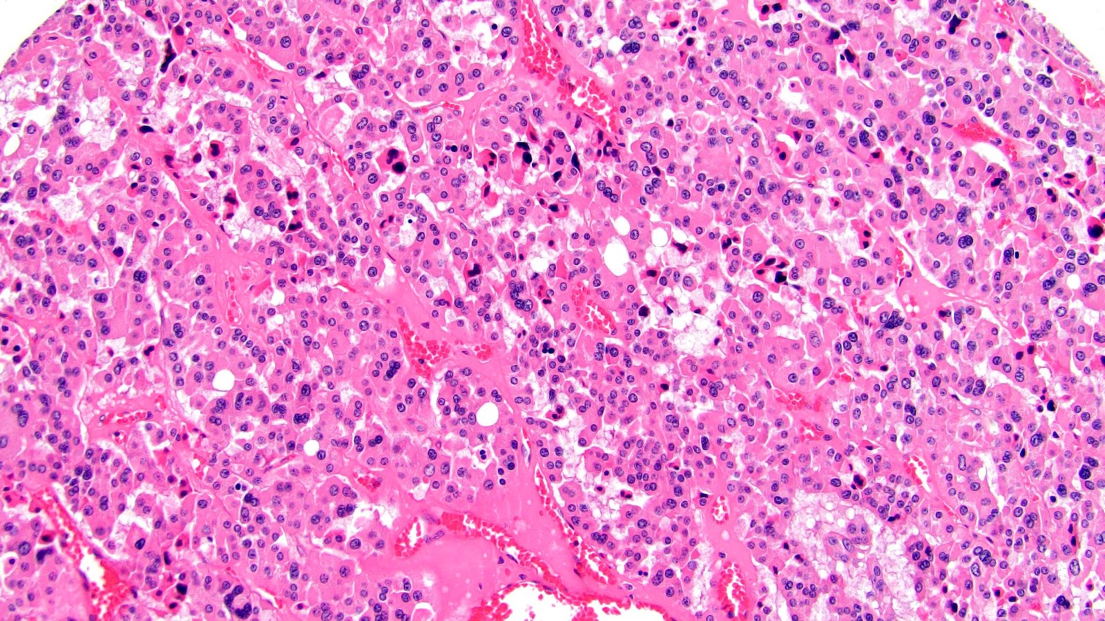
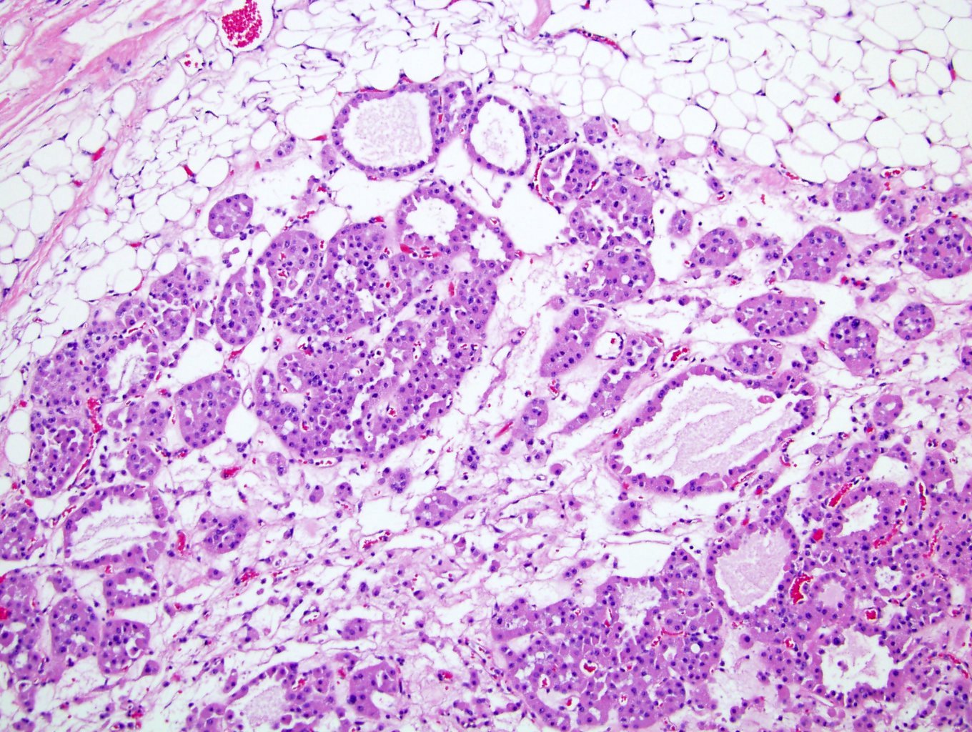
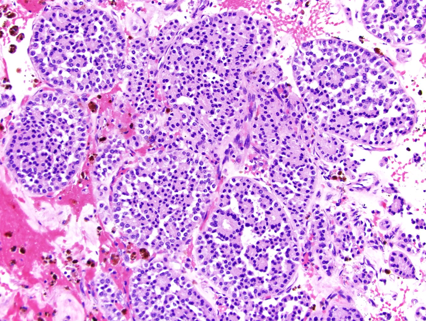
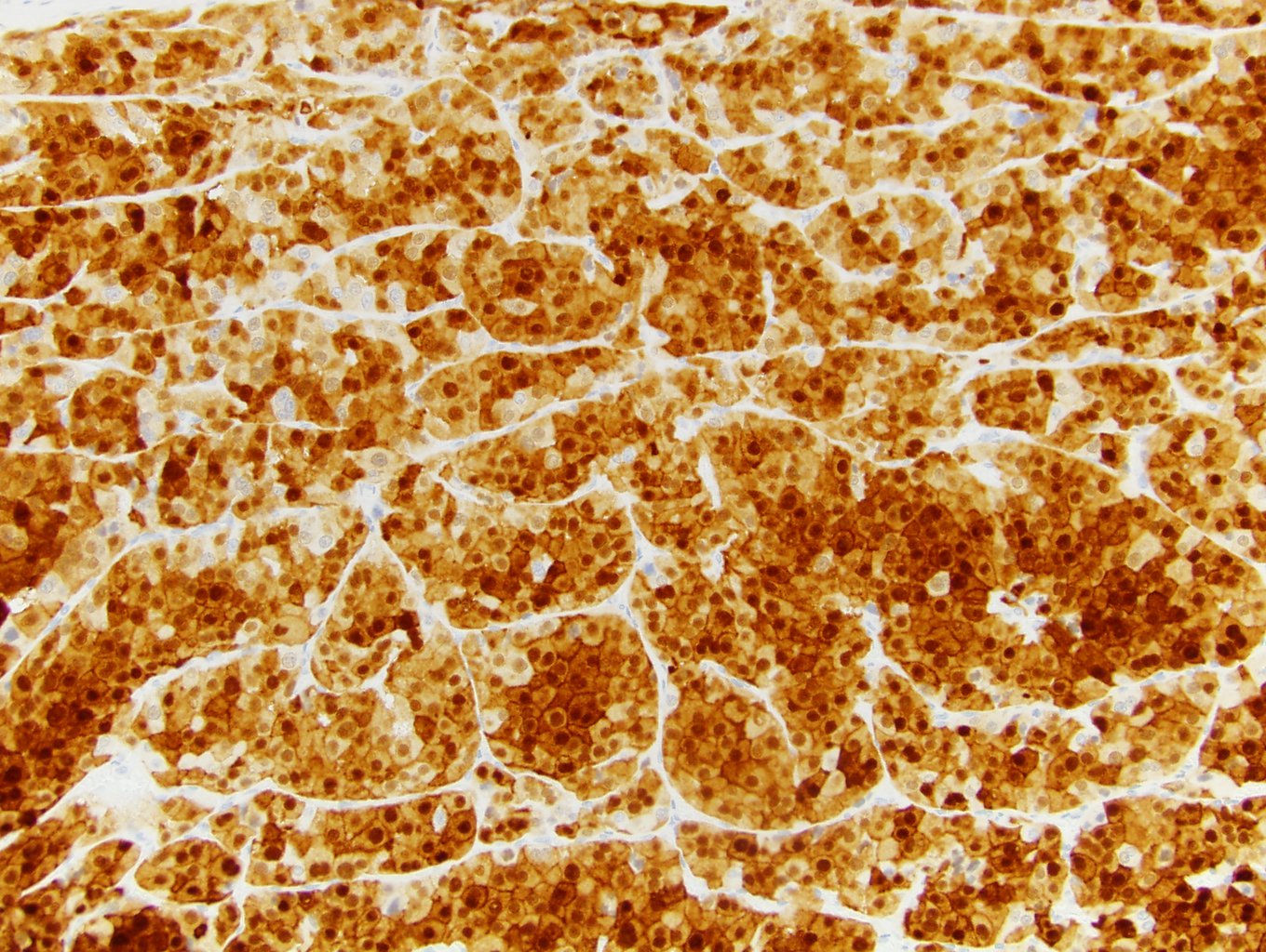
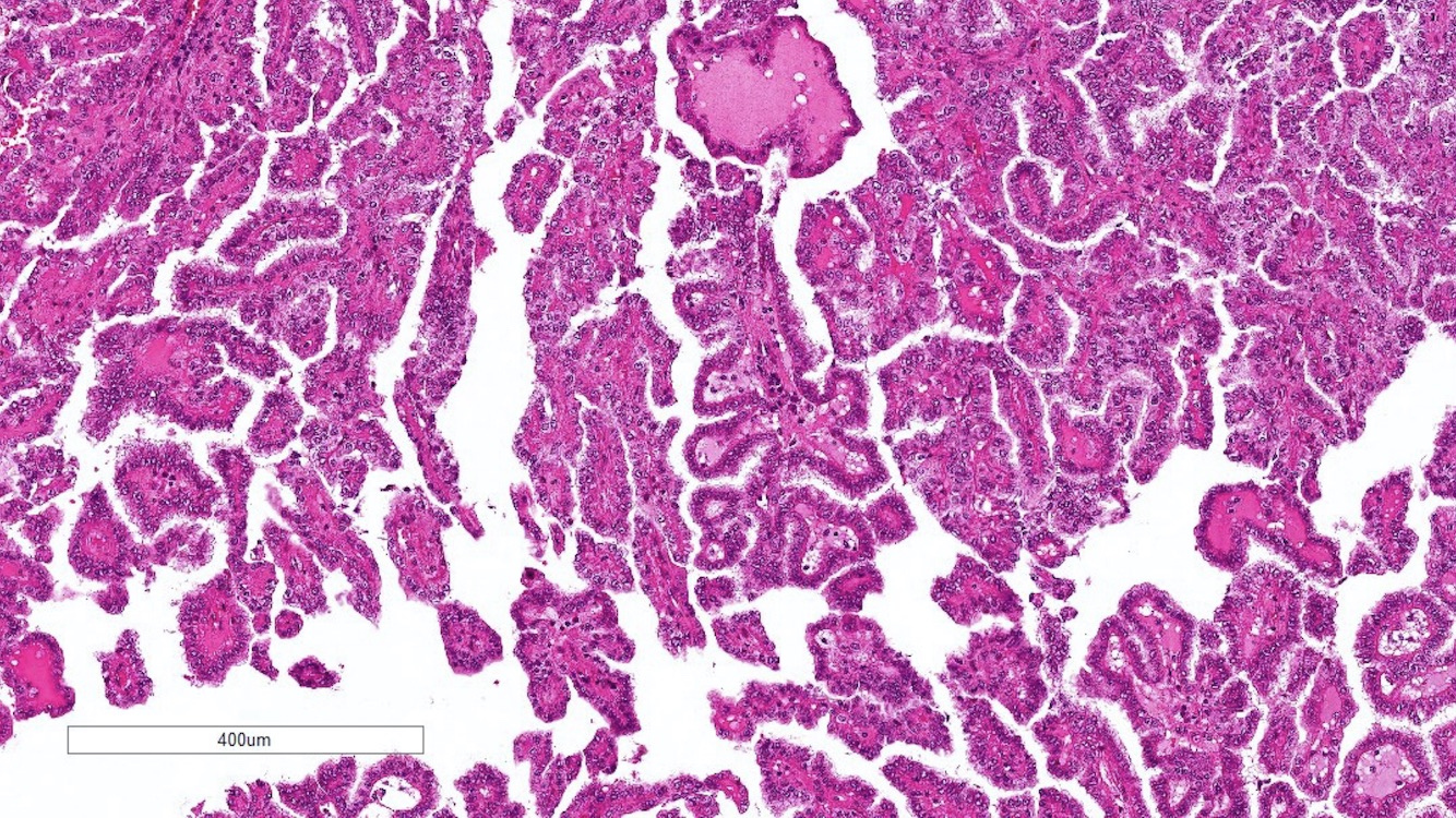
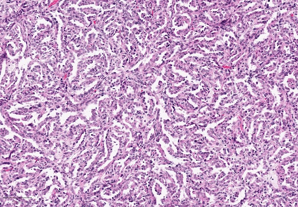
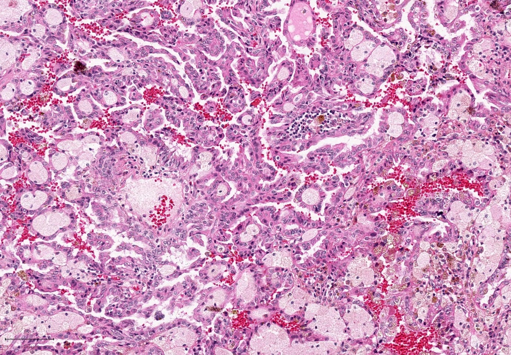
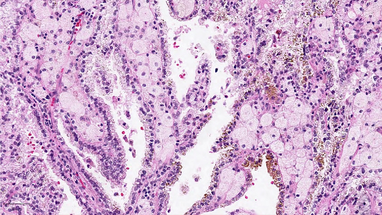
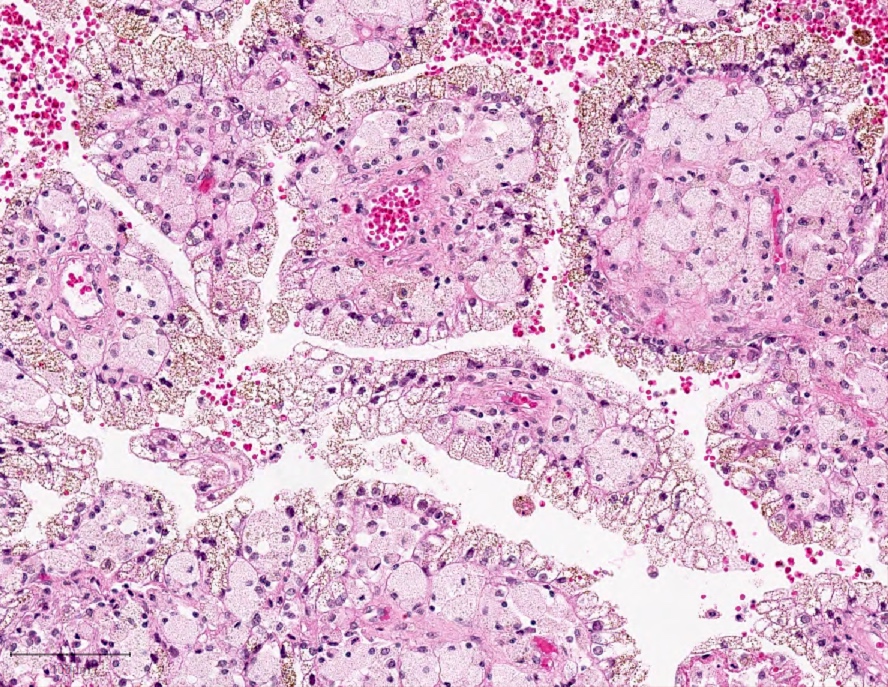
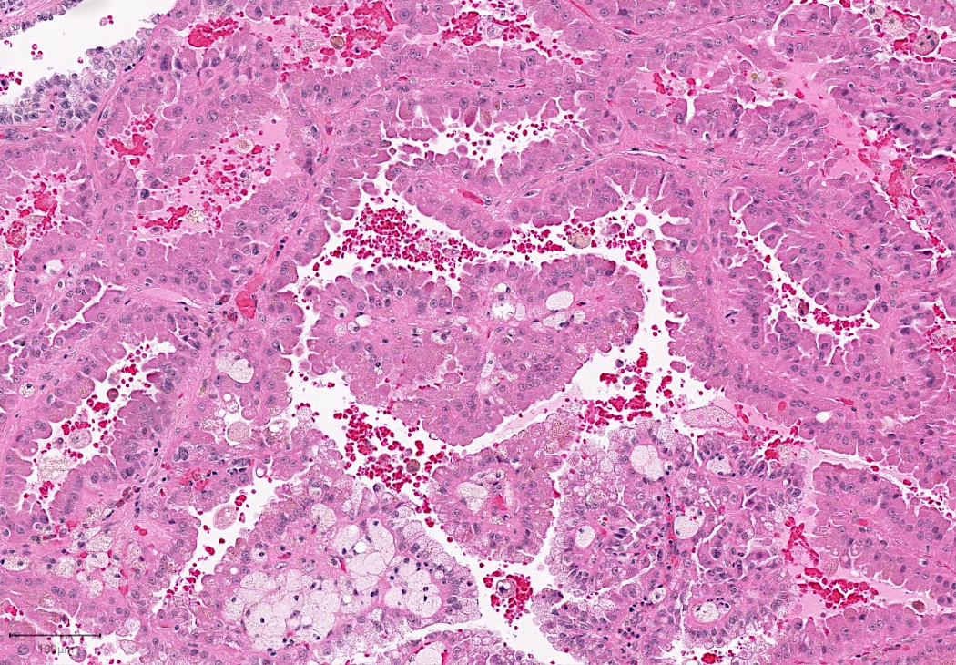
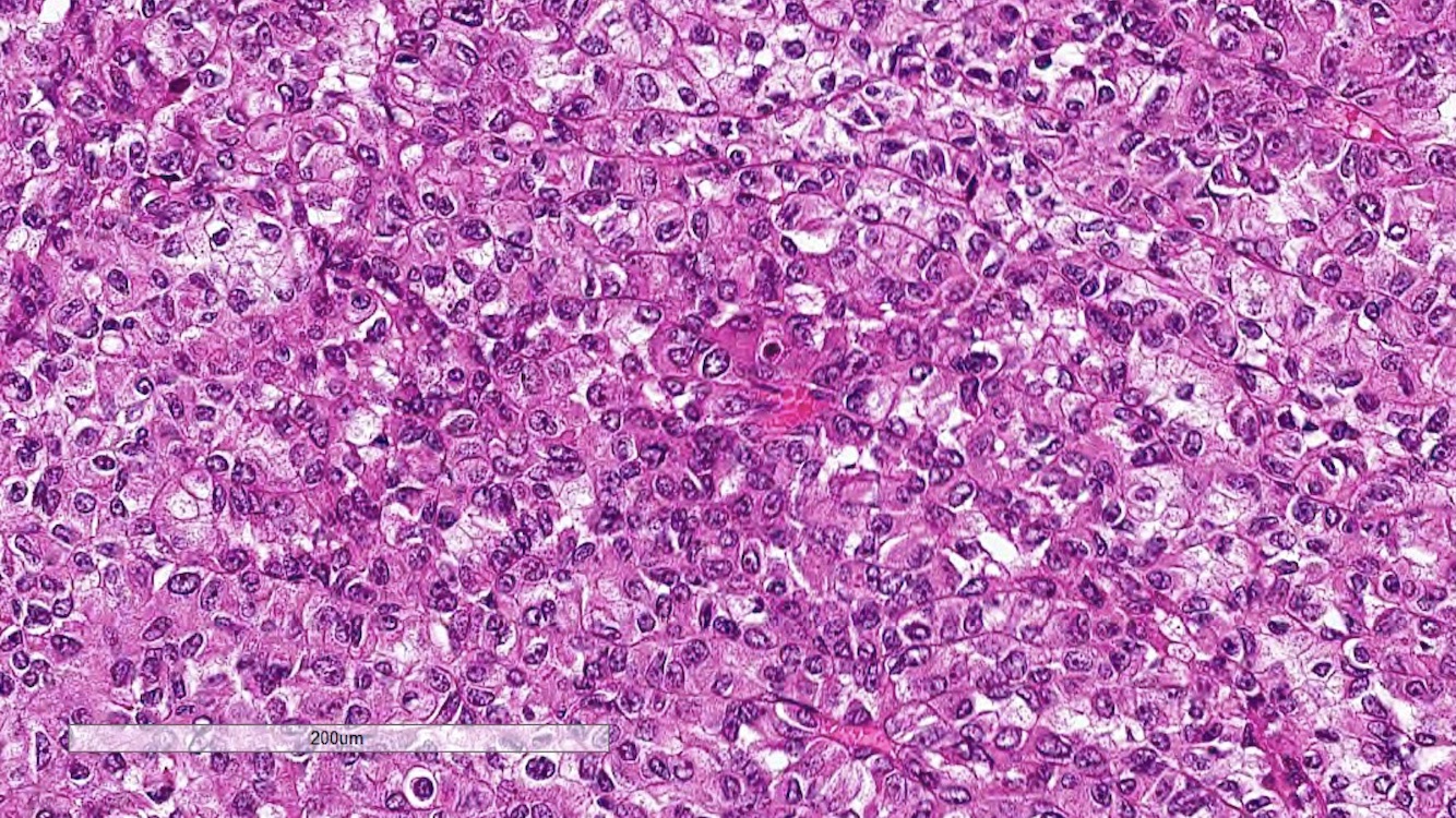
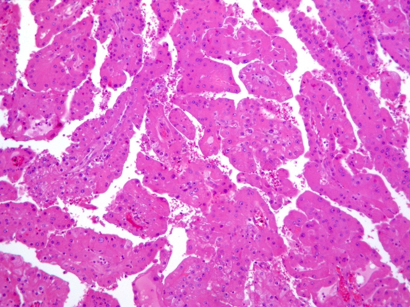
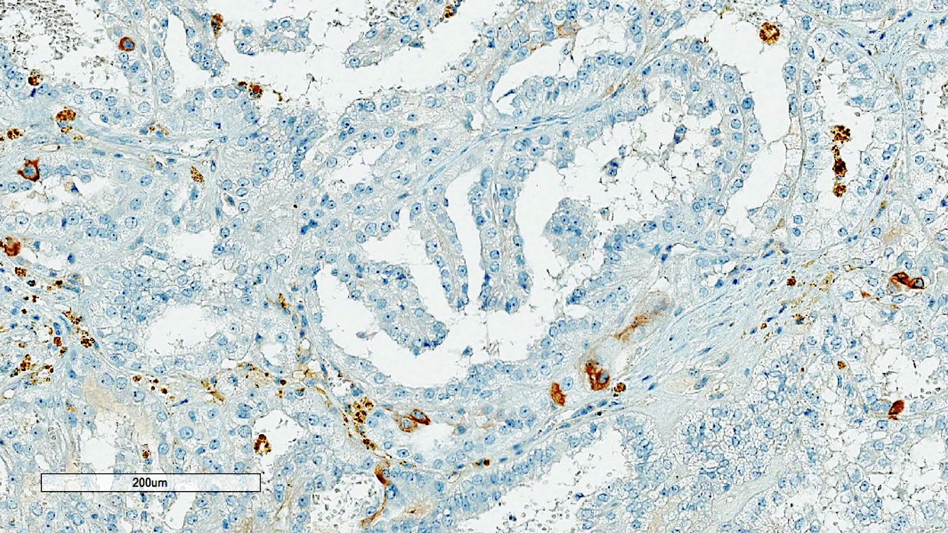
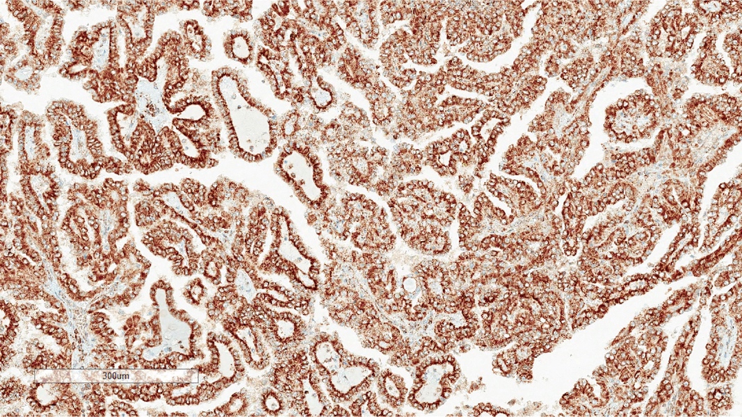
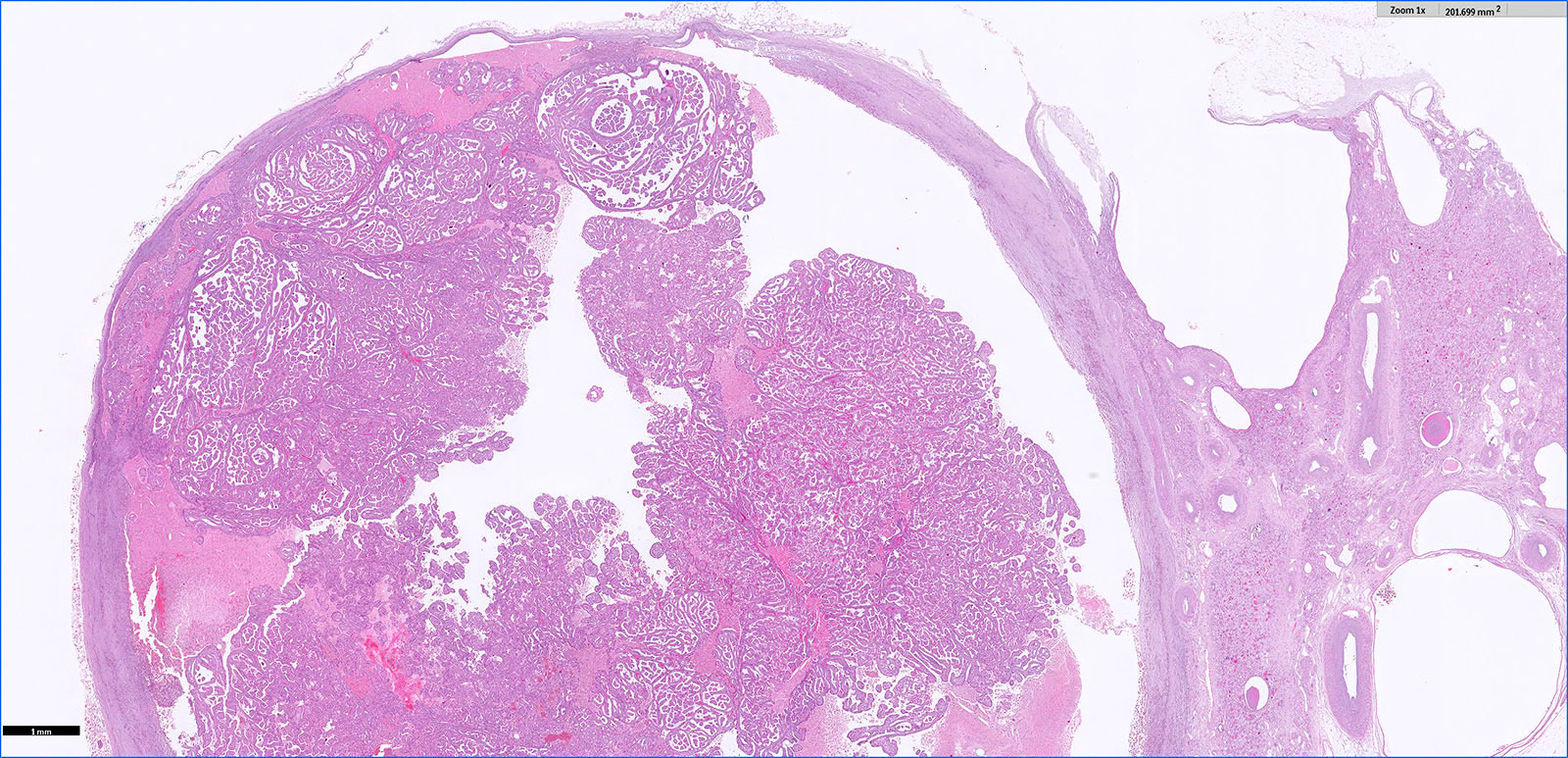
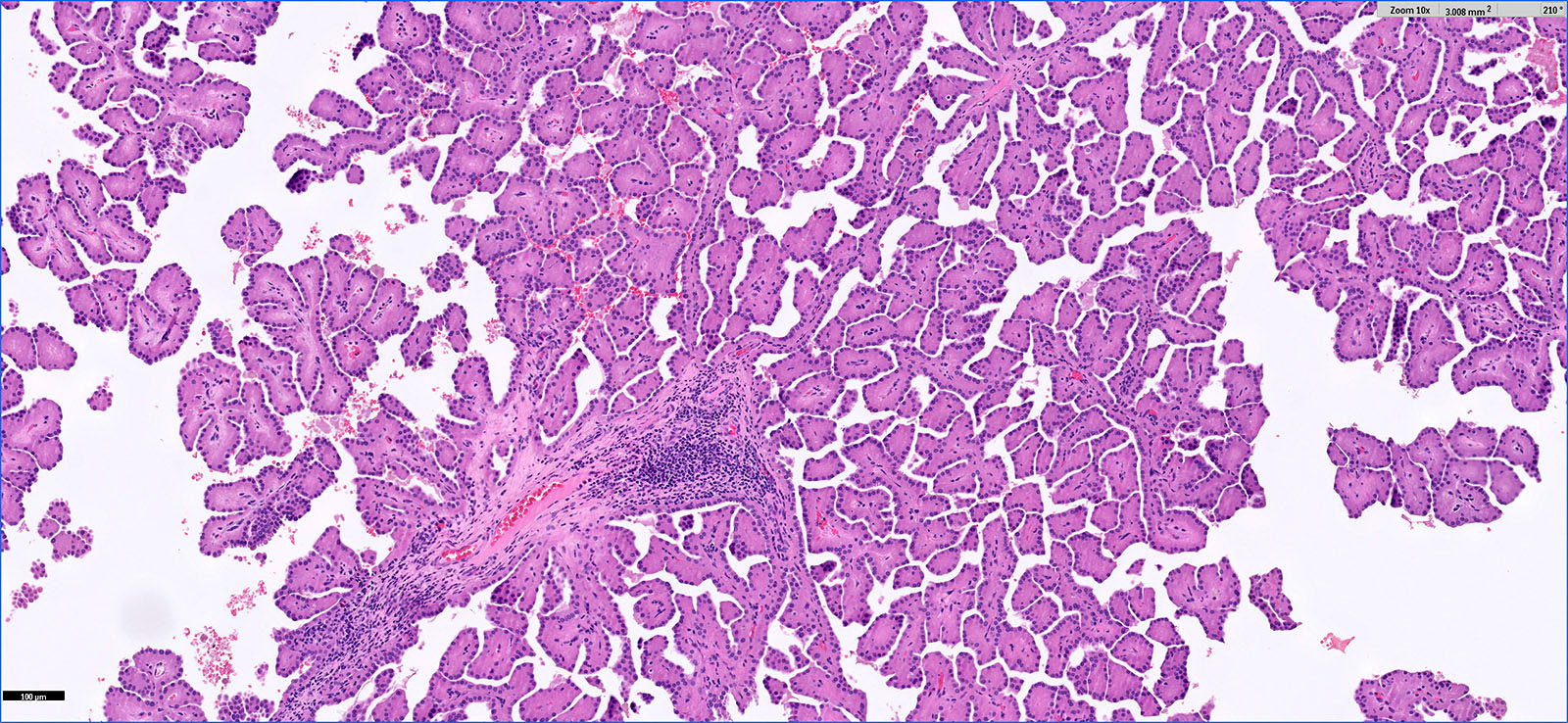
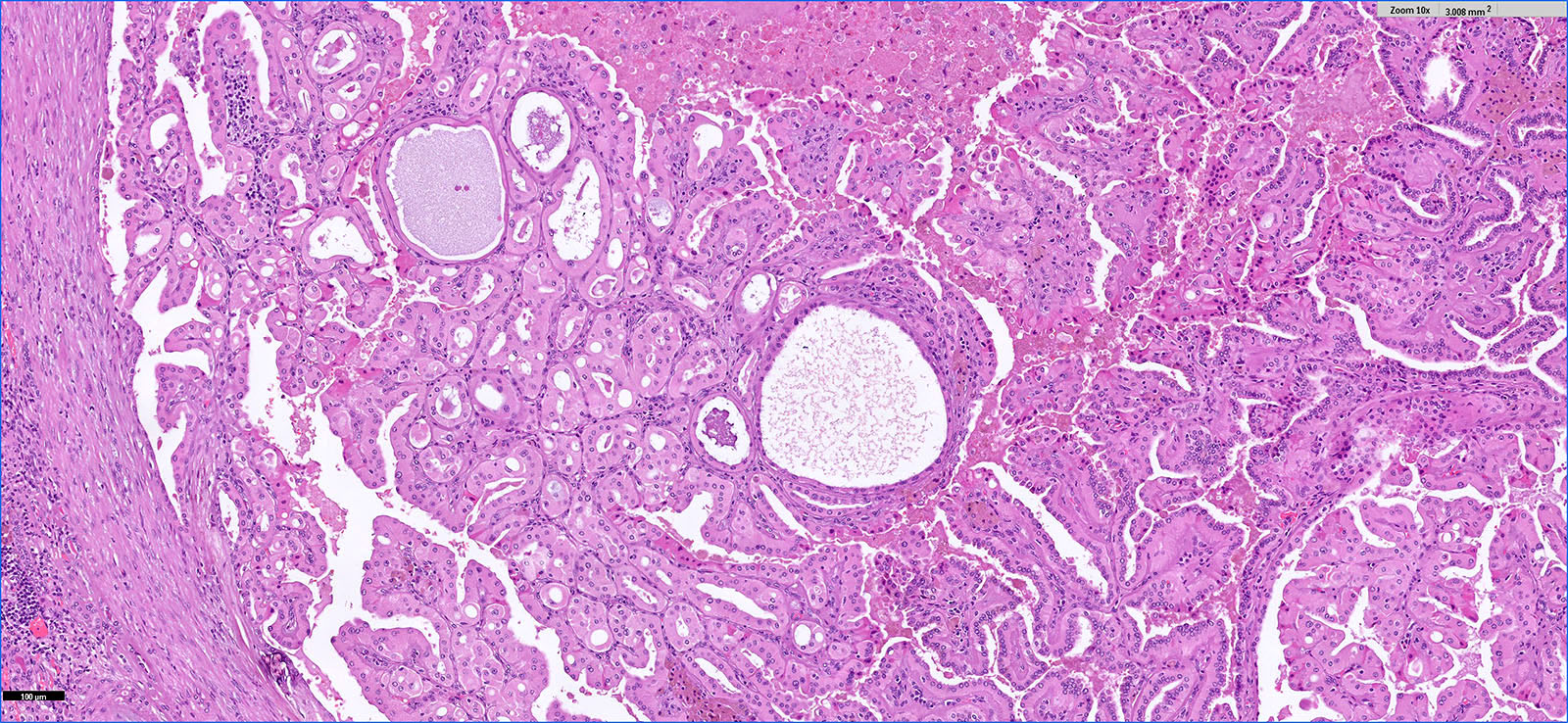
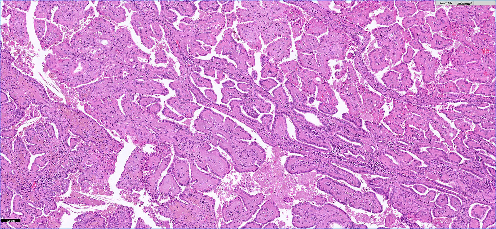
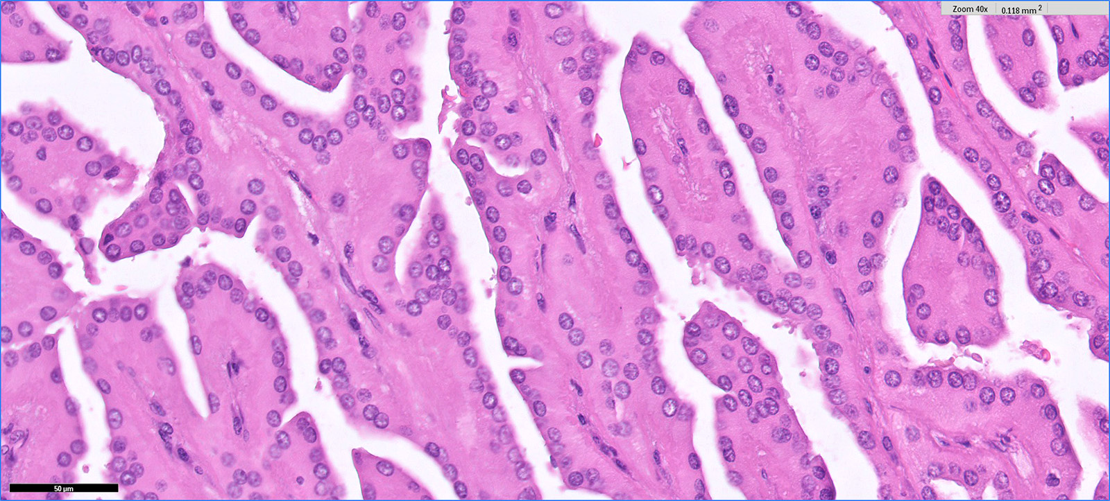
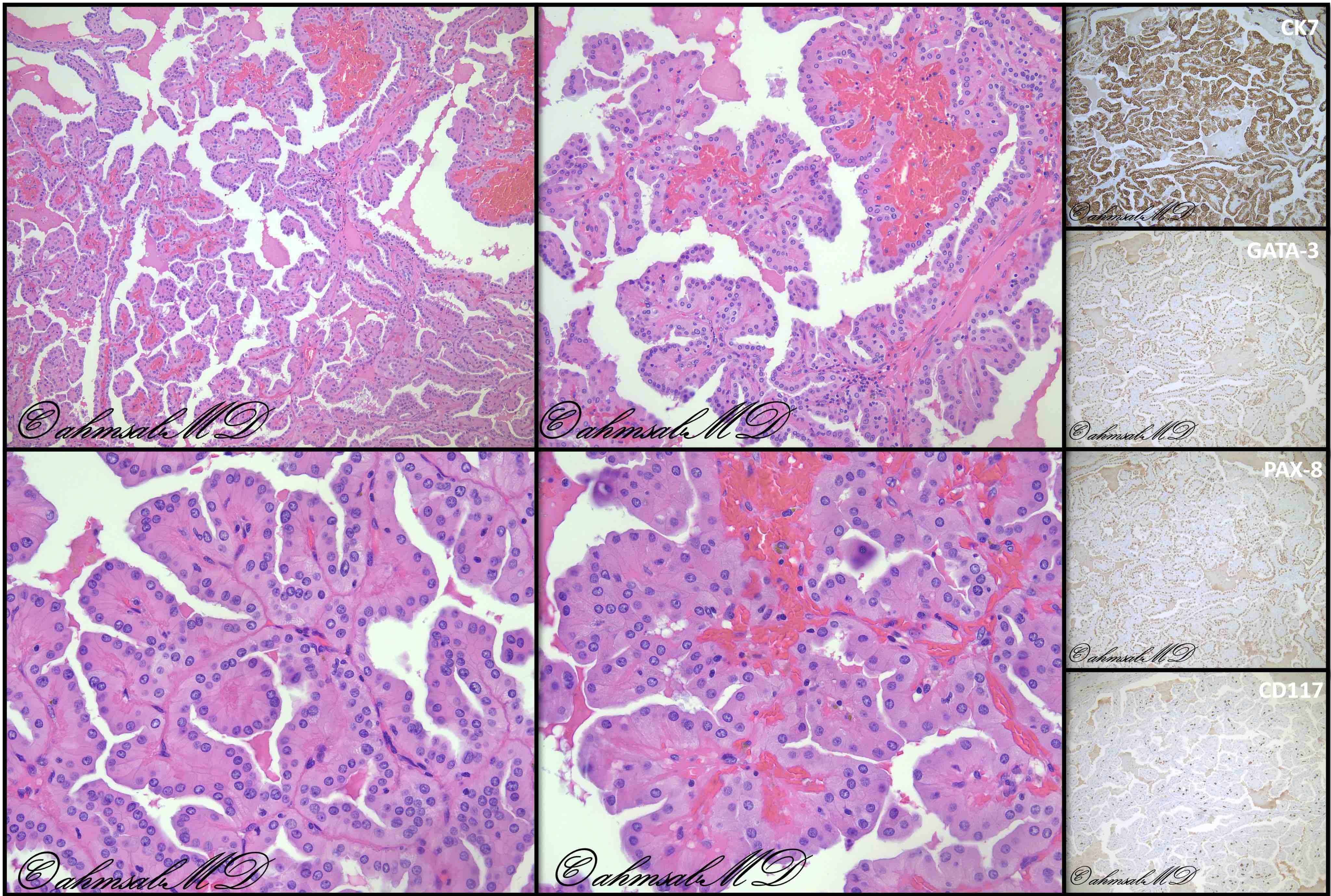
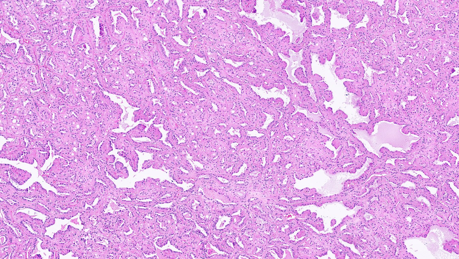
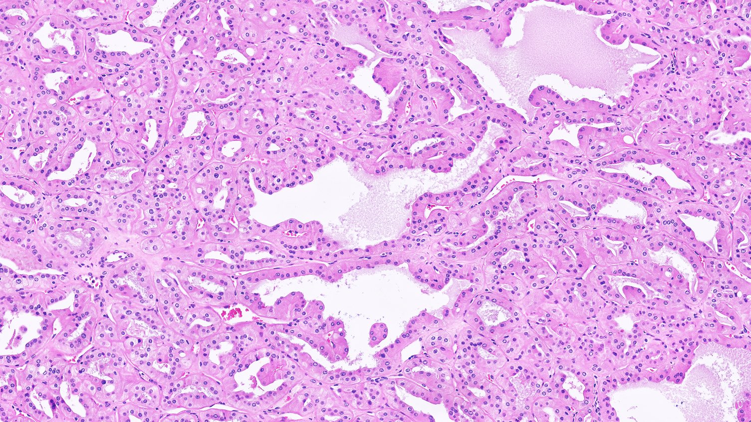
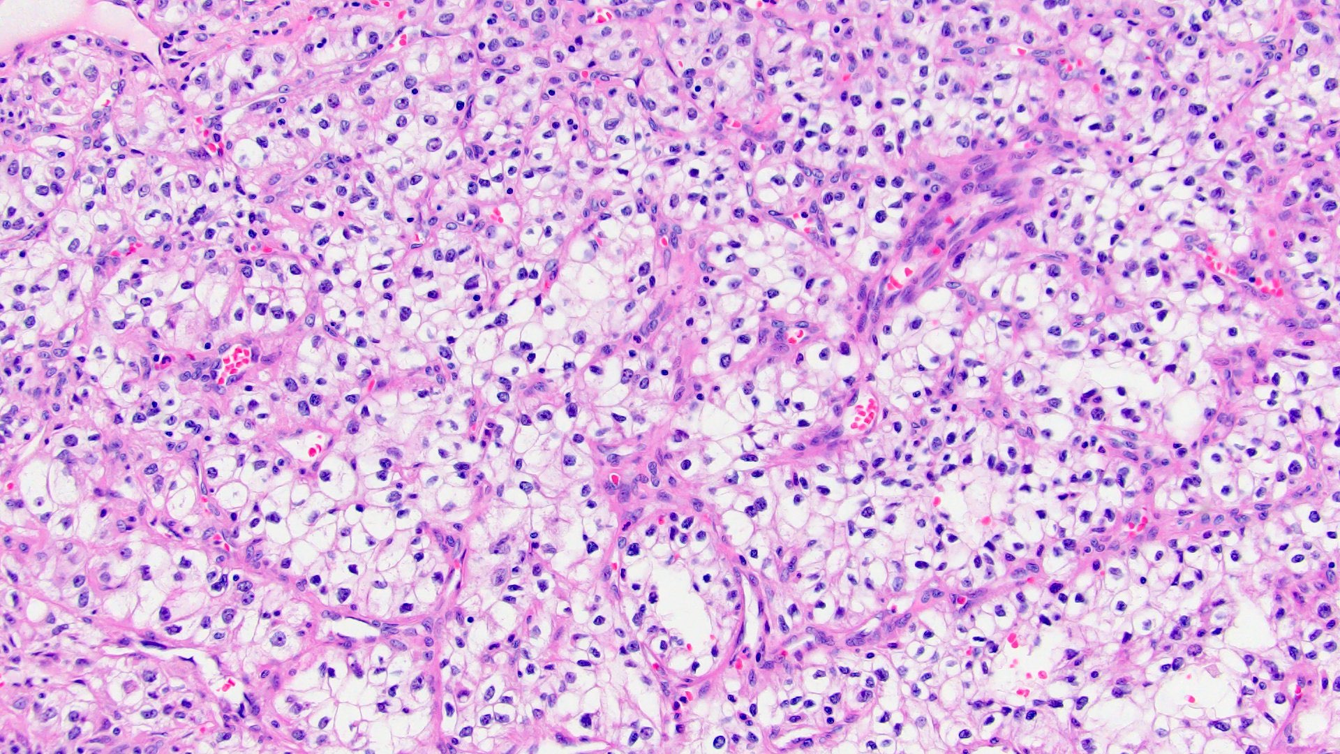
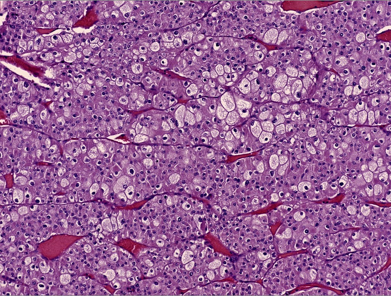
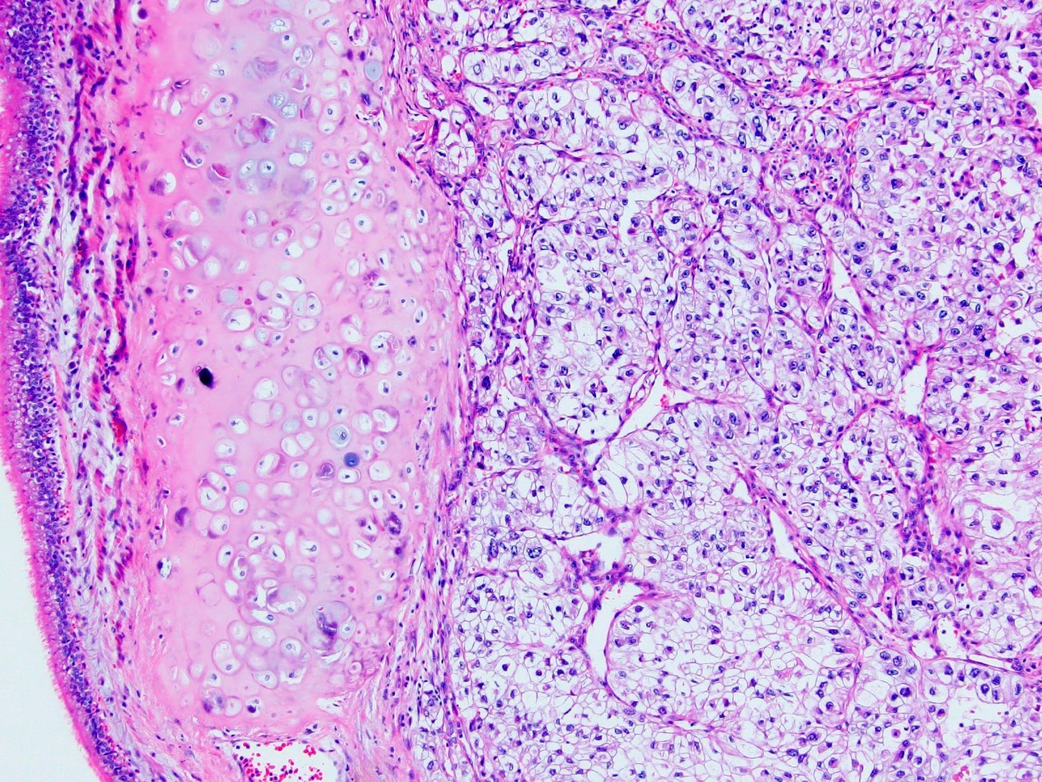
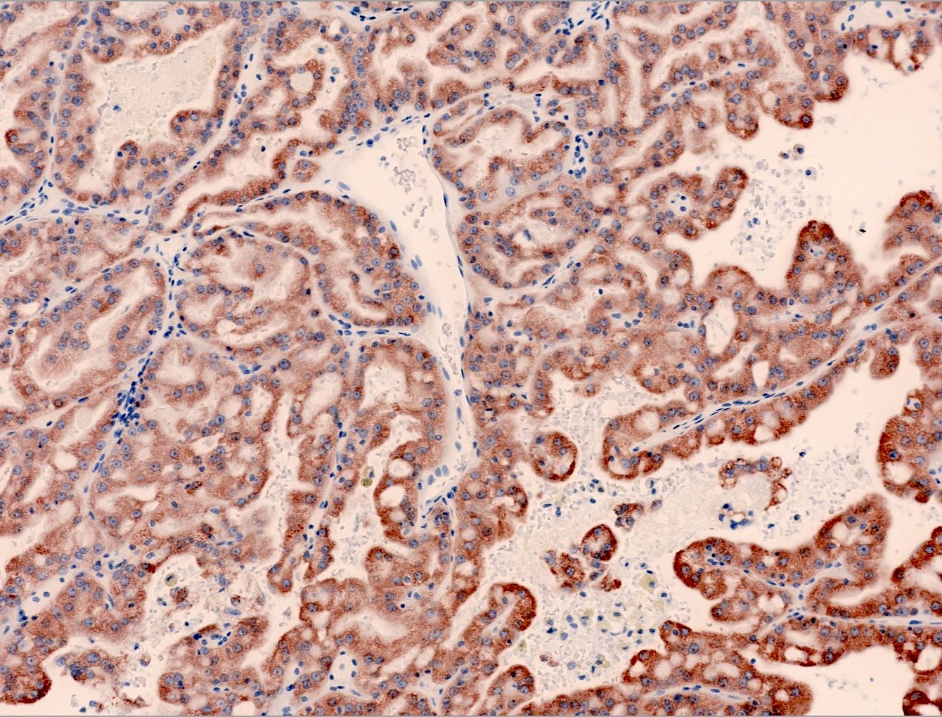
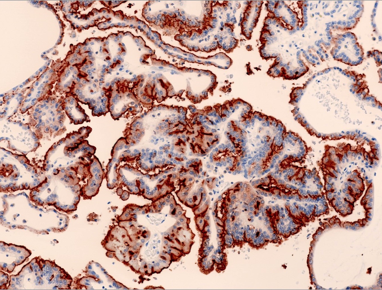
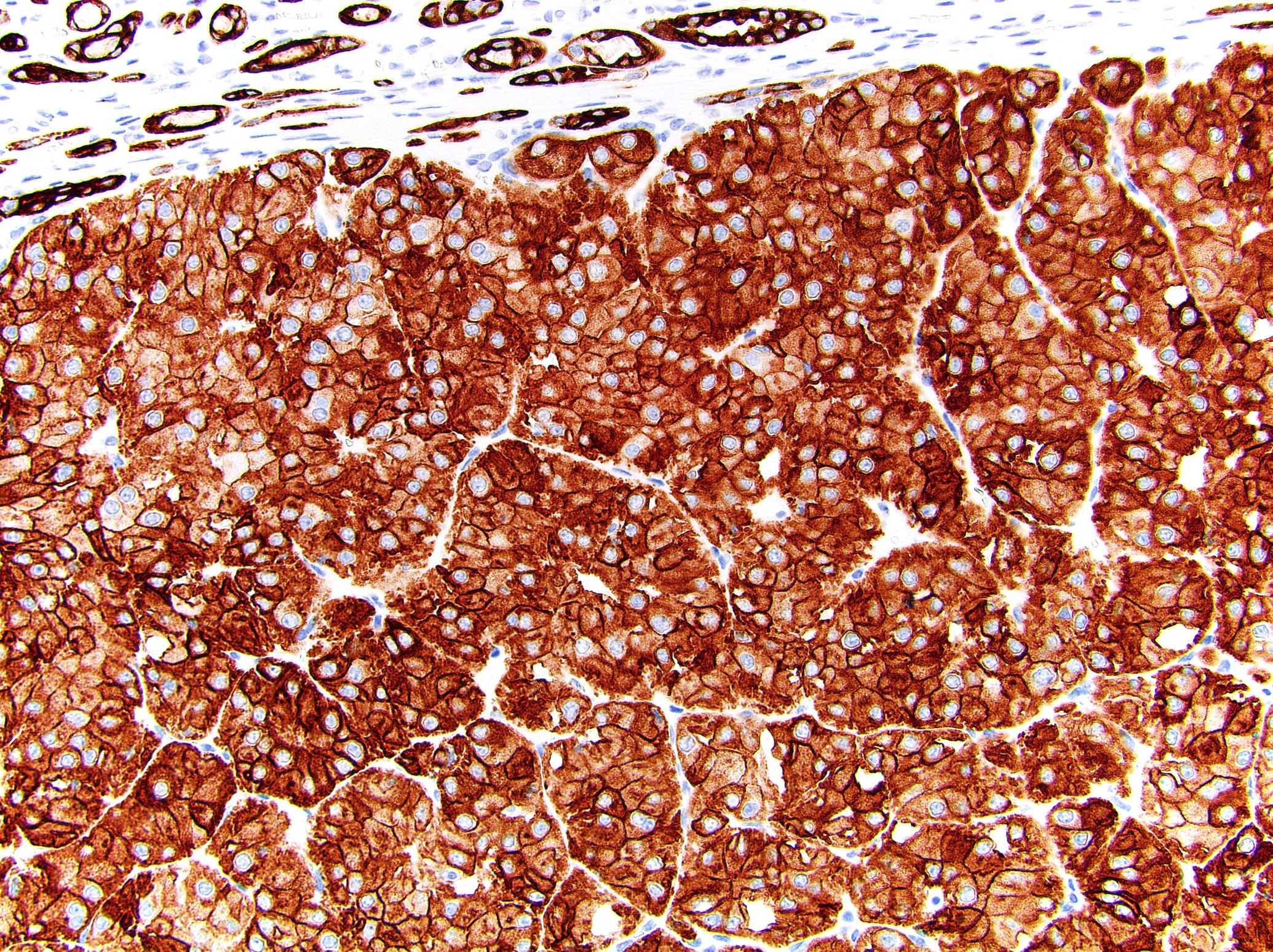
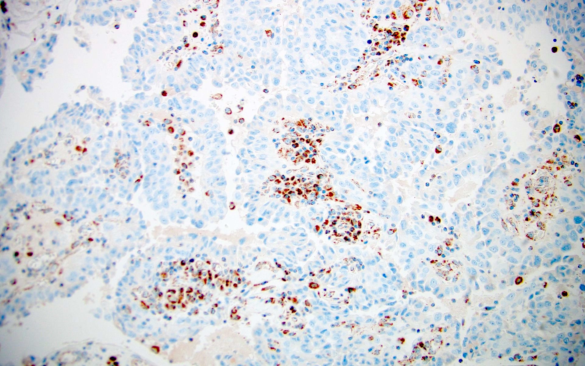
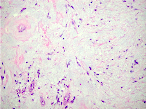
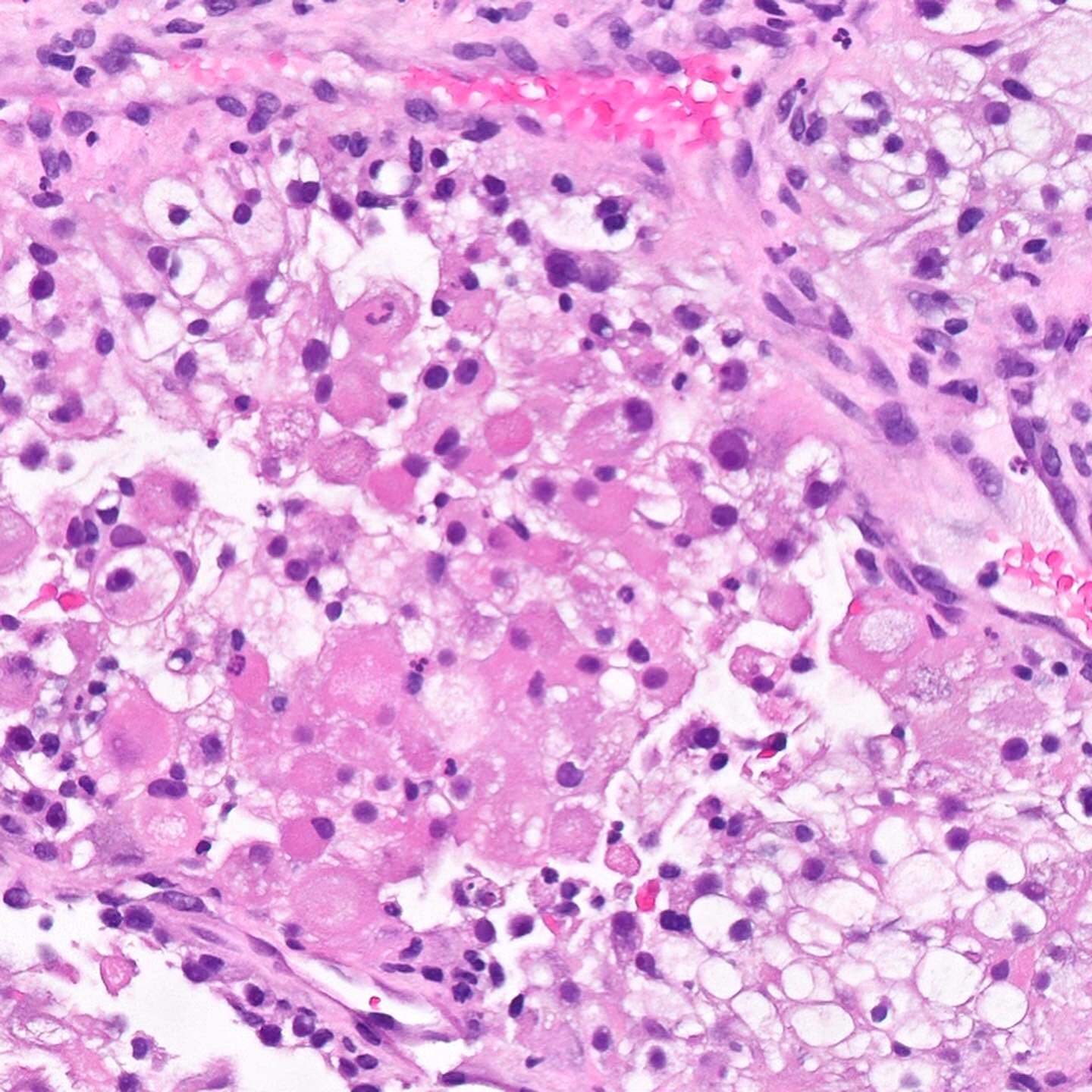
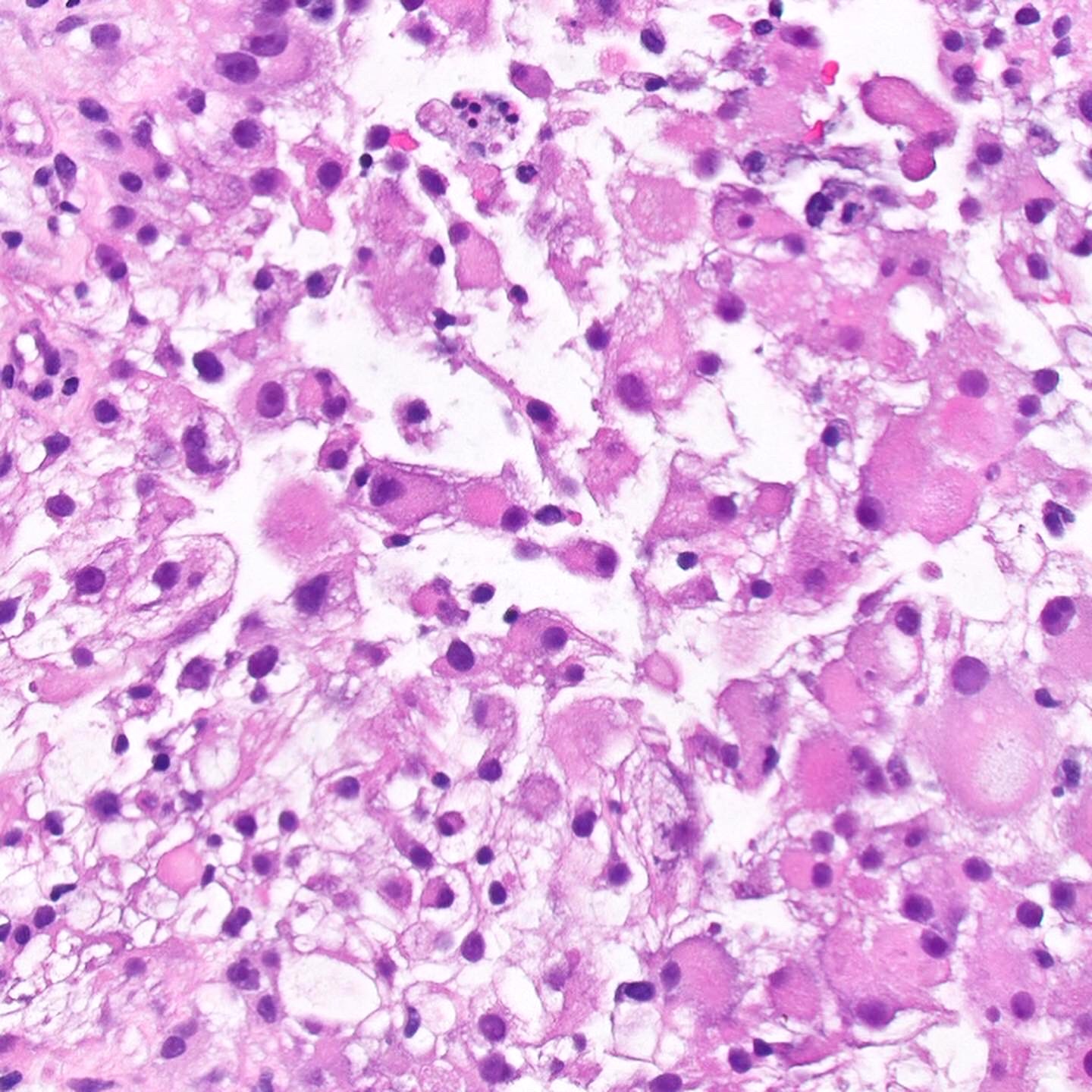
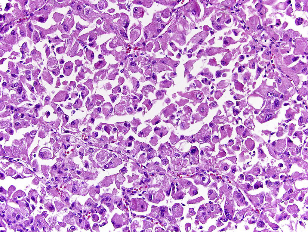
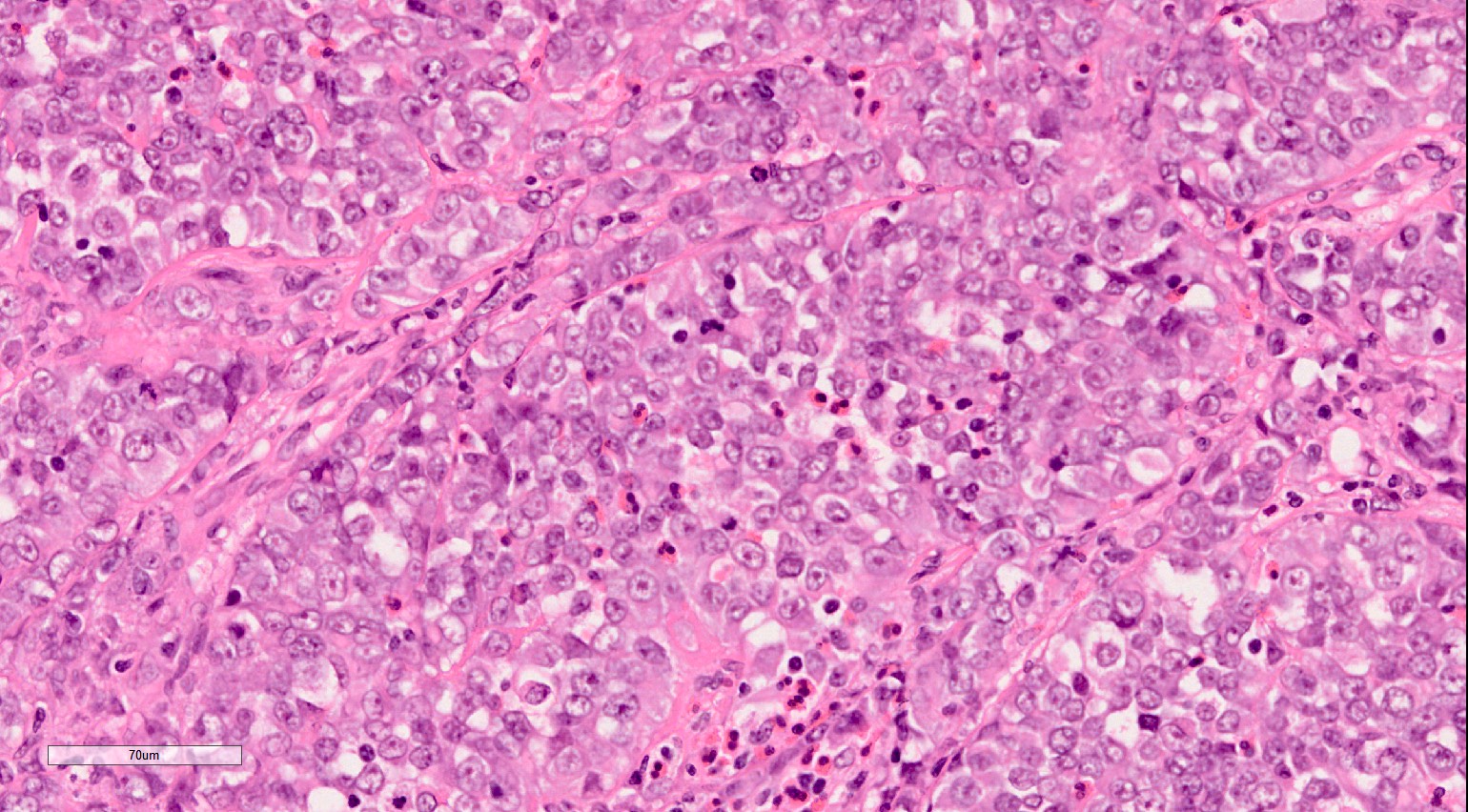
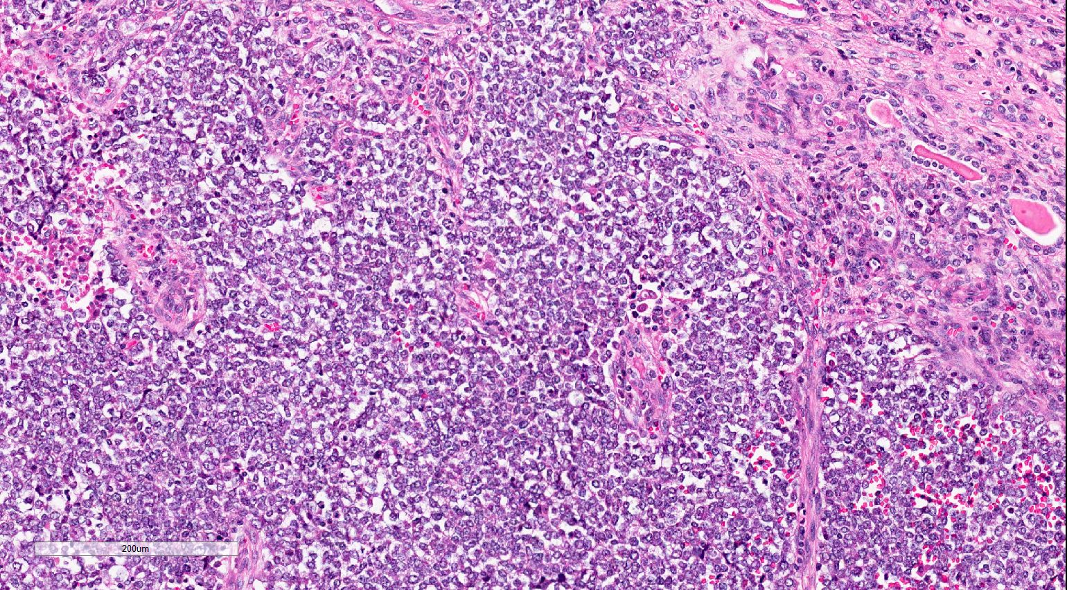
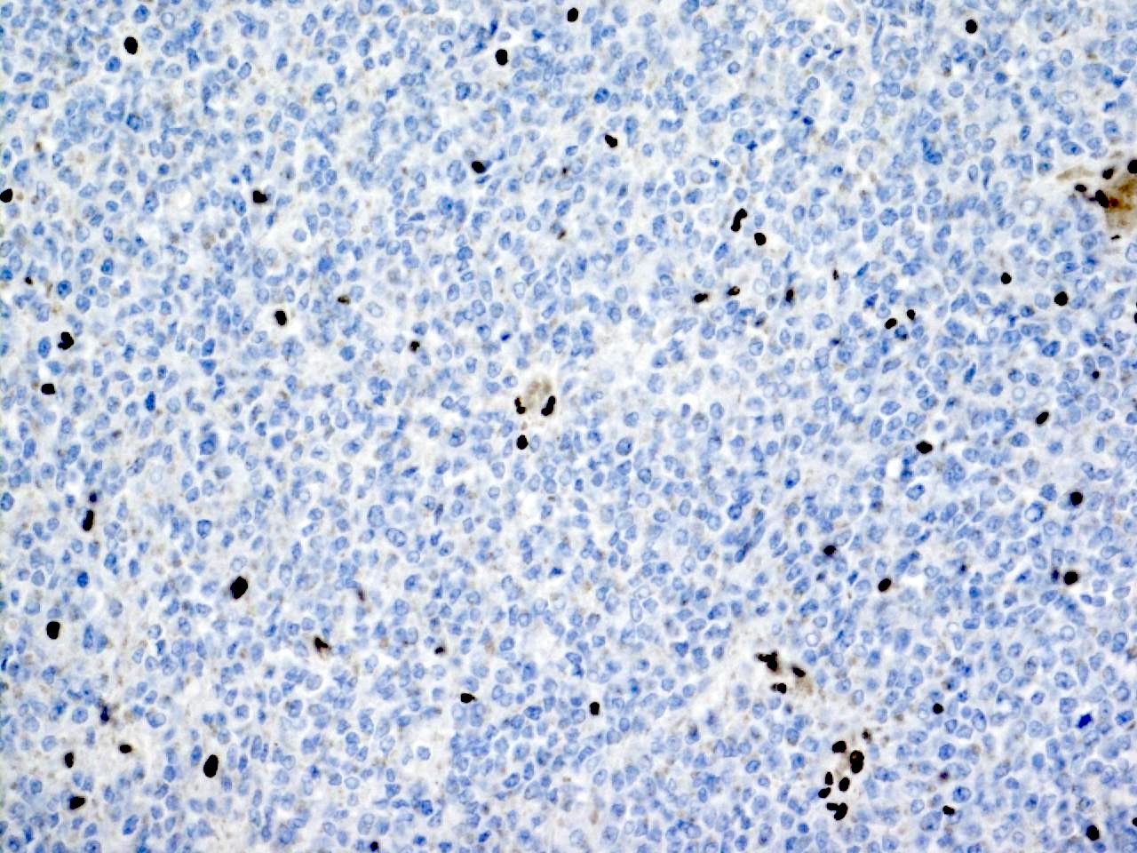
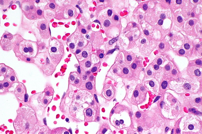
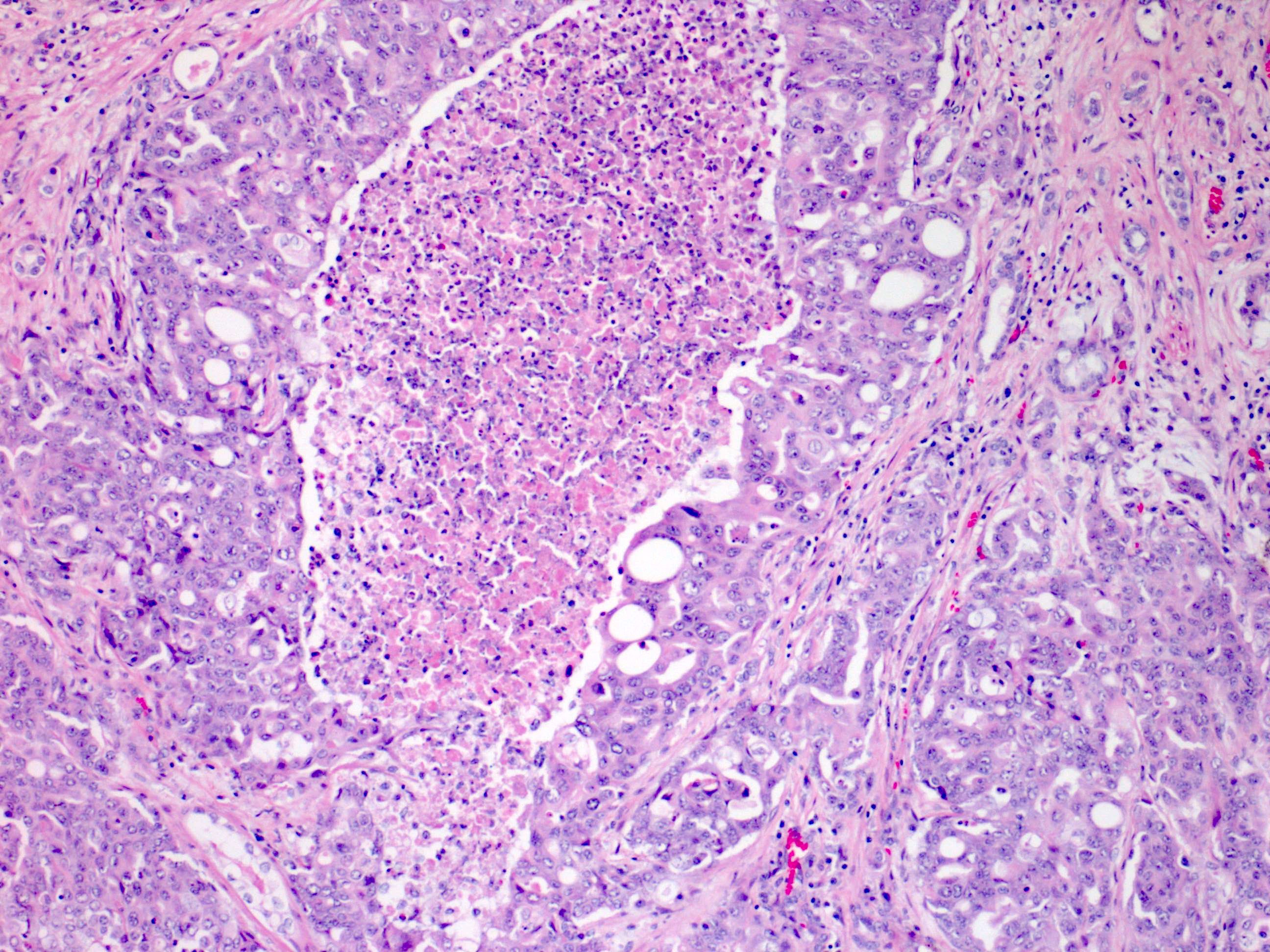
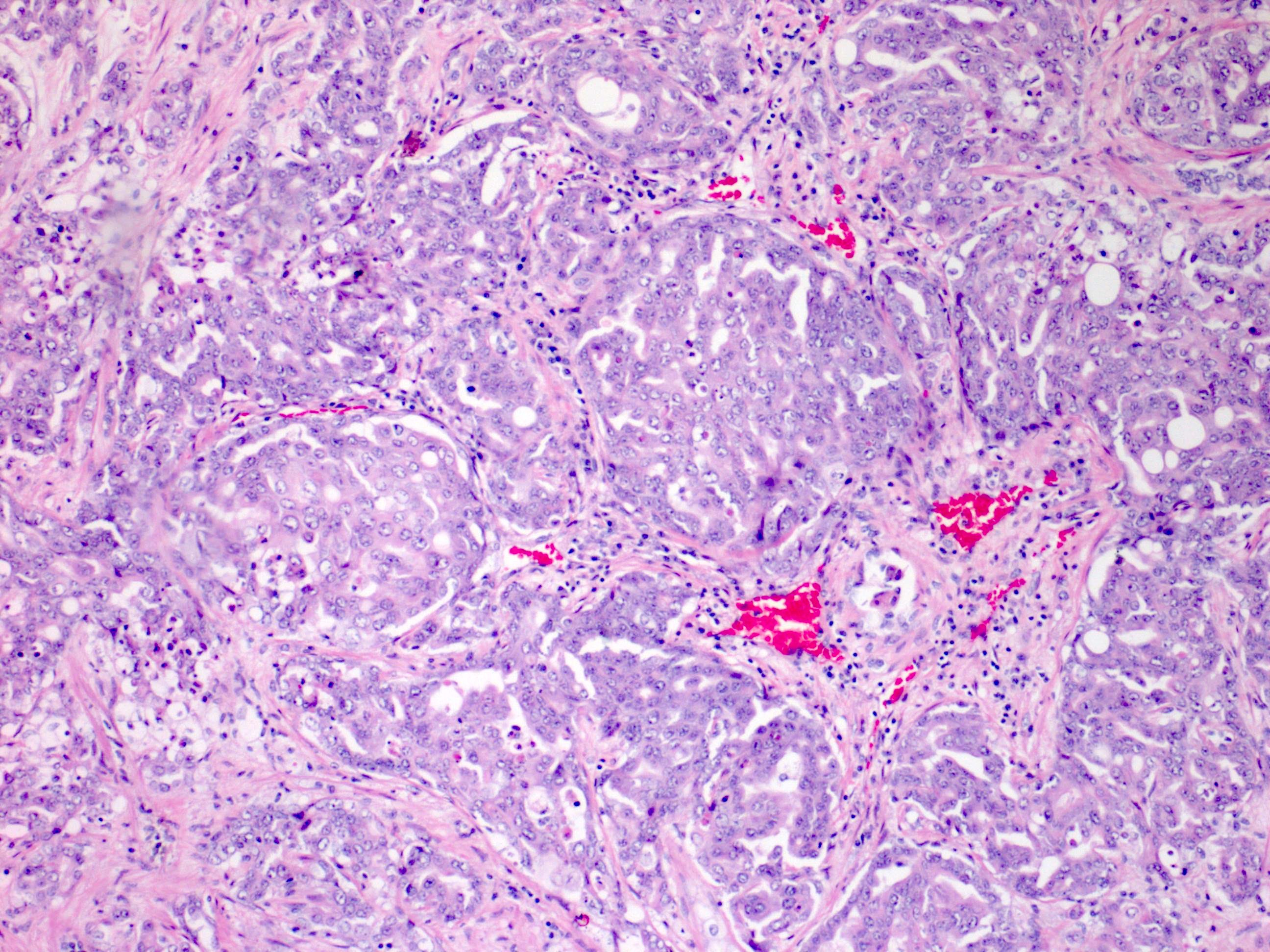
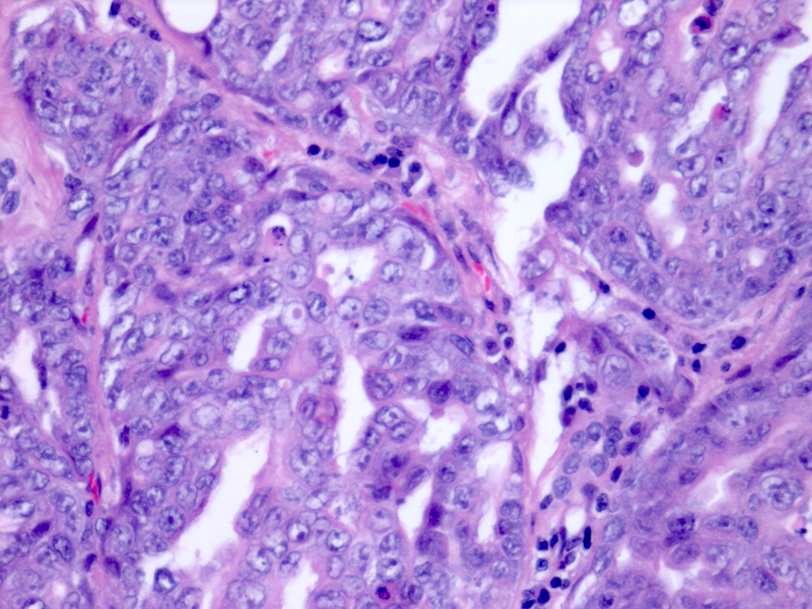
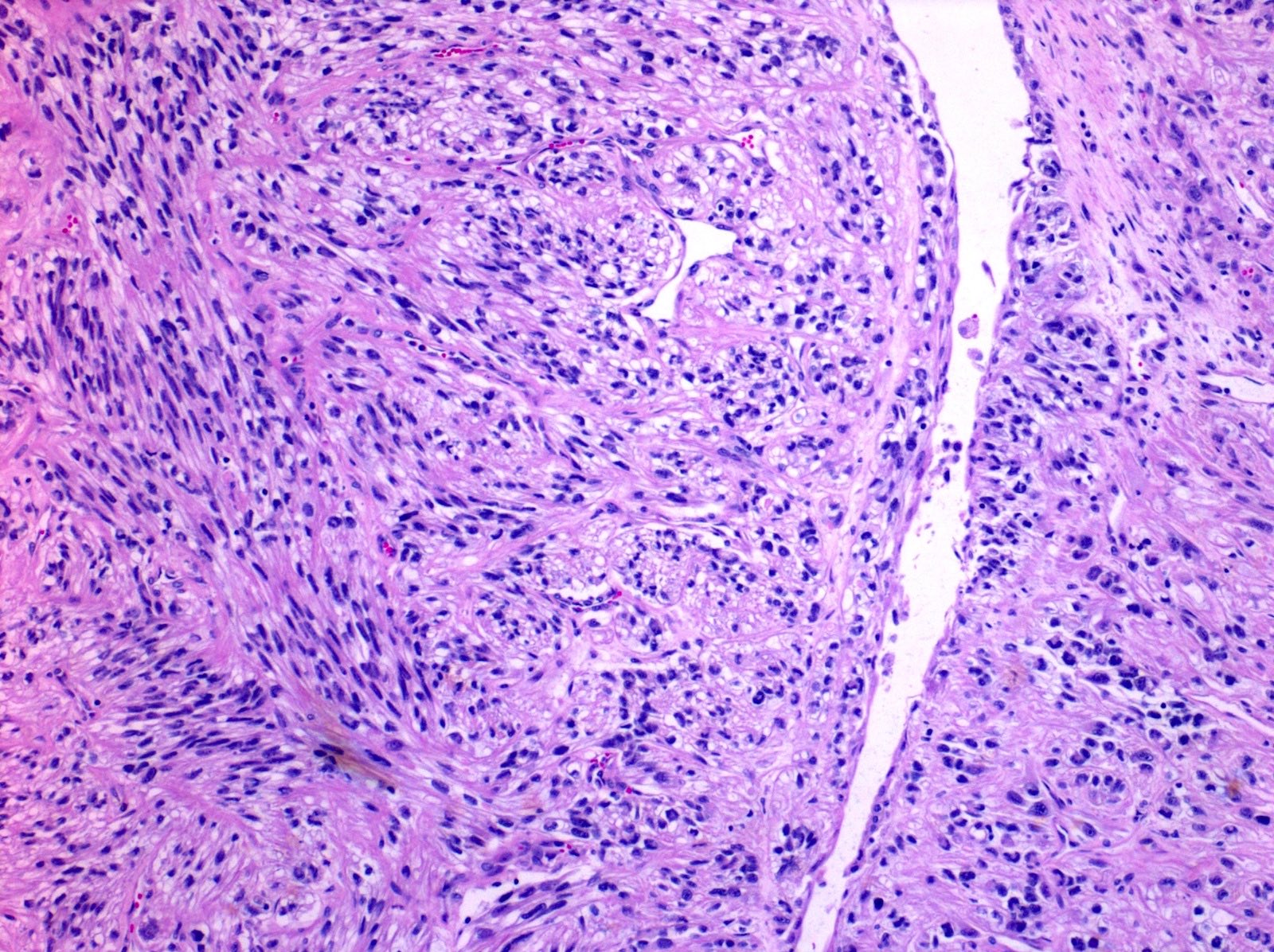
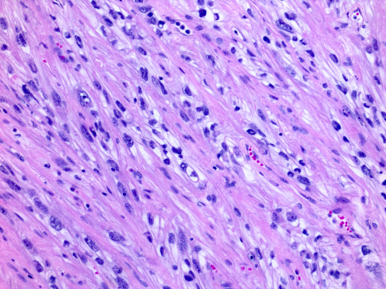
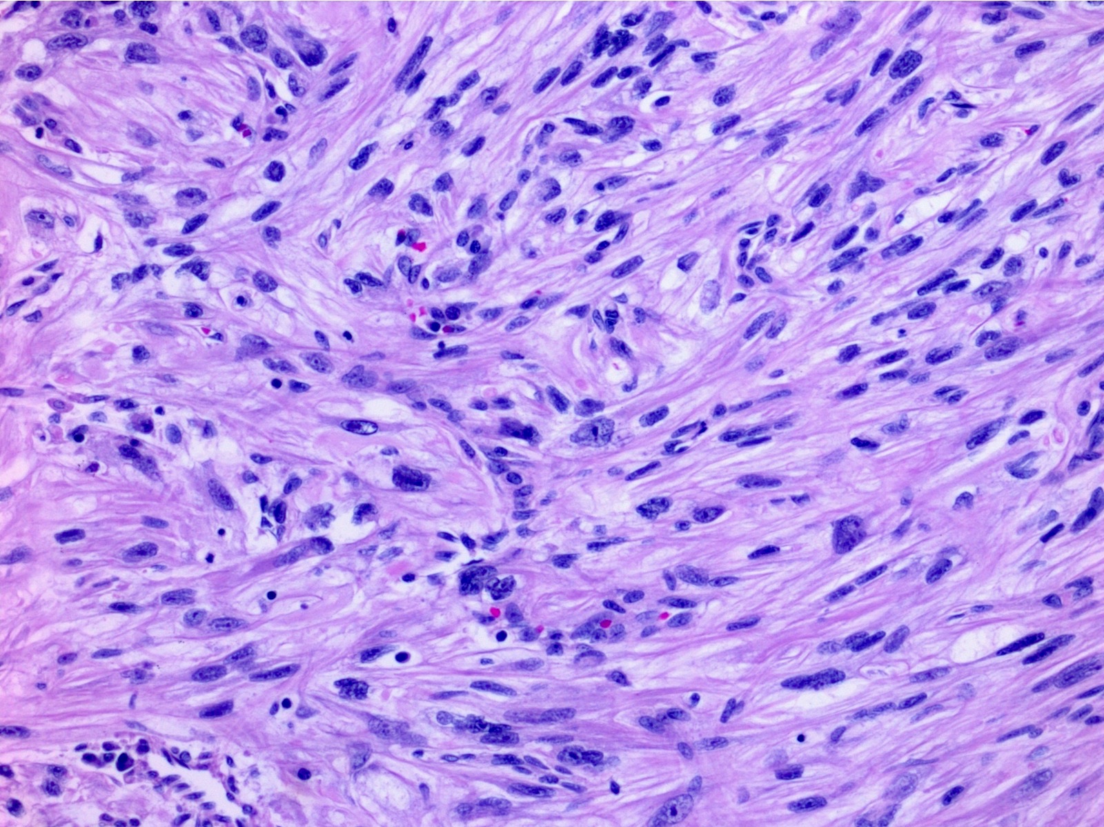
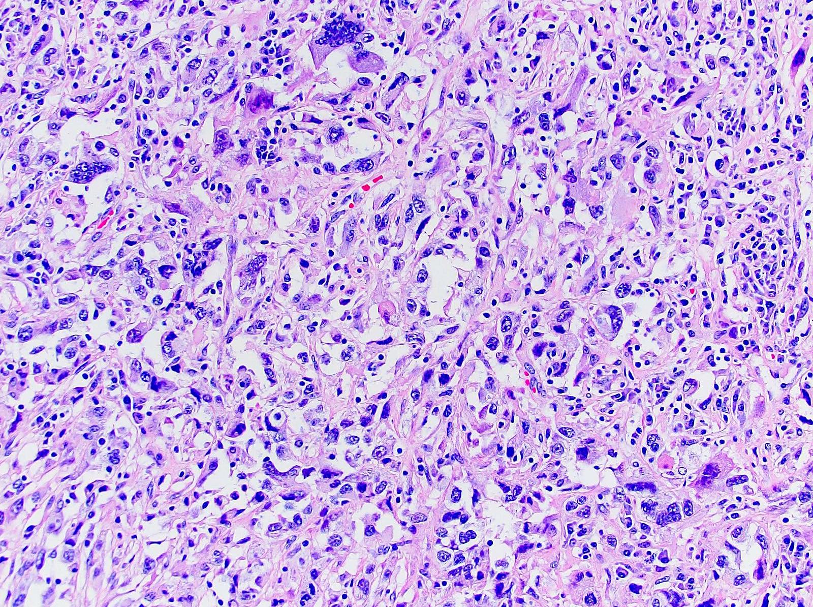
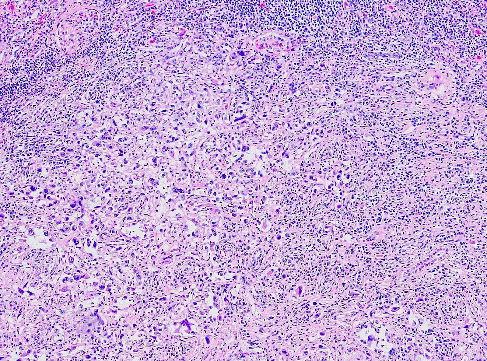
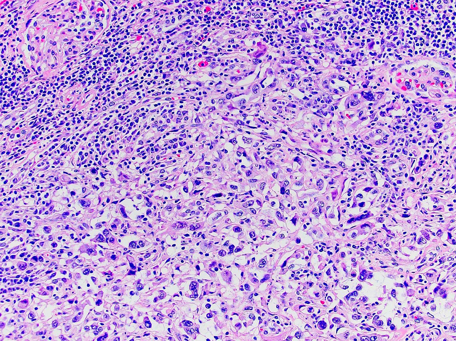
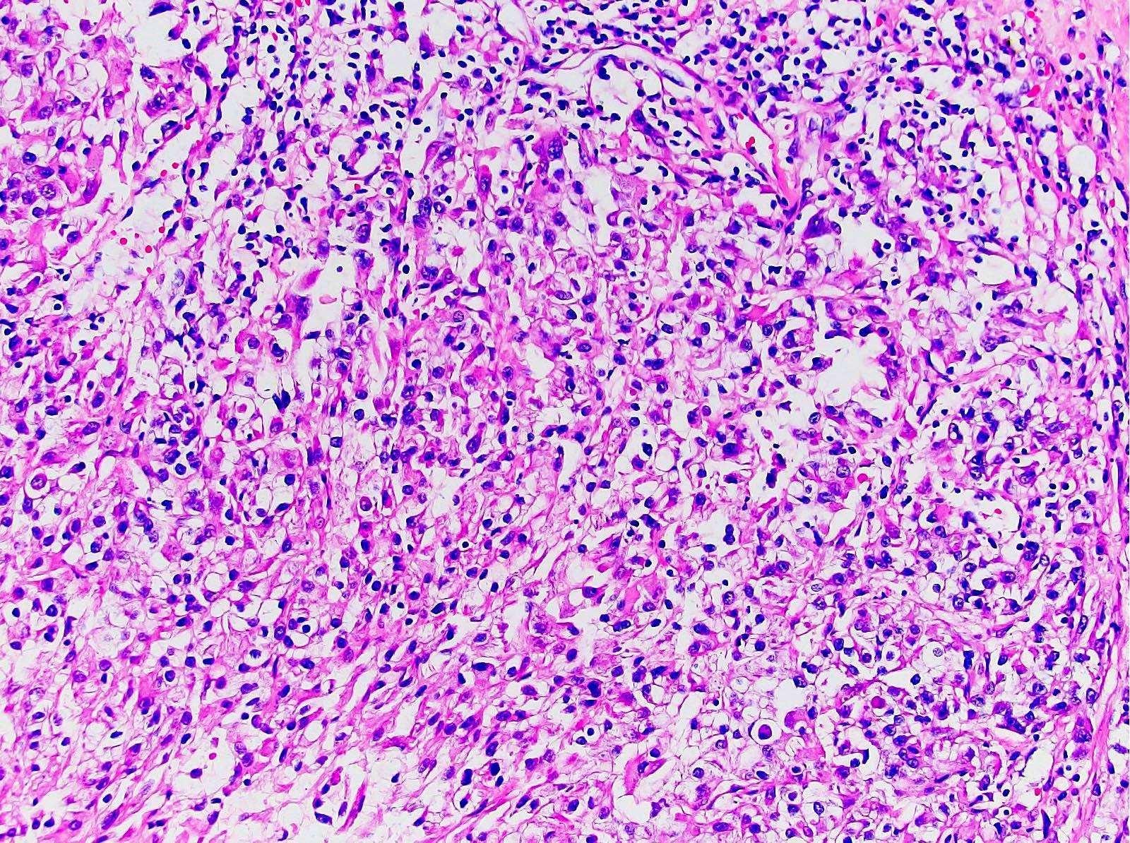
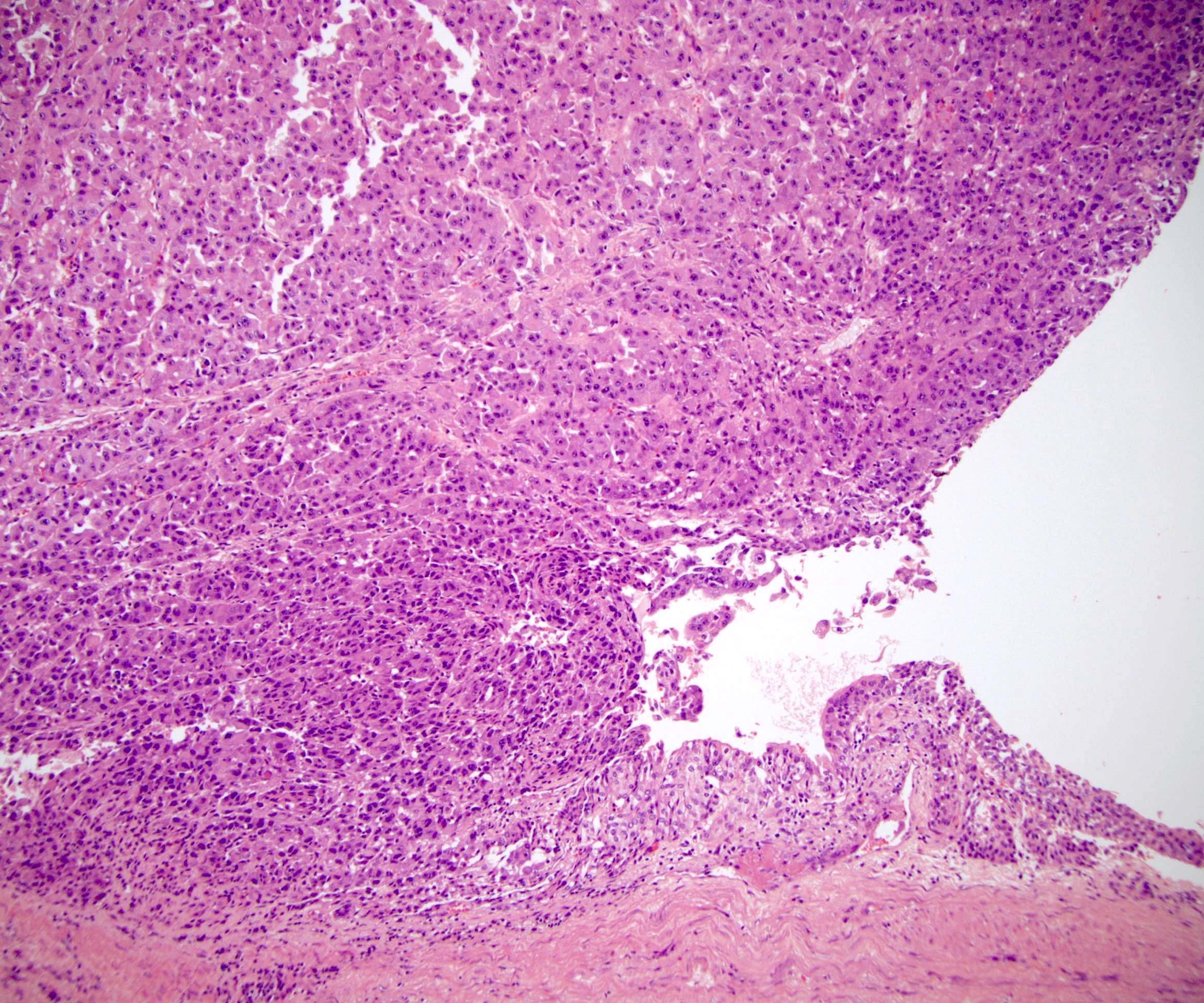
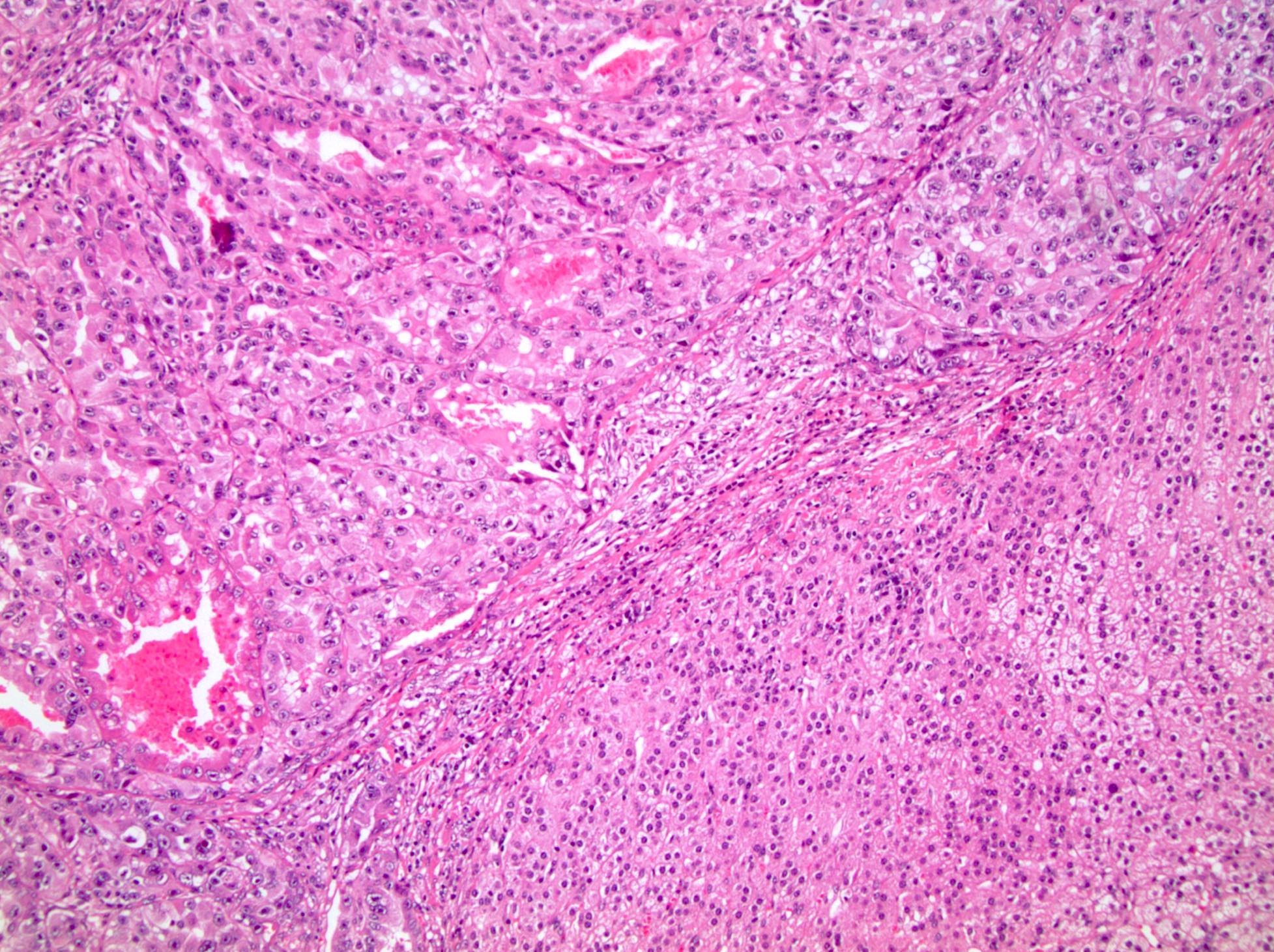
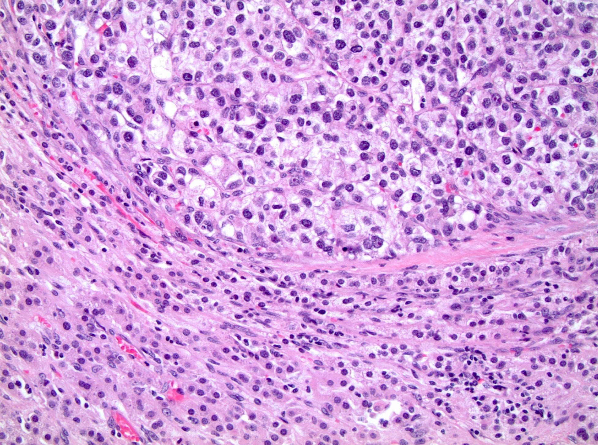
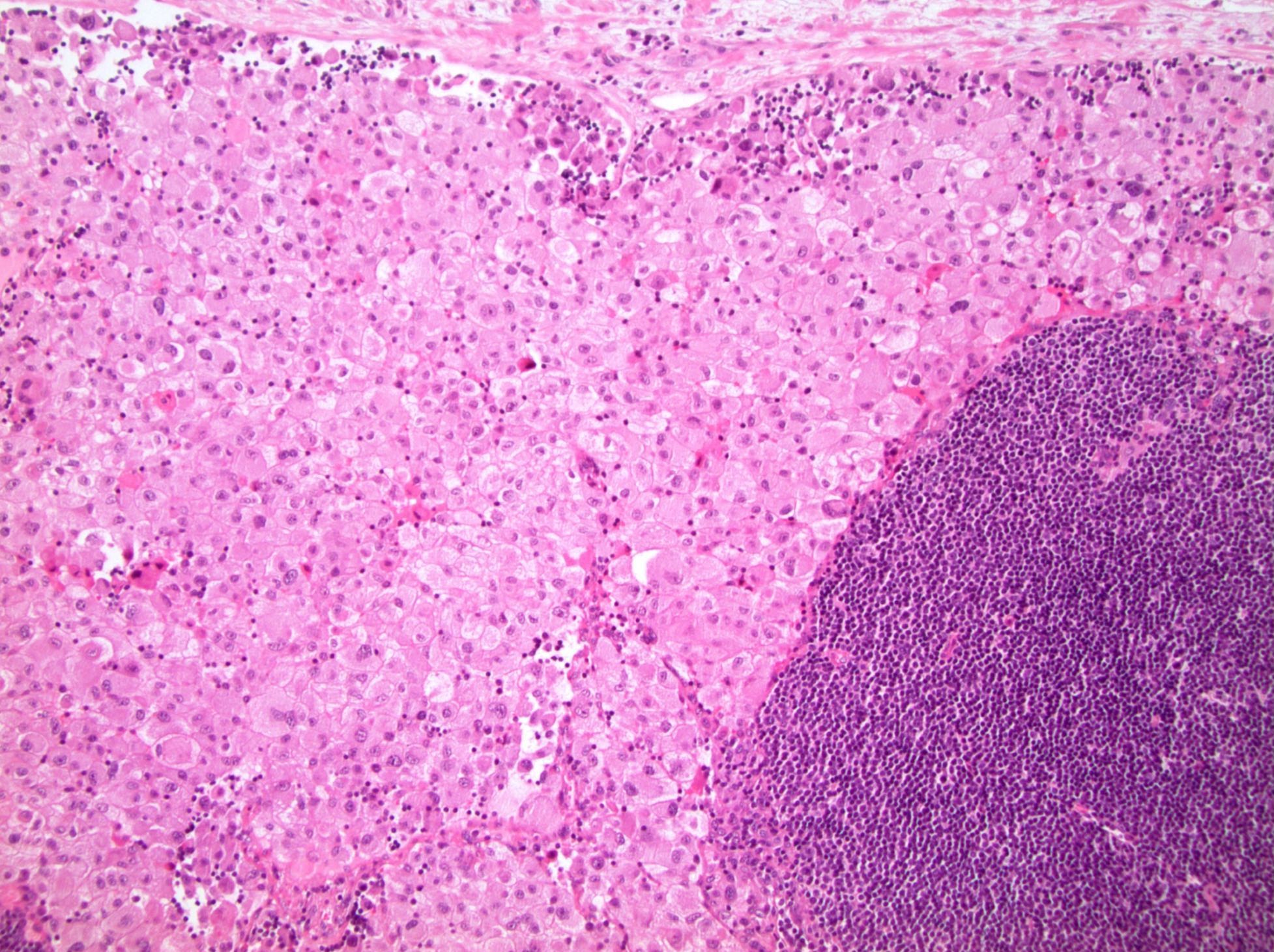
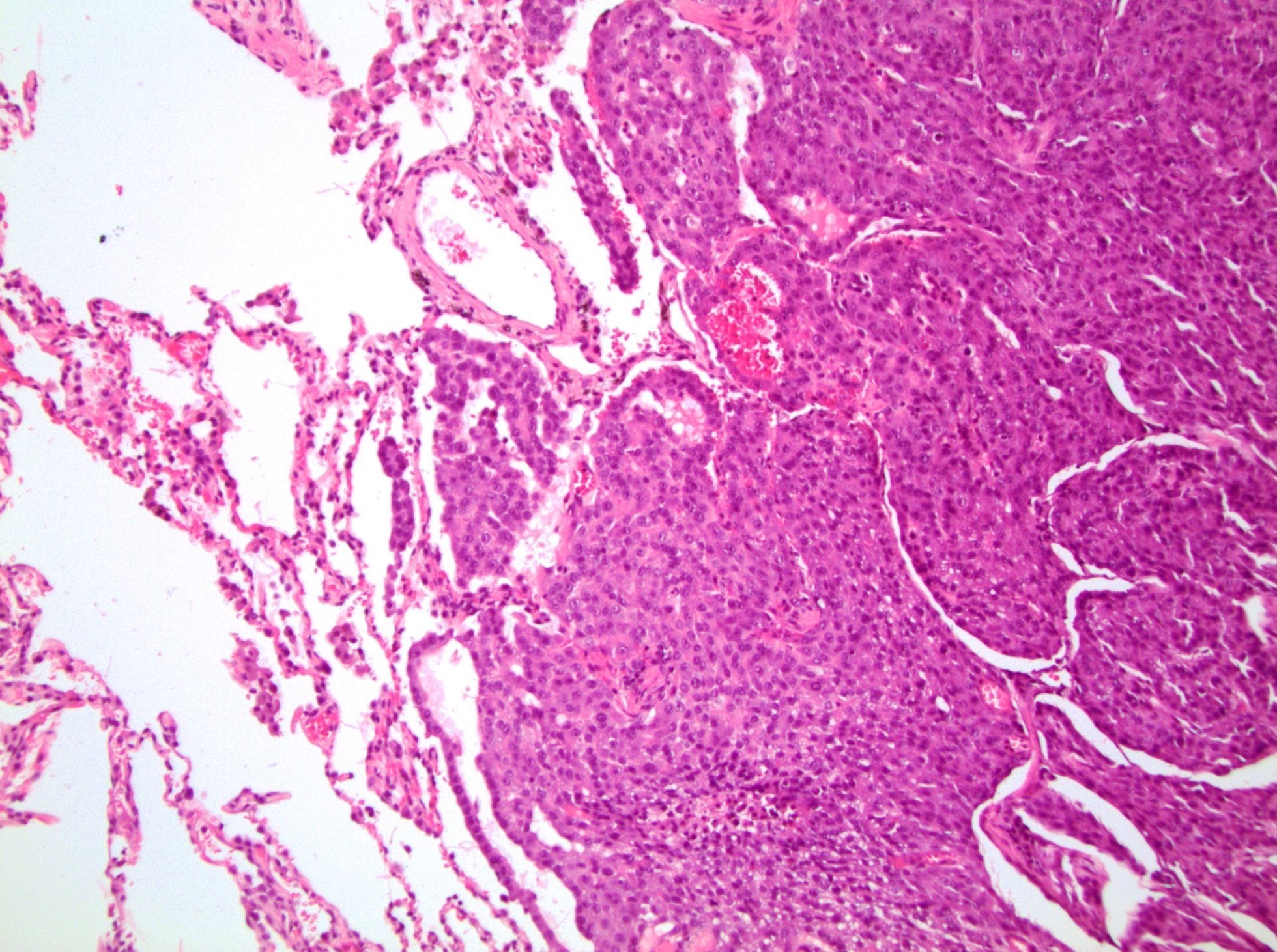
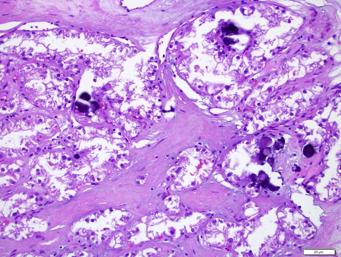
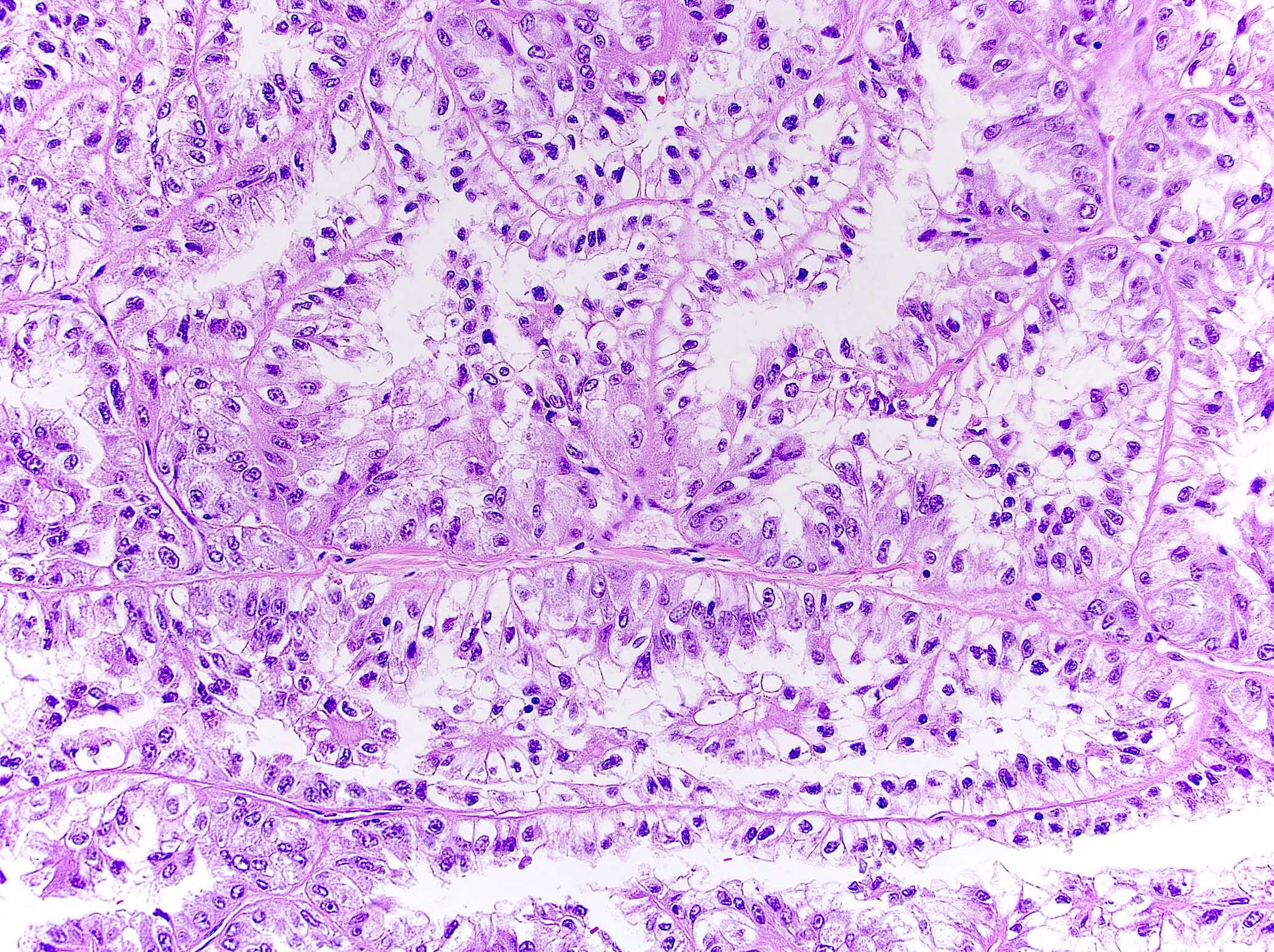
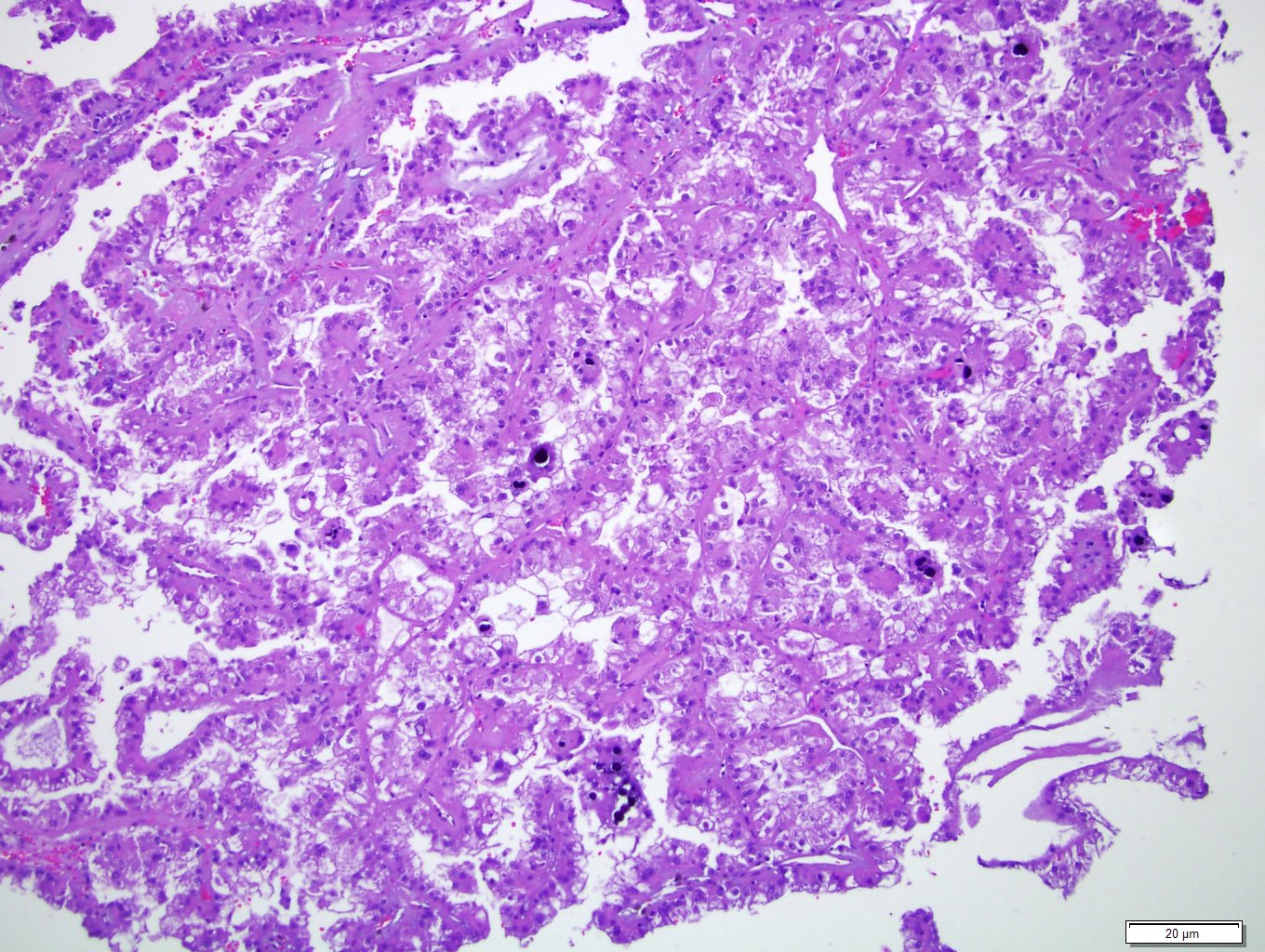
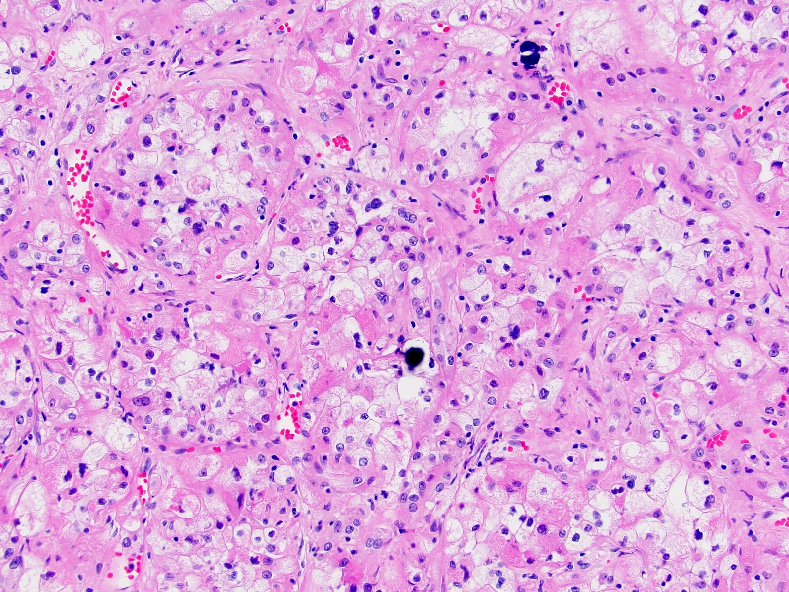
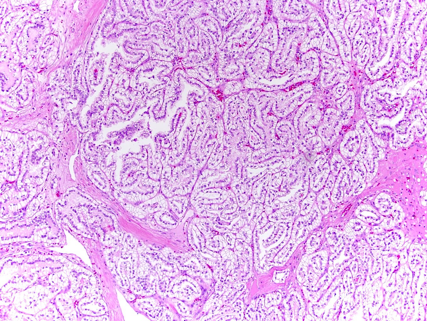
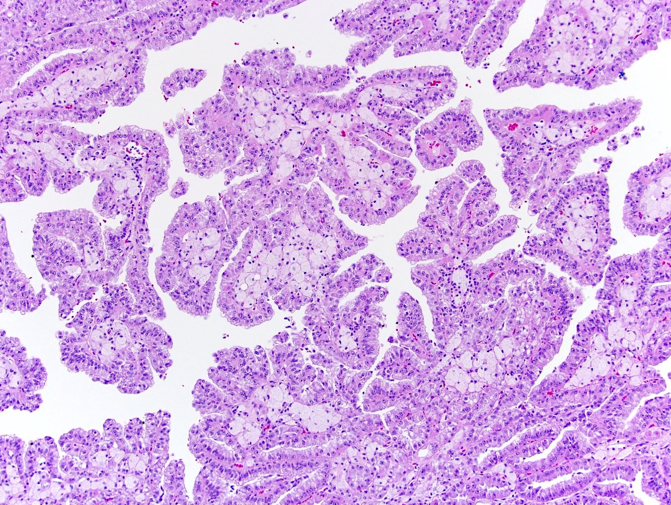
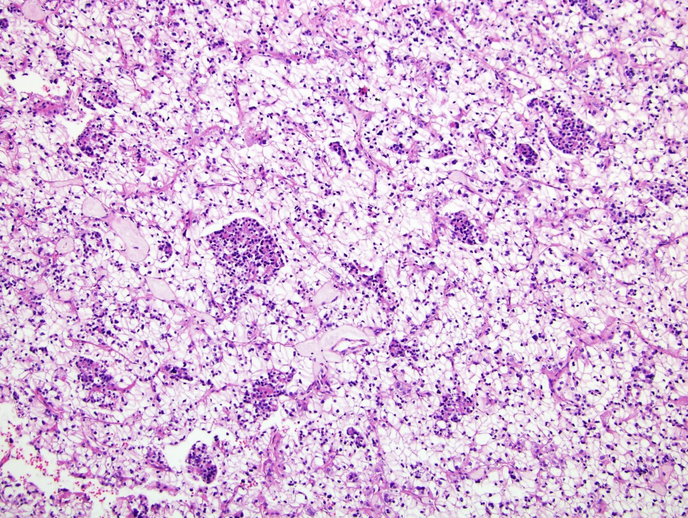
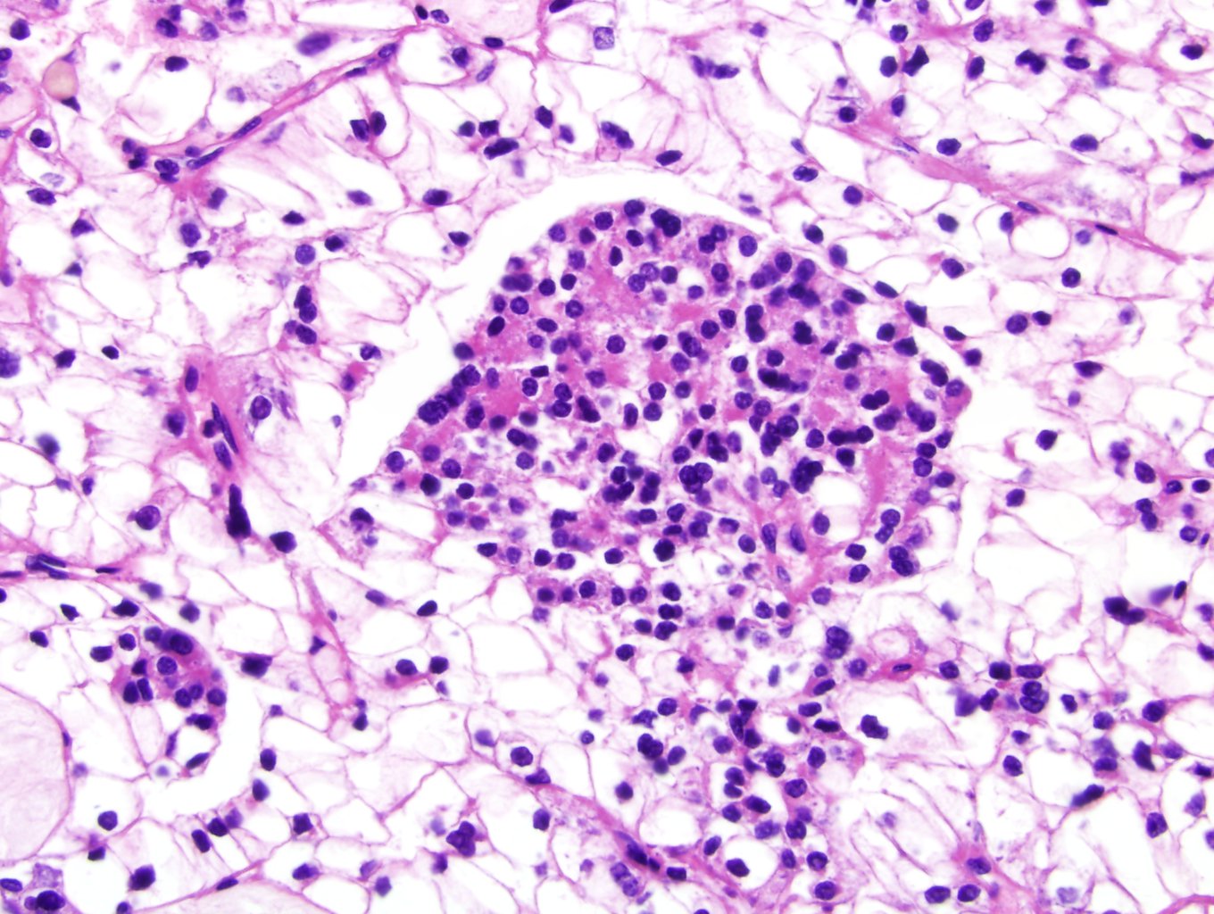
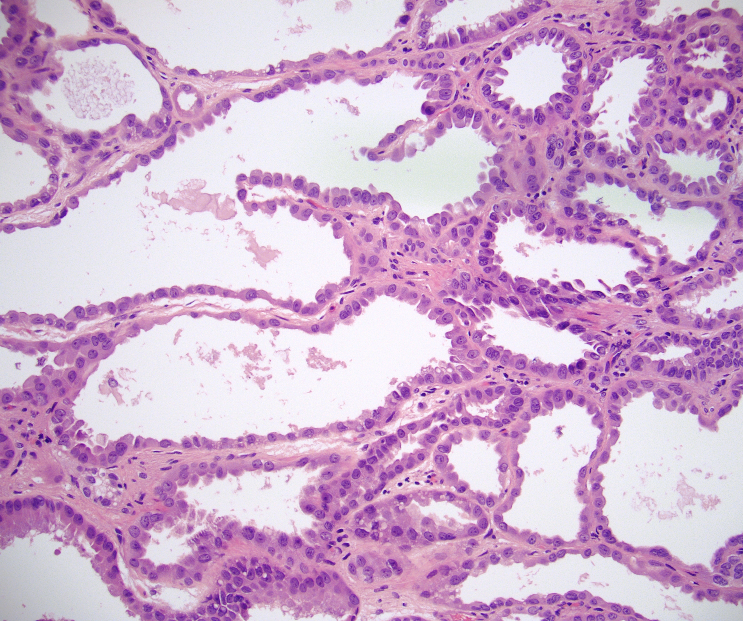
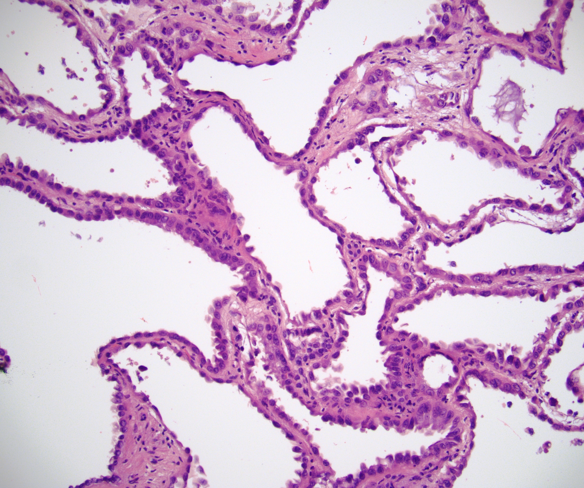
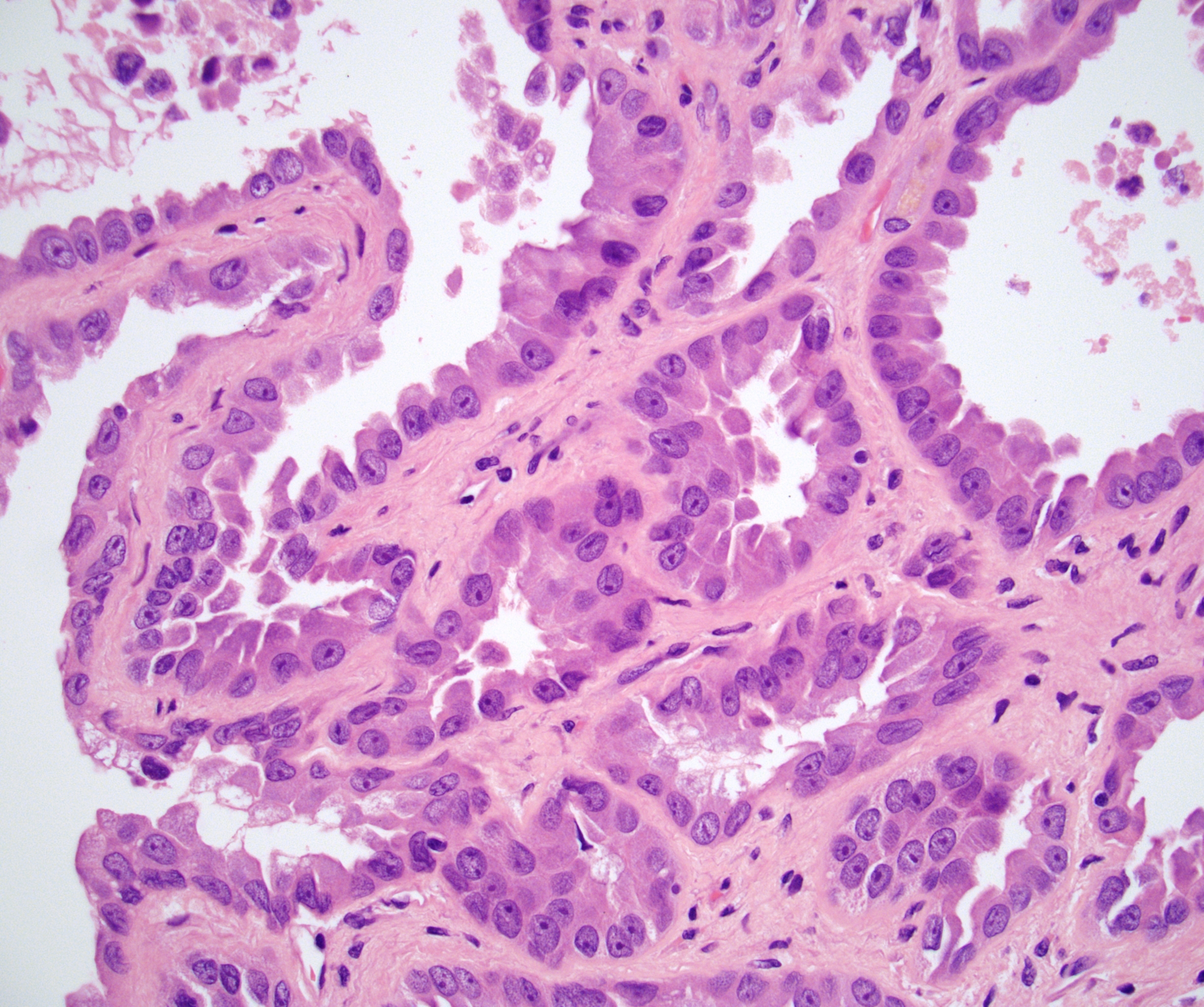
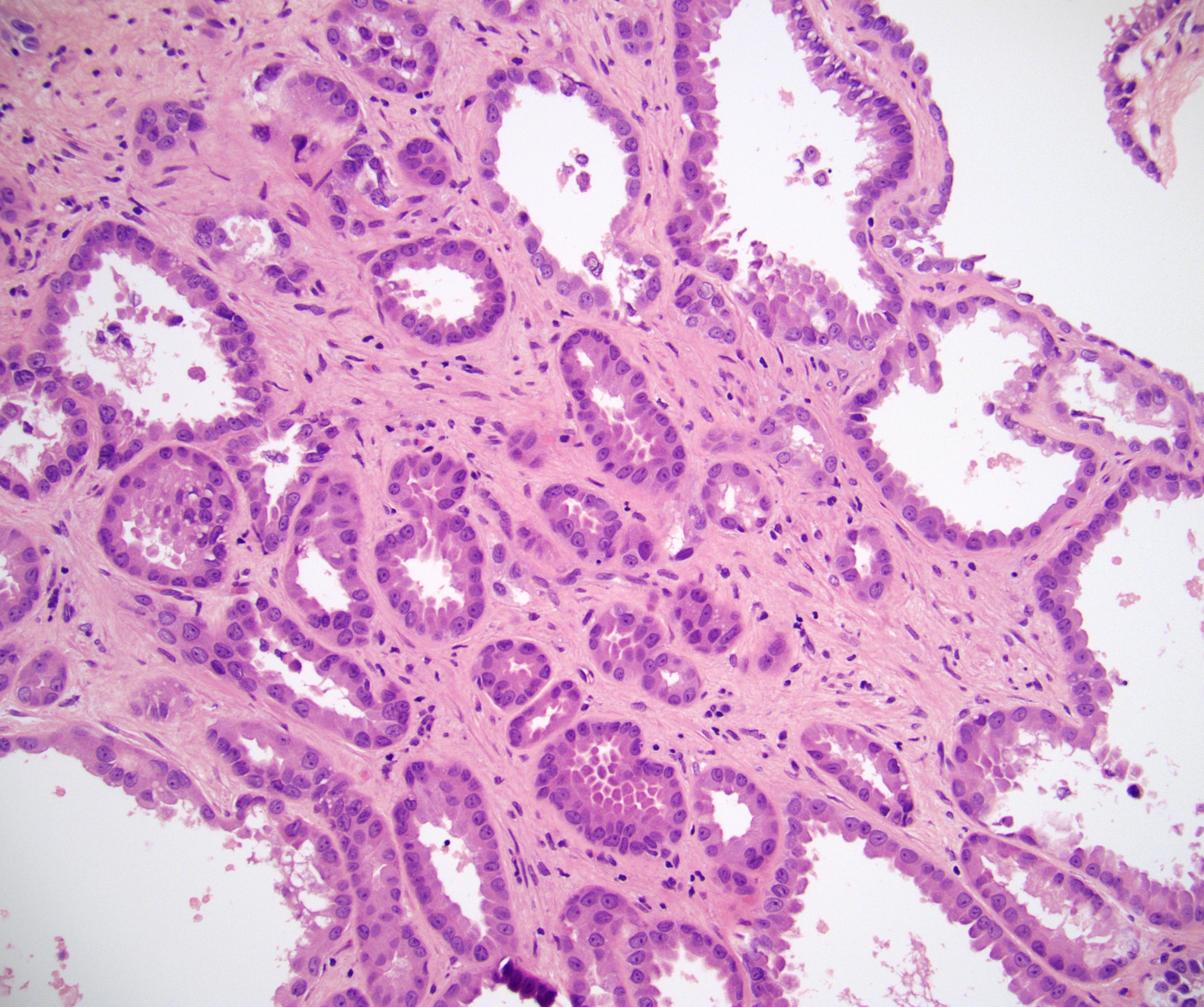
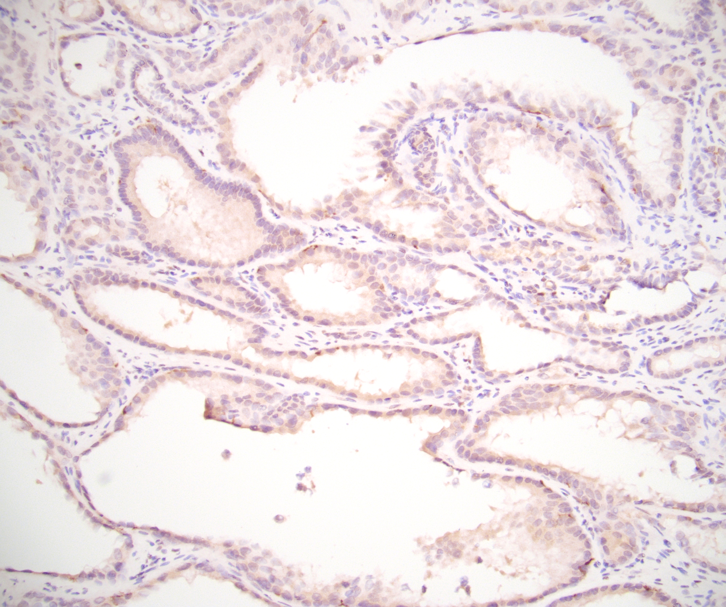
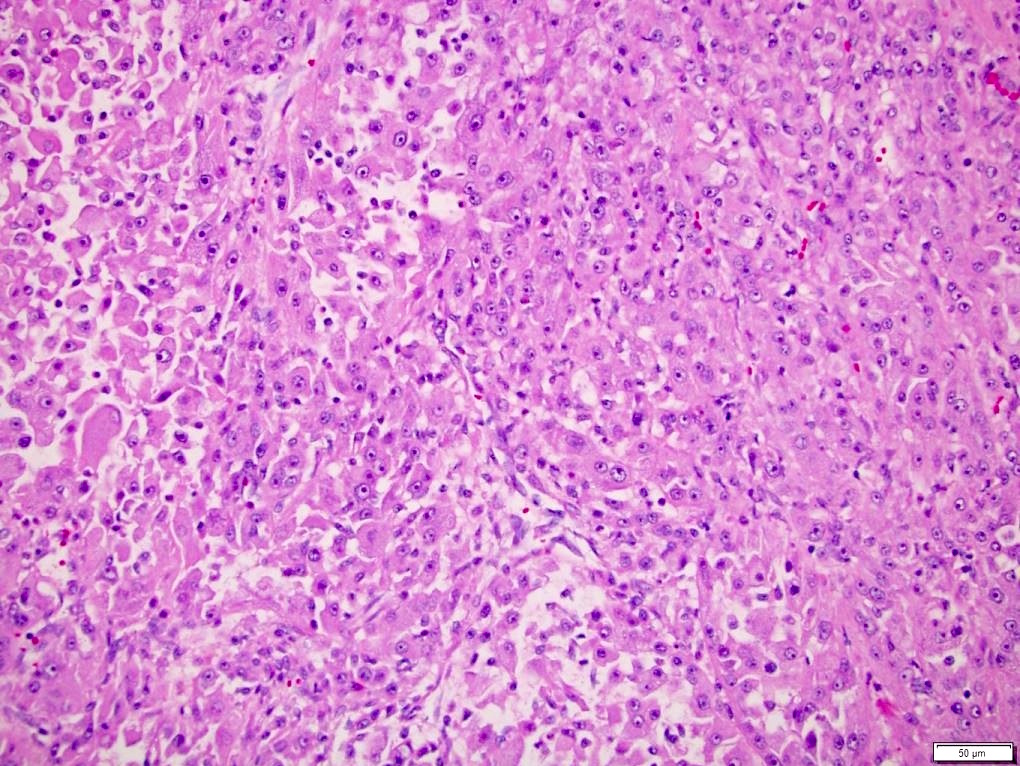
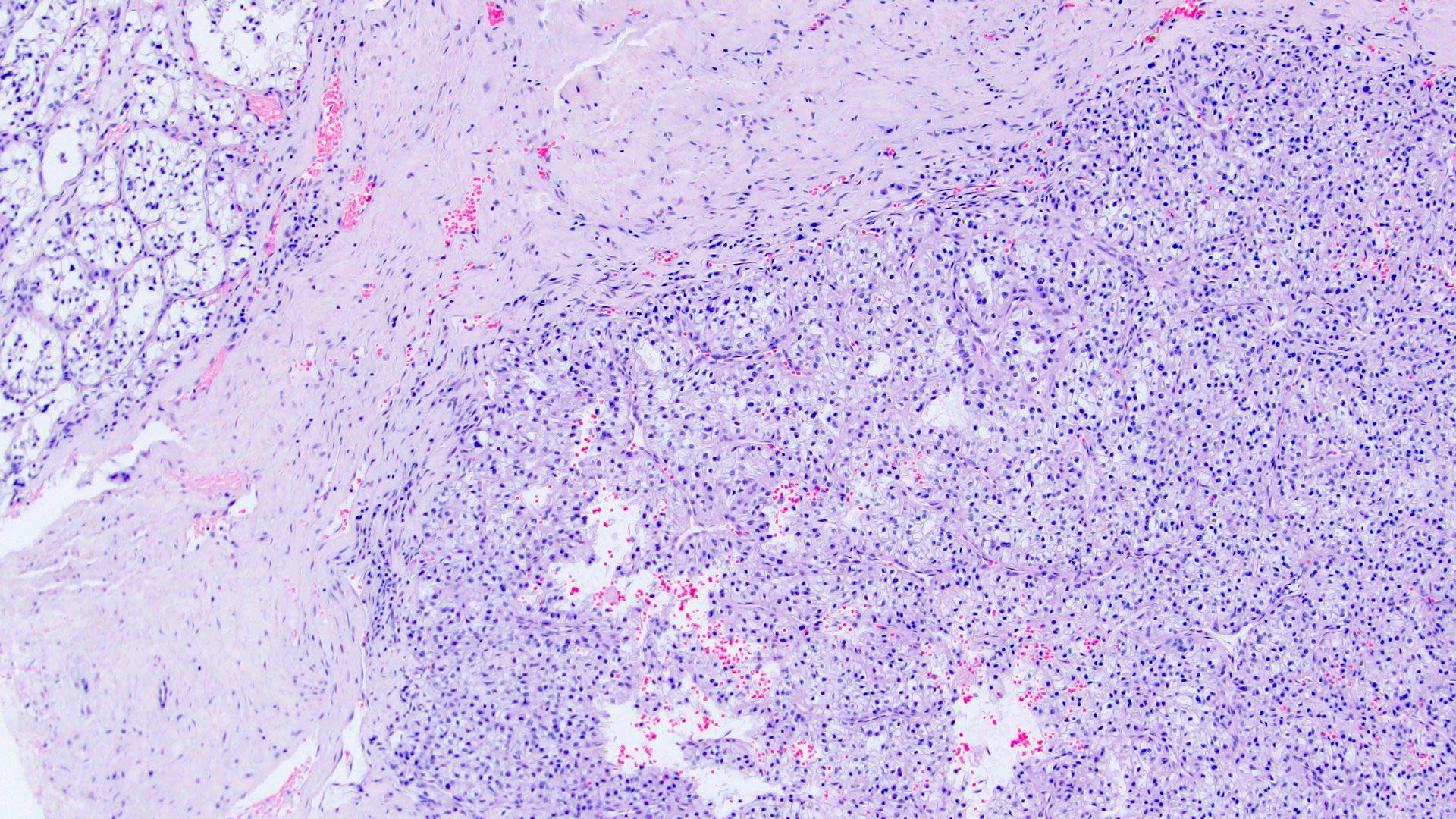
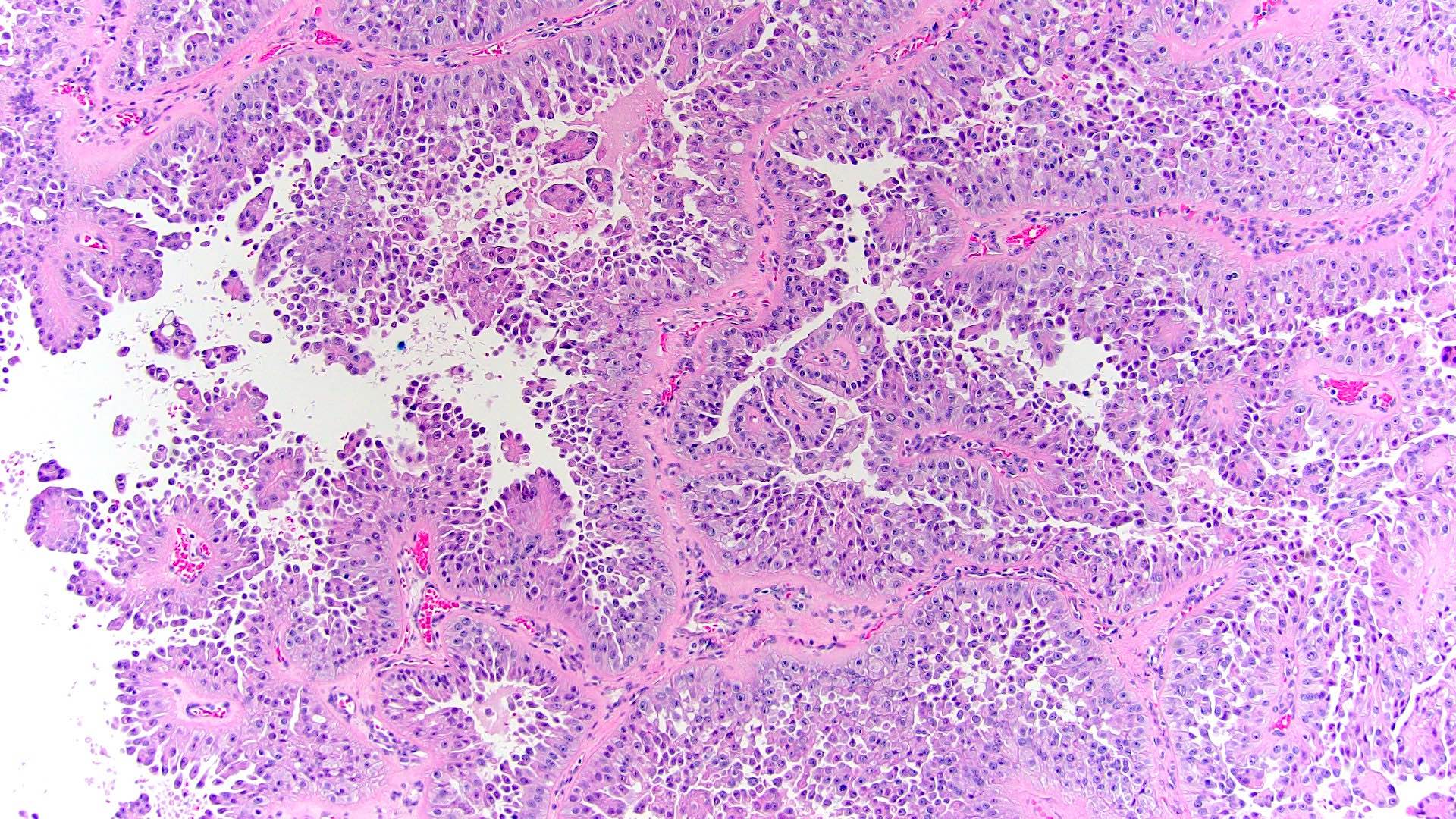
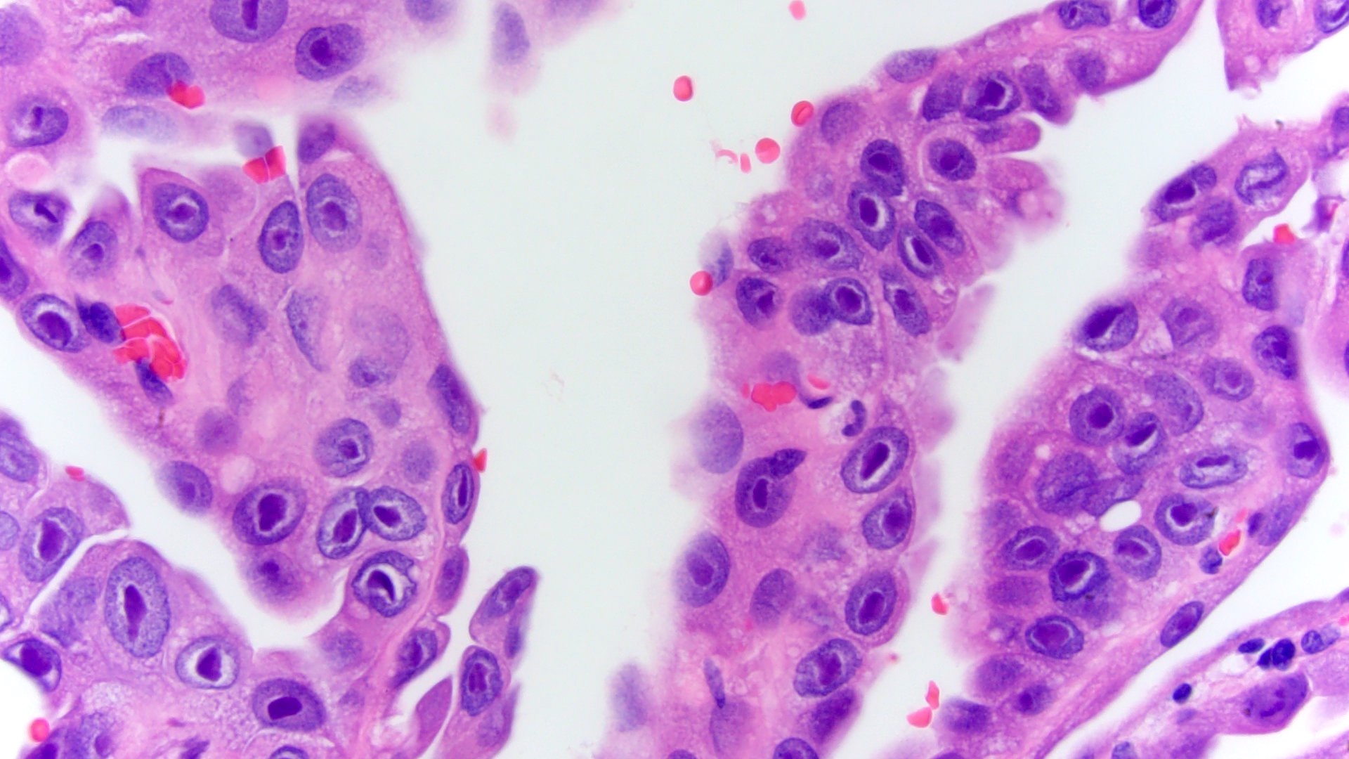
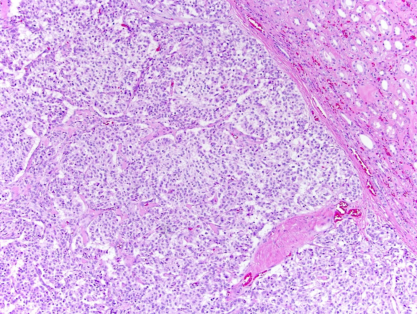
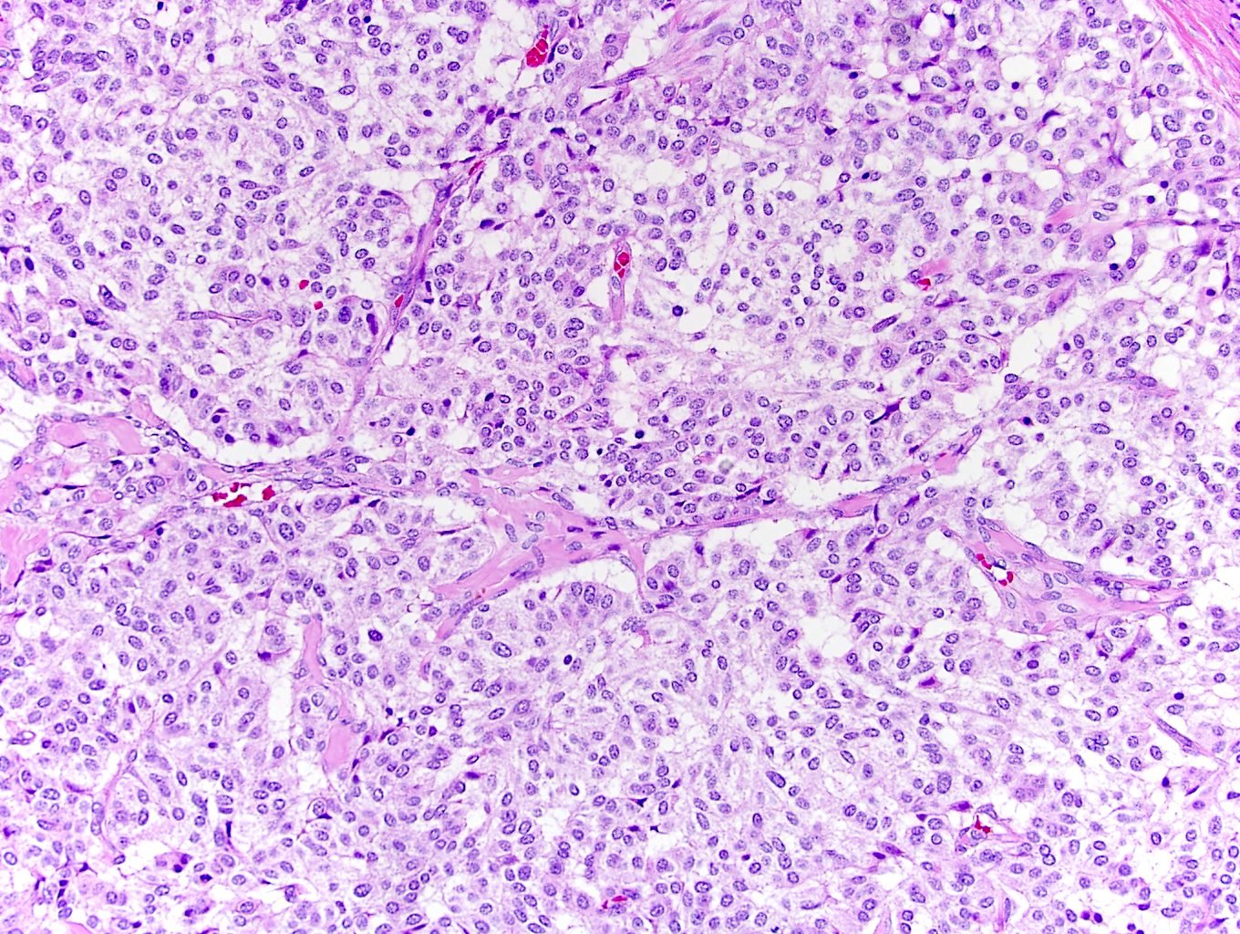
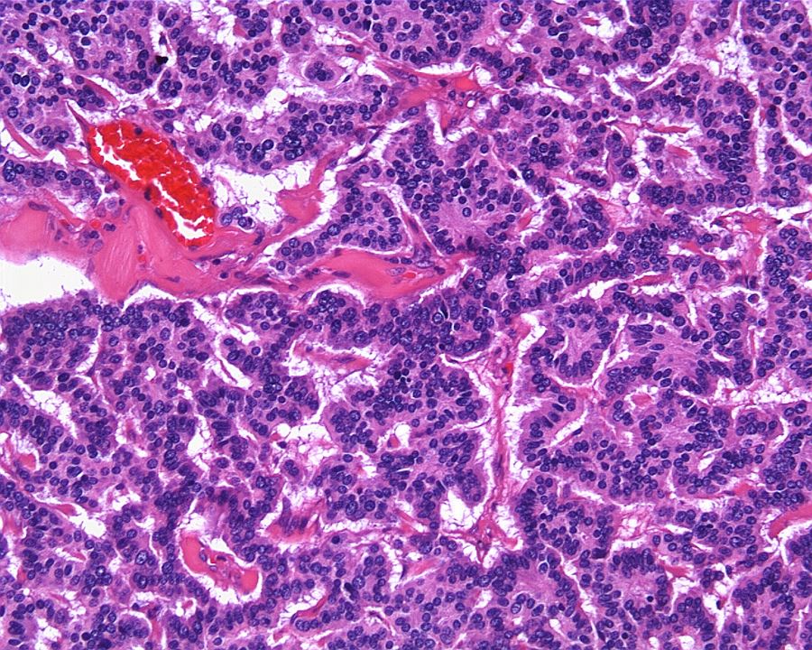
ALK-RCC in adult patient
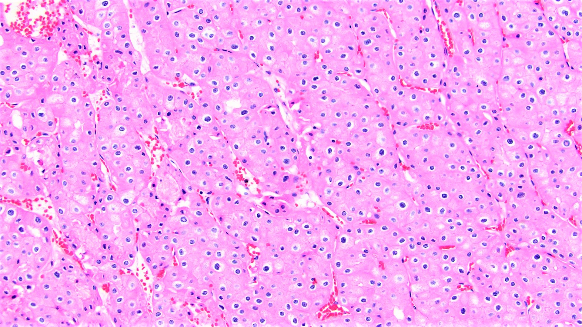
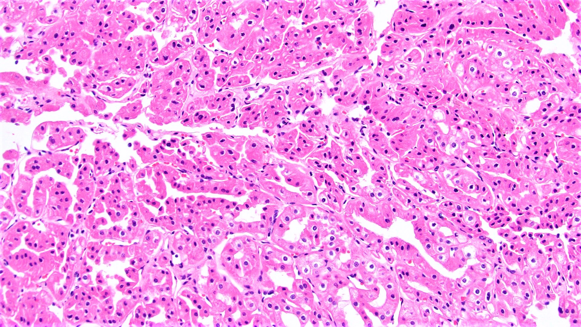
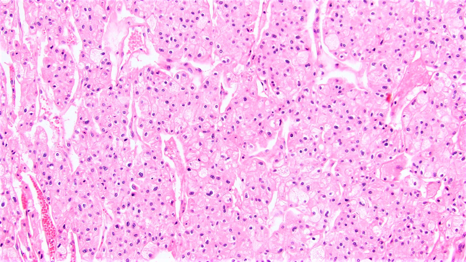
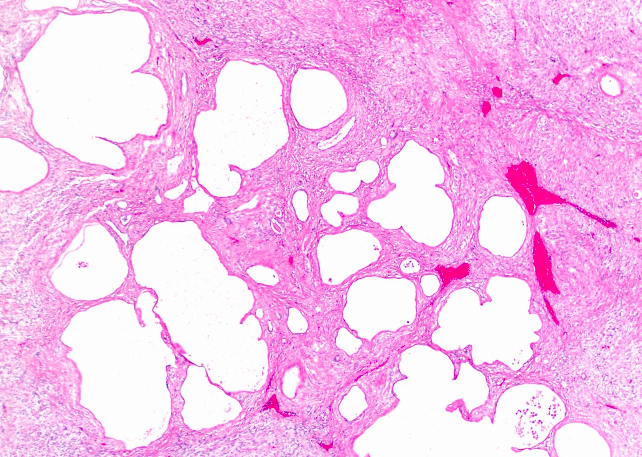
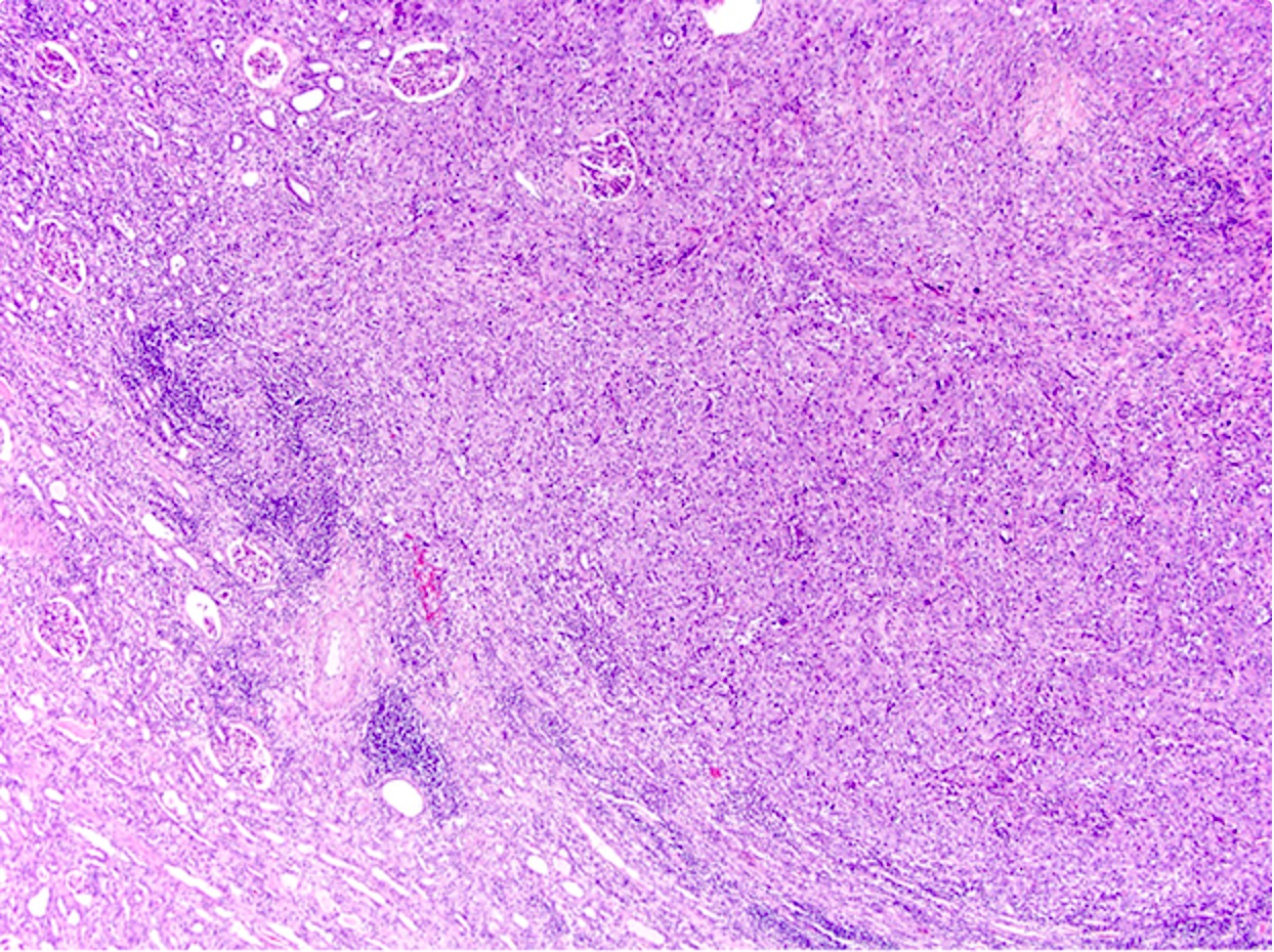
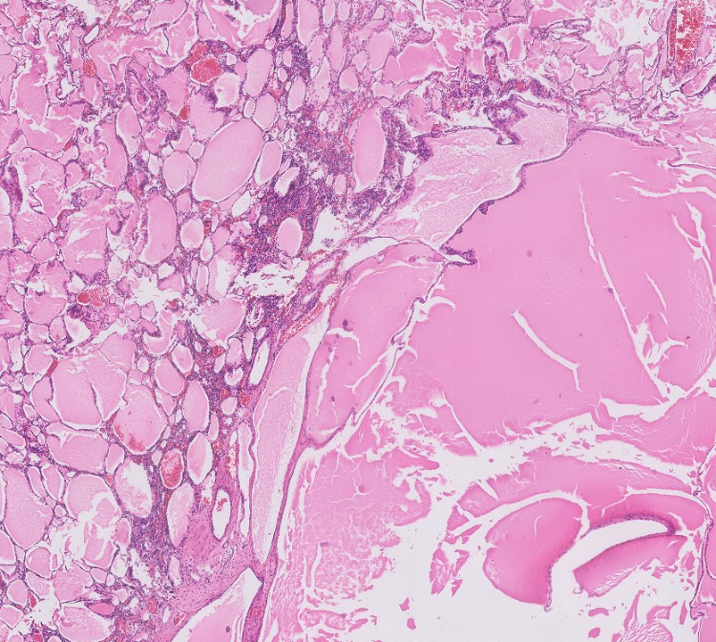
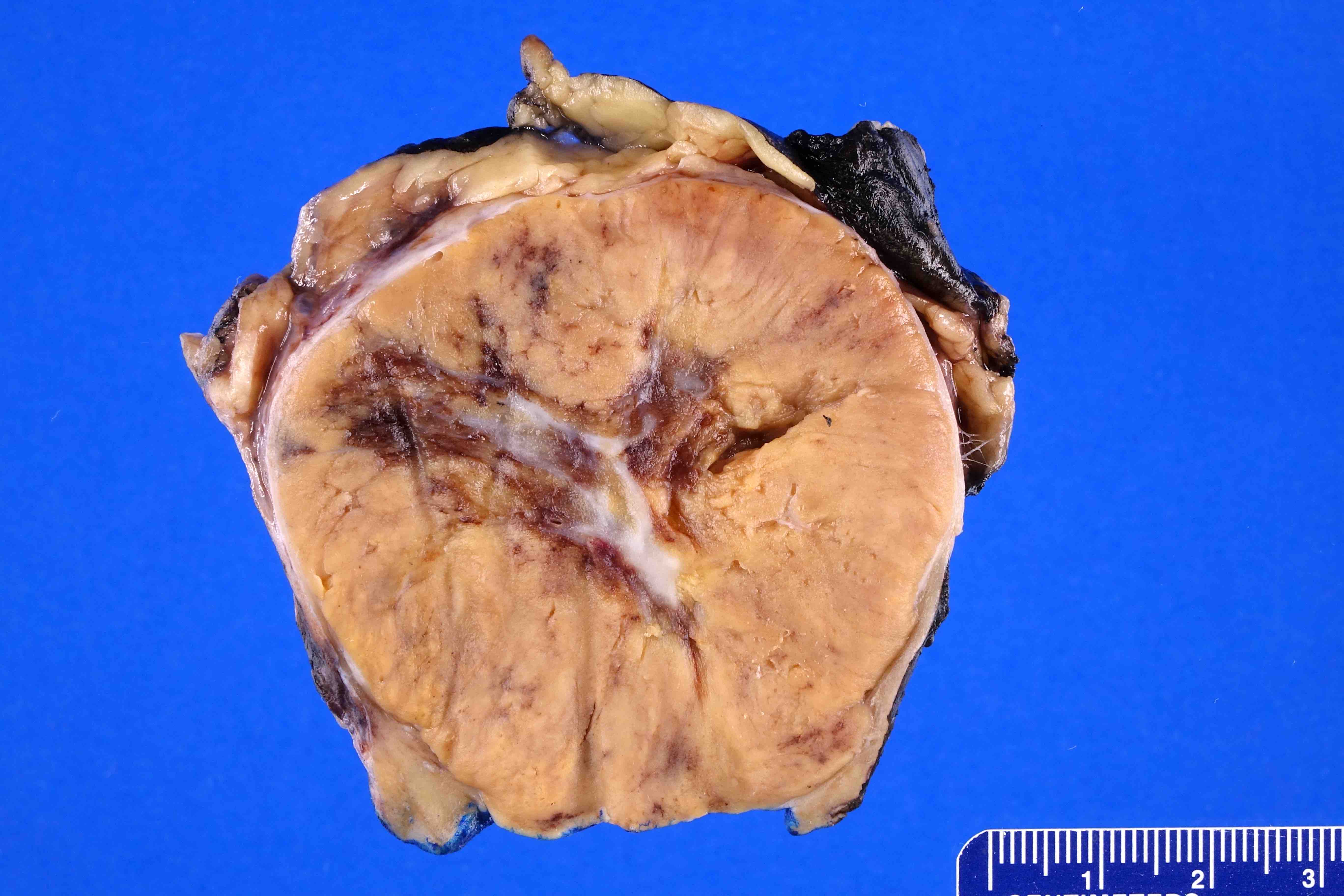
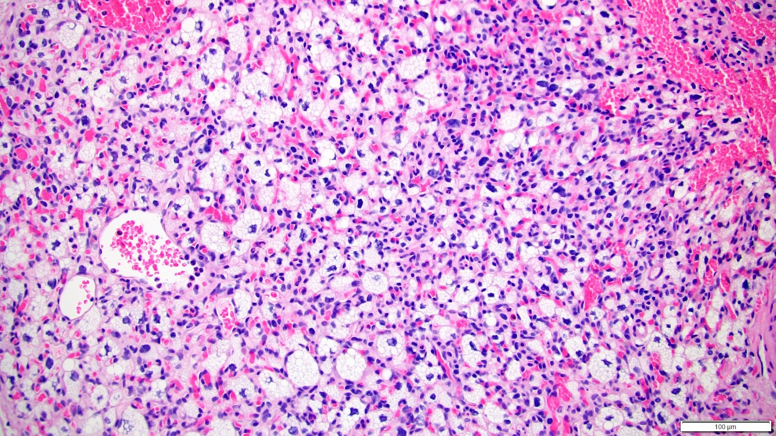
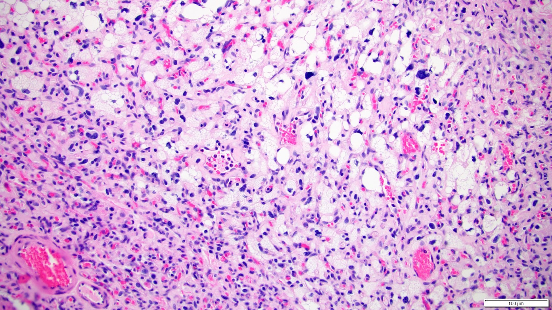
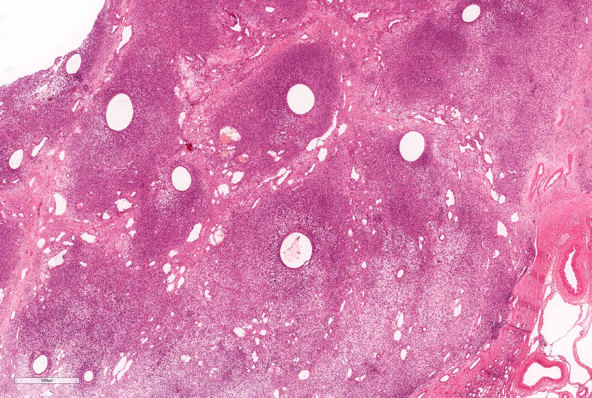
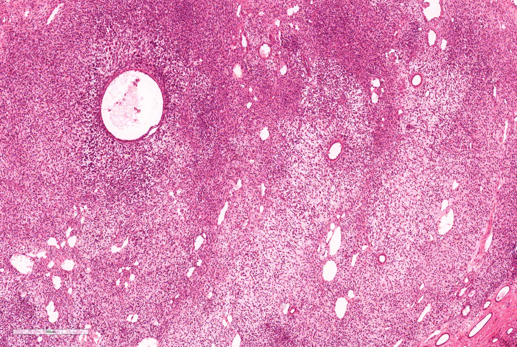
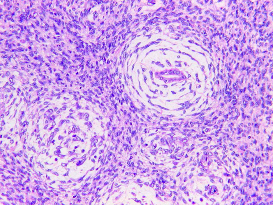
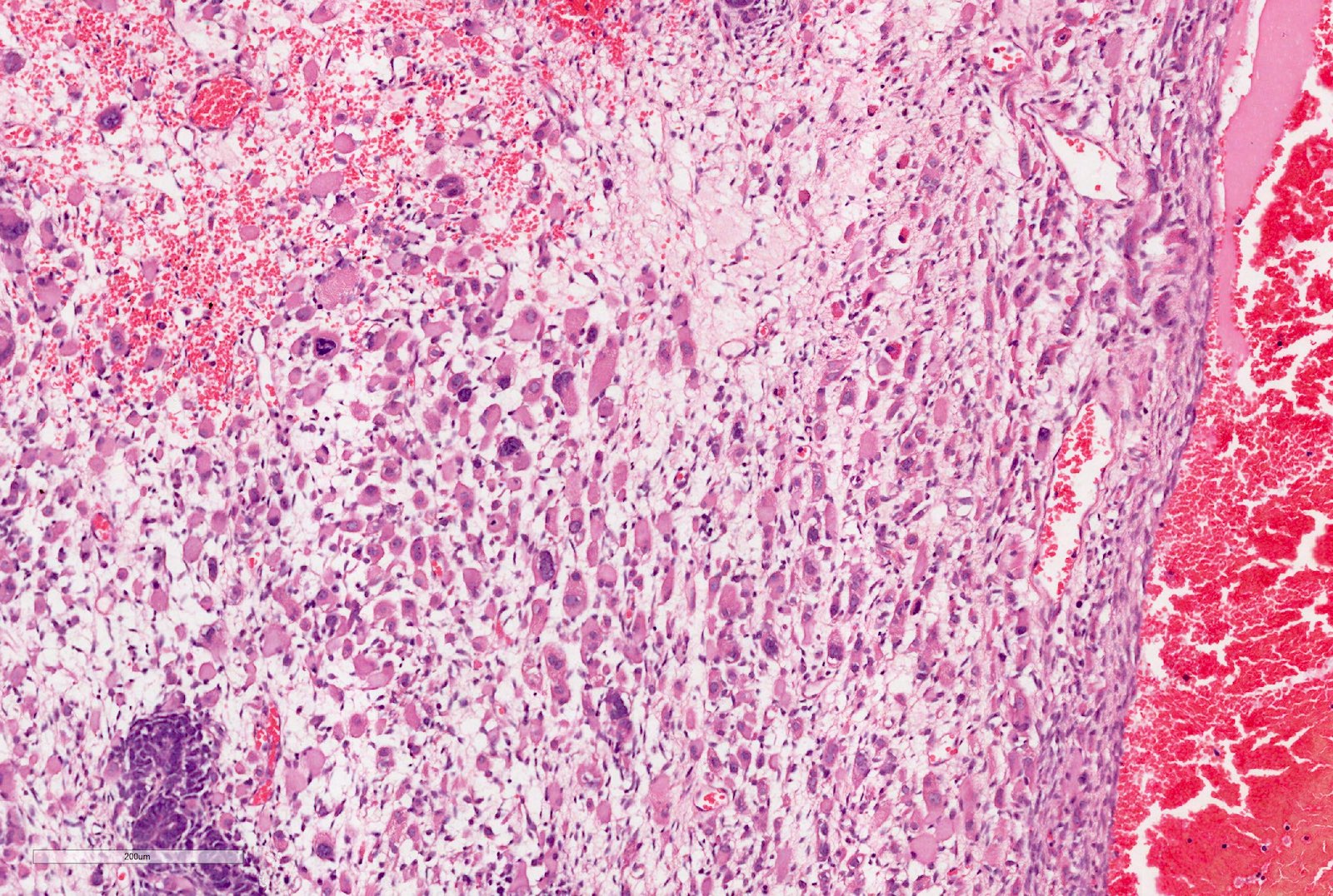
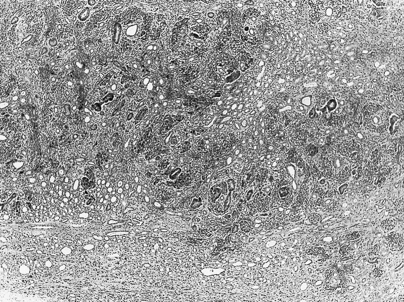
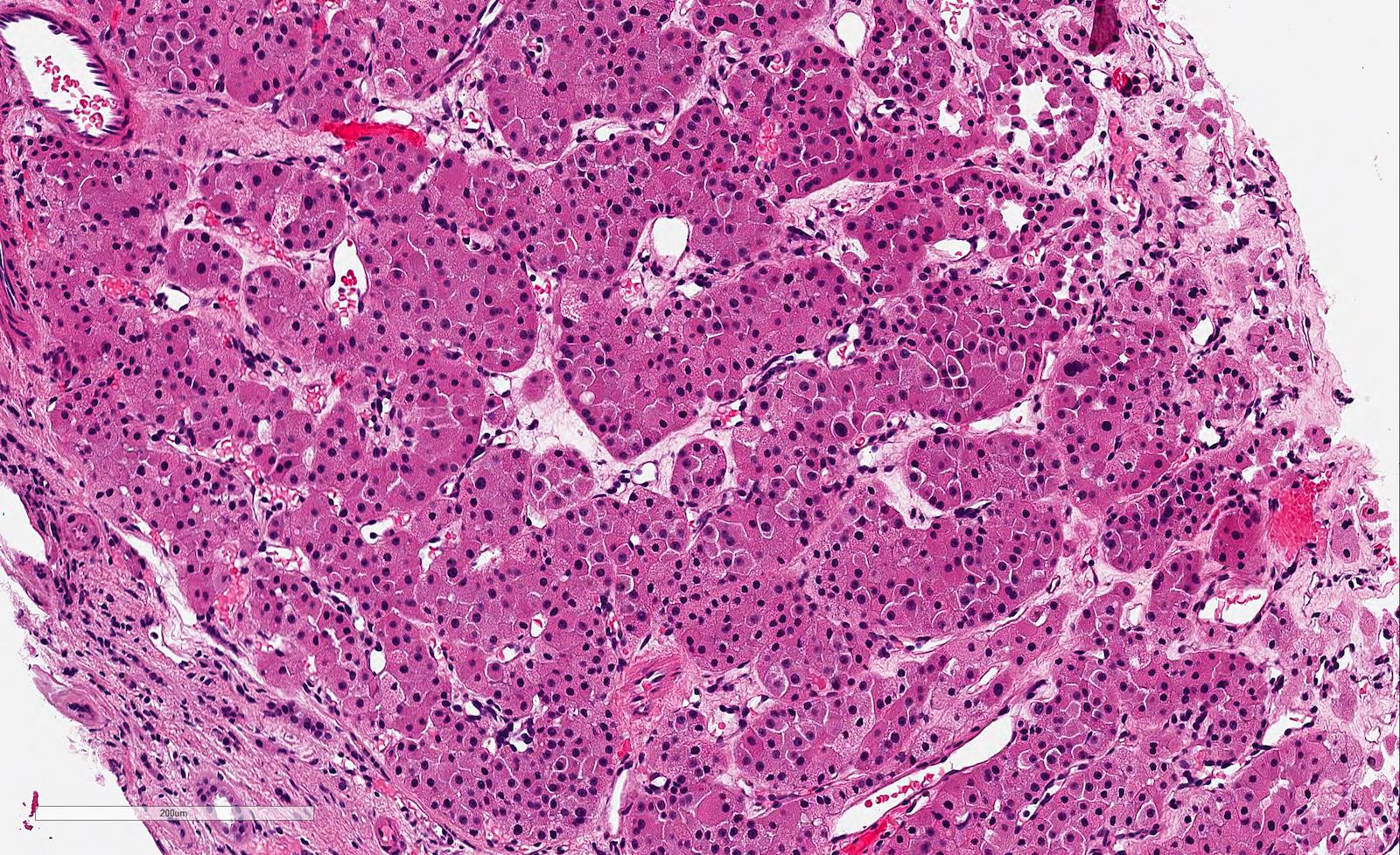
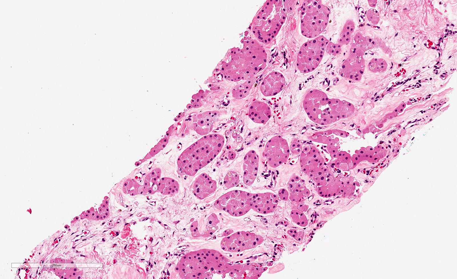
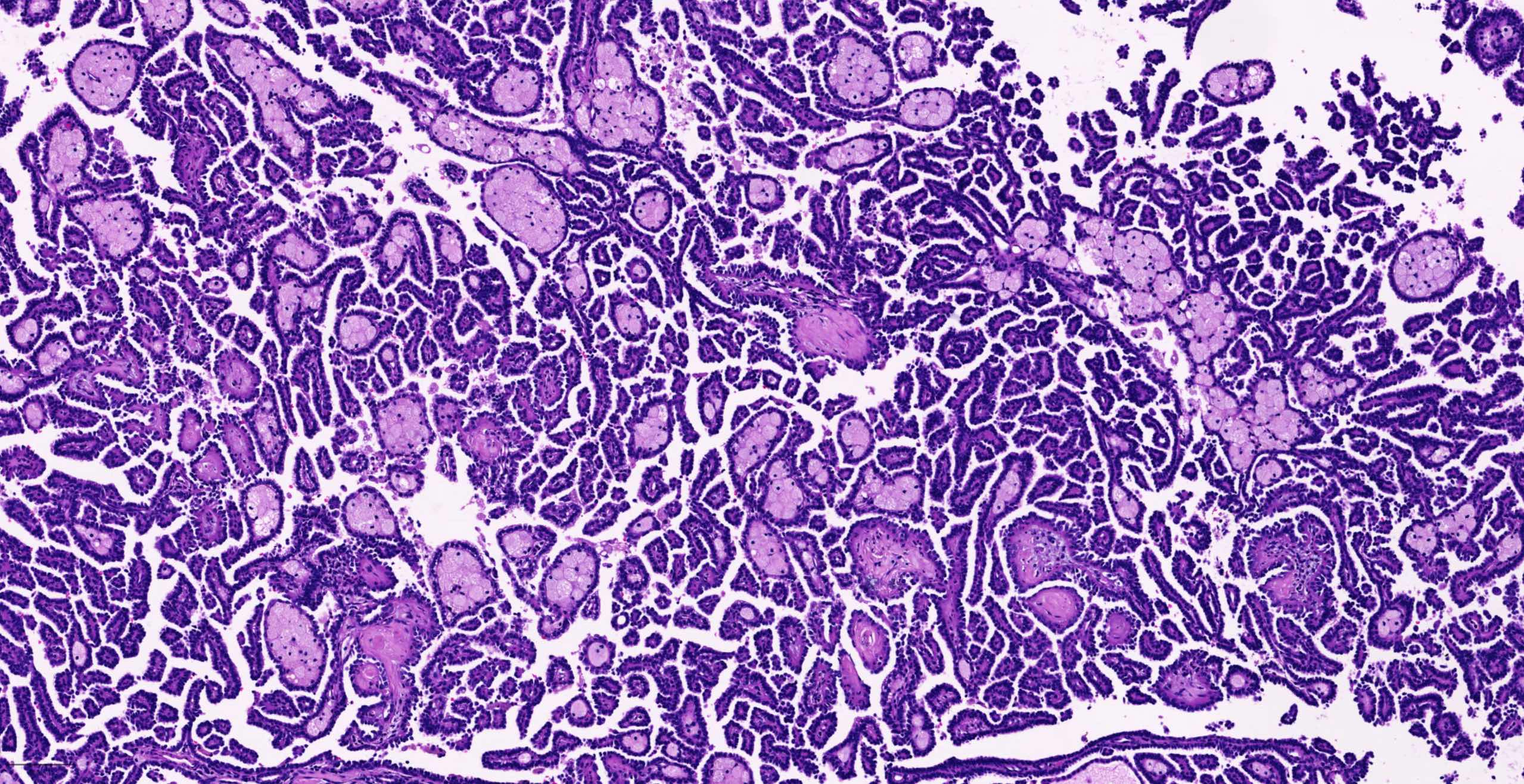
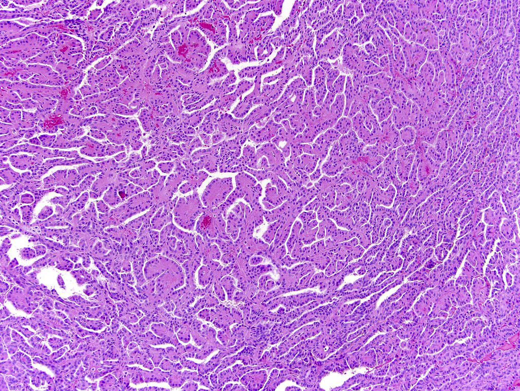
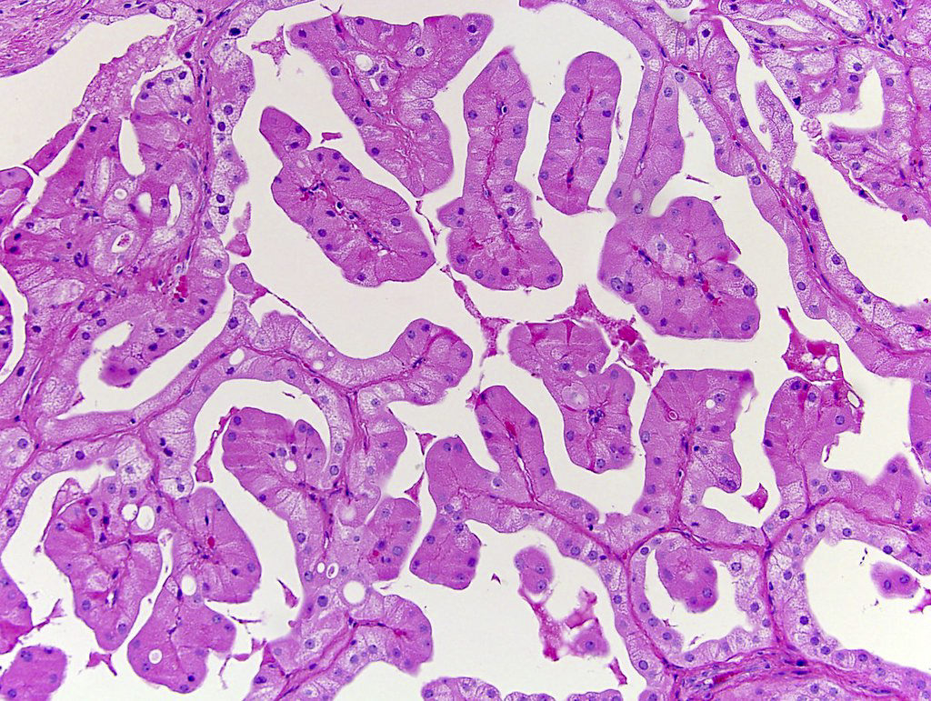
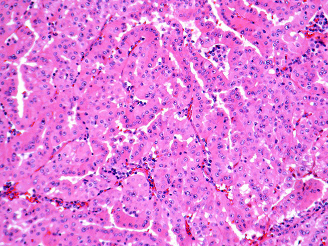
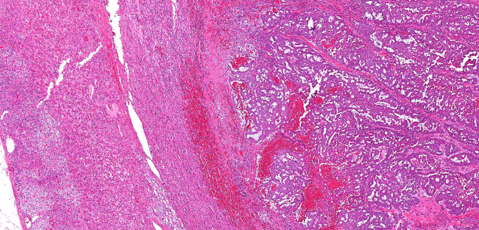
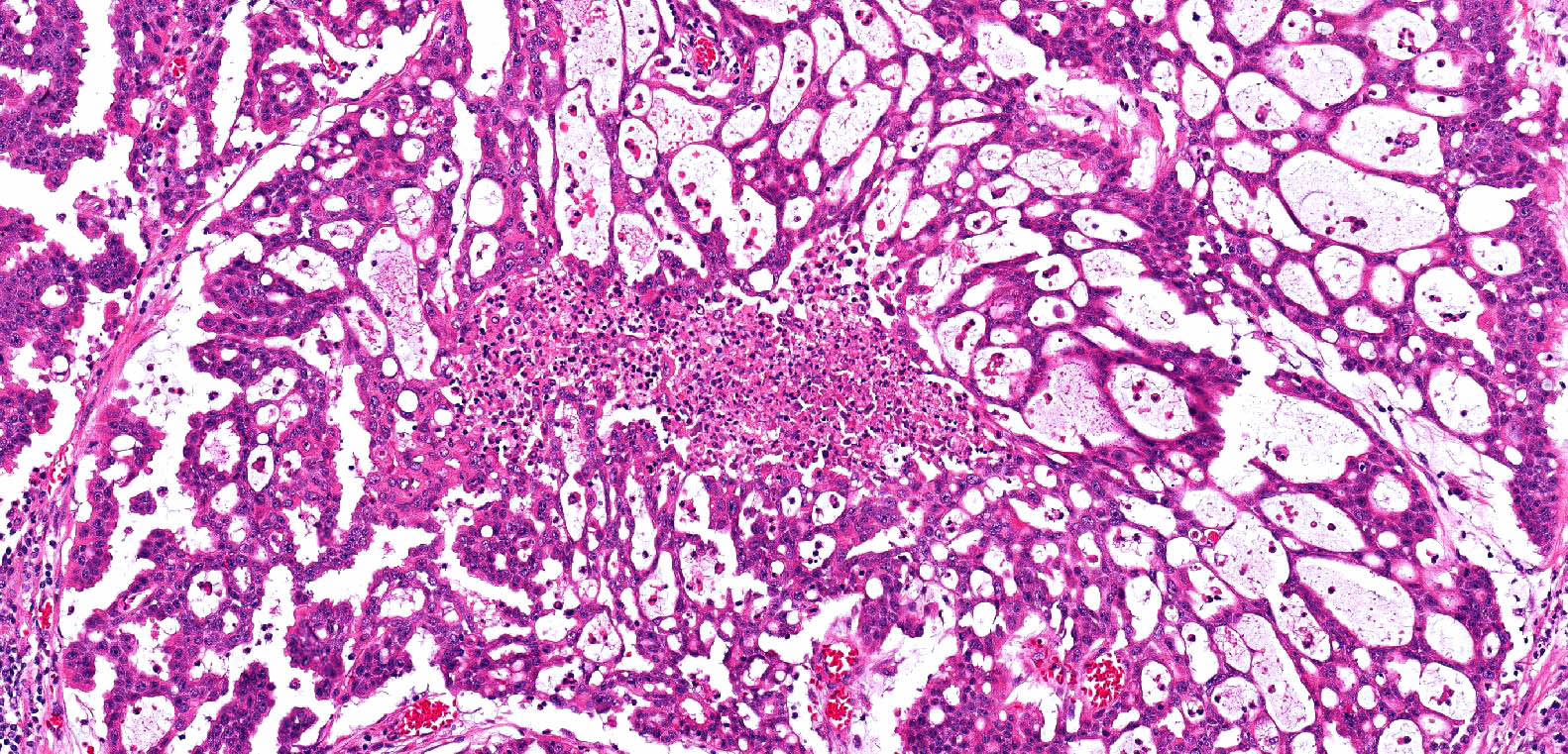
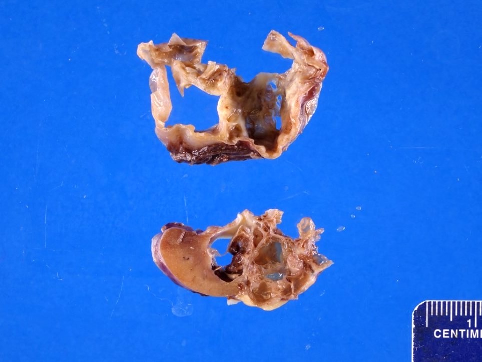
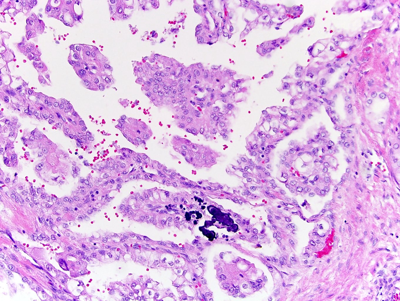
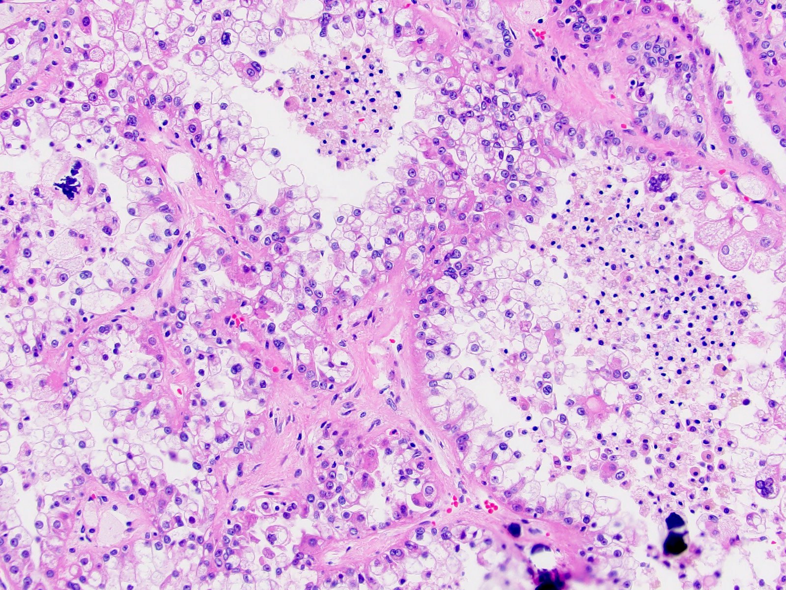
Mixed solid and papillary areas with abundant mucin
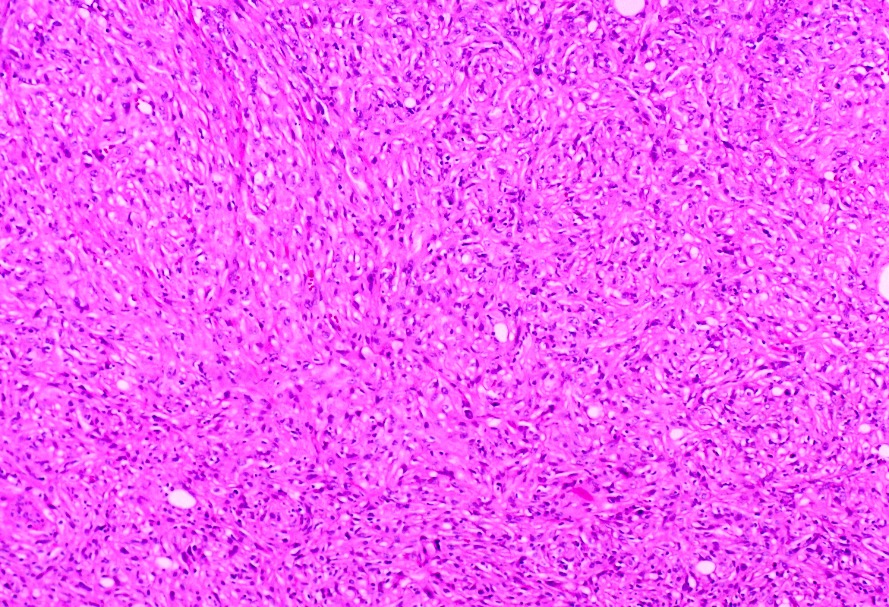
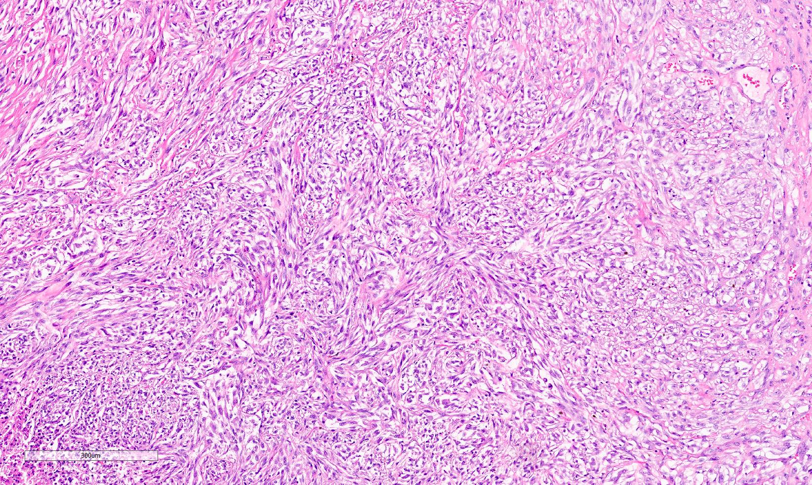
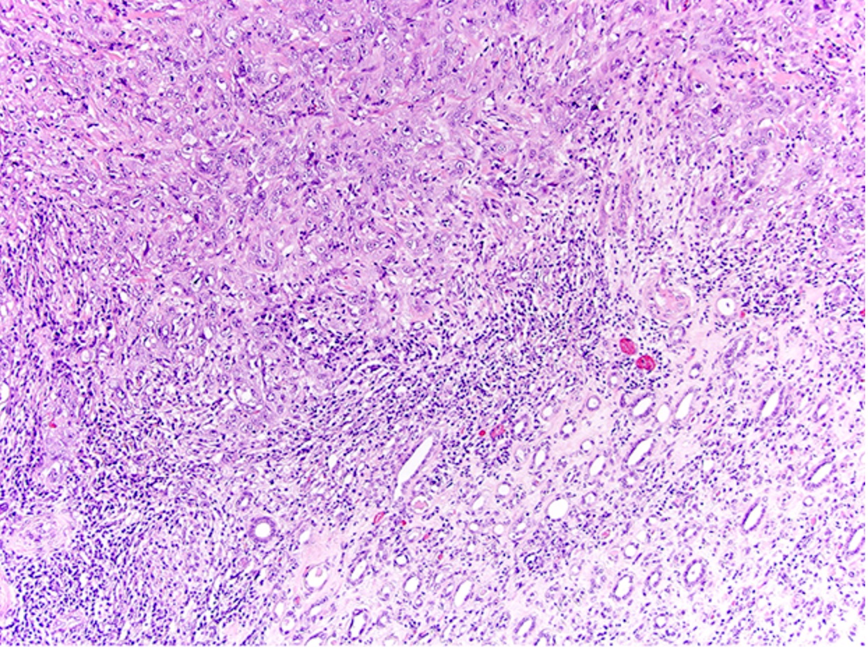
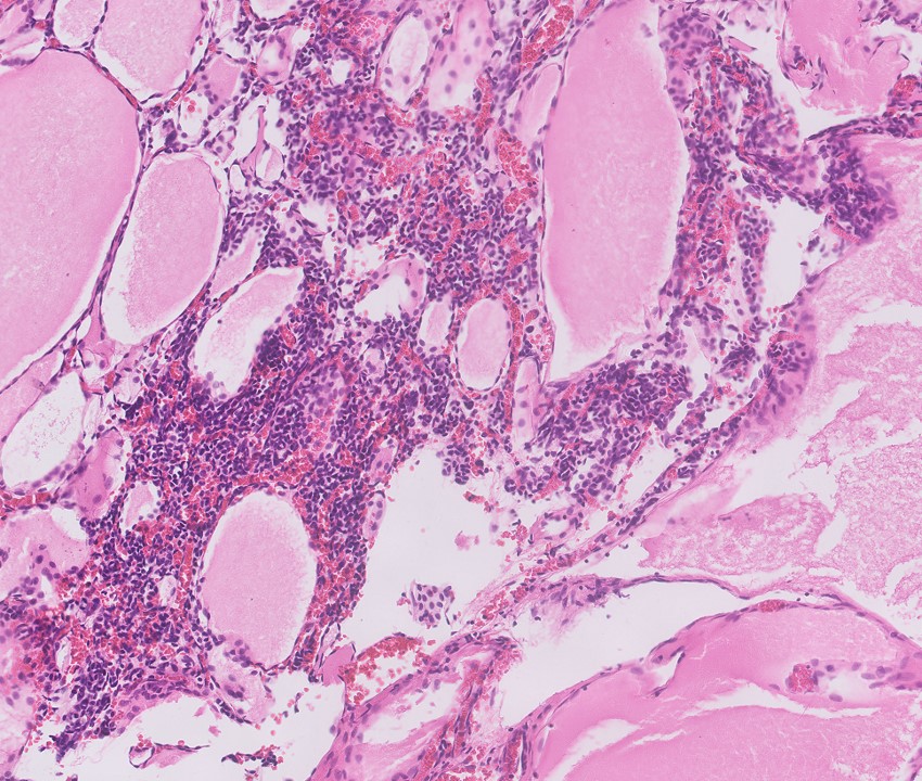
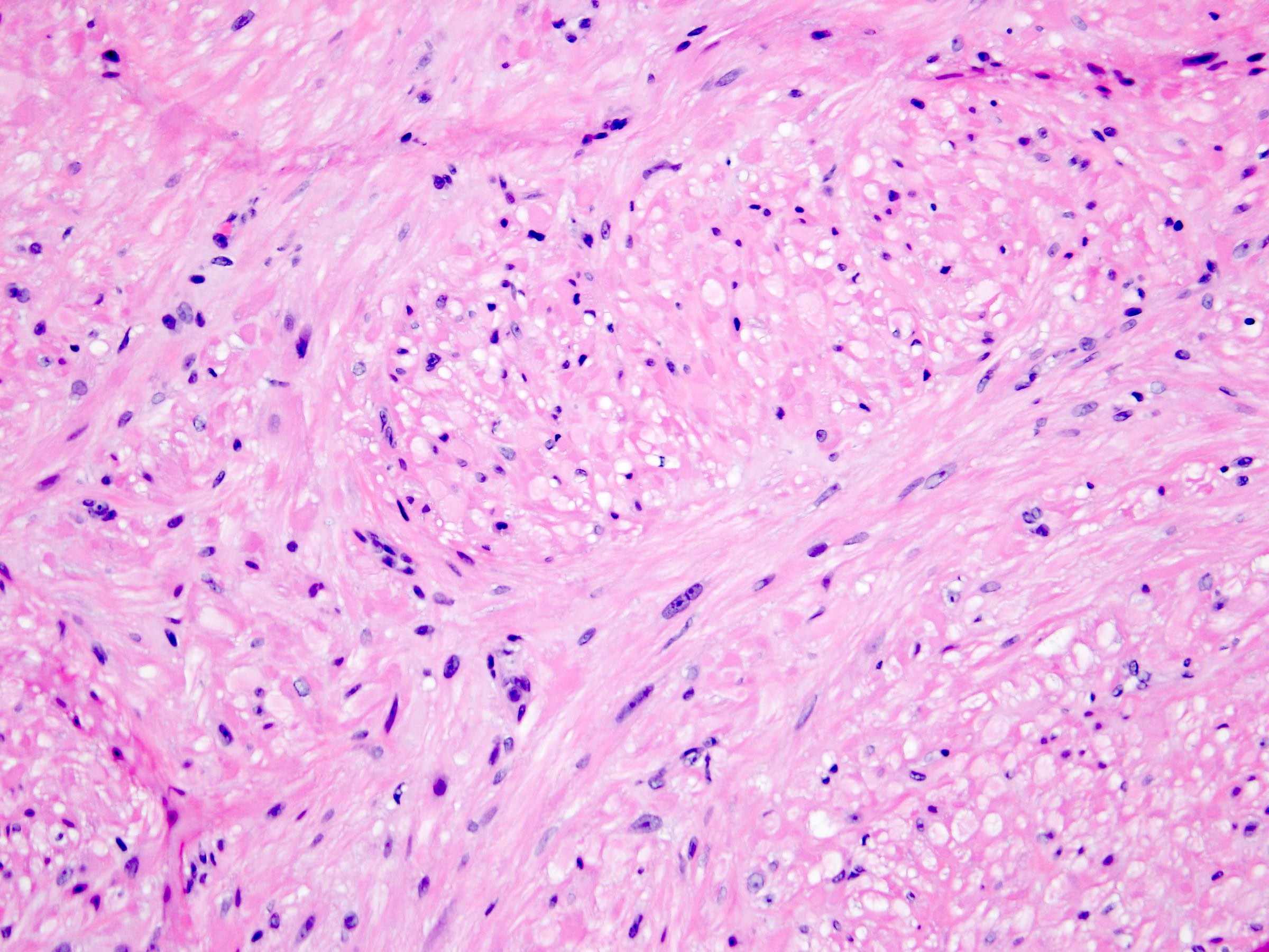
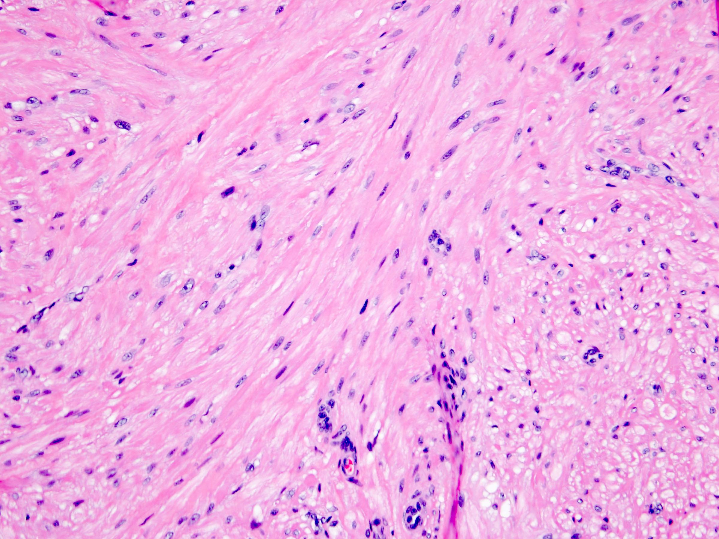
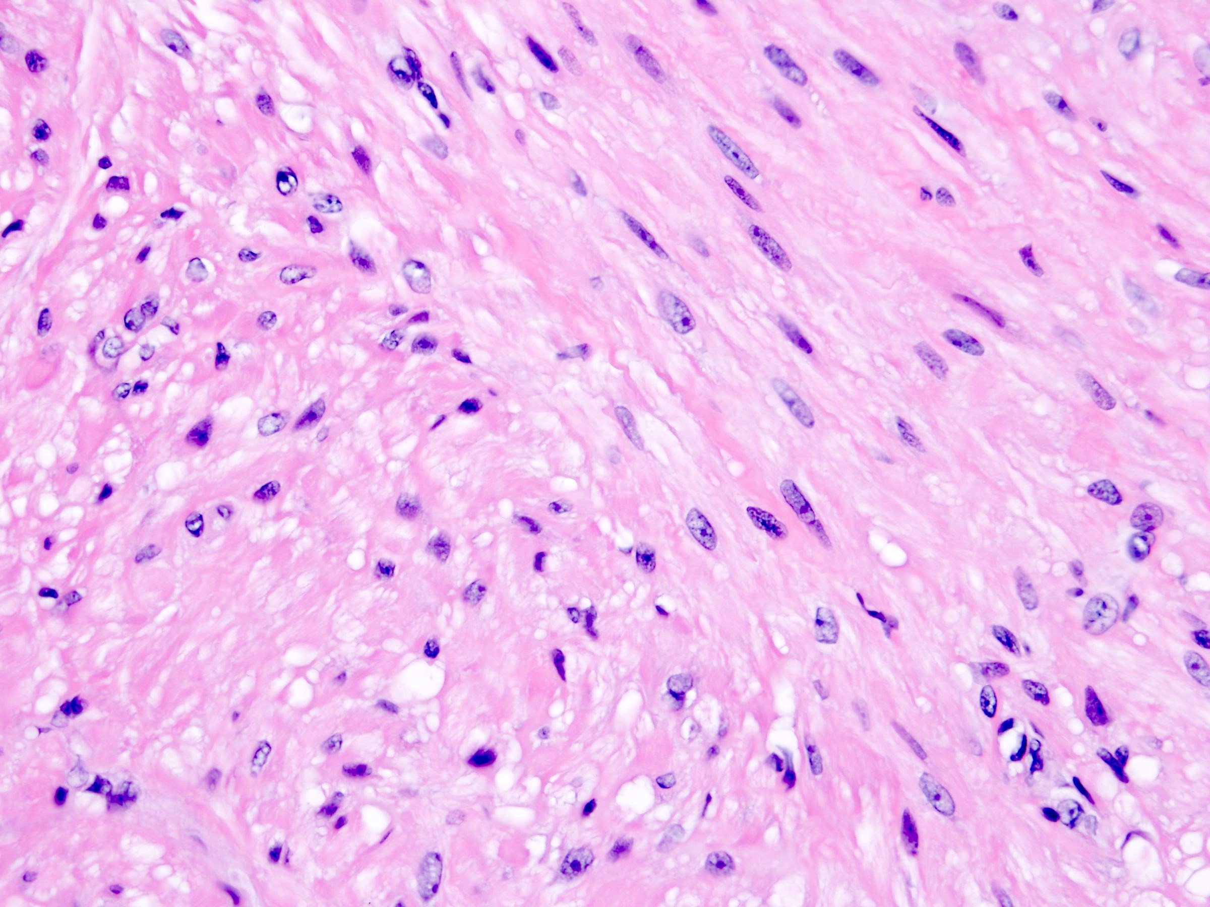
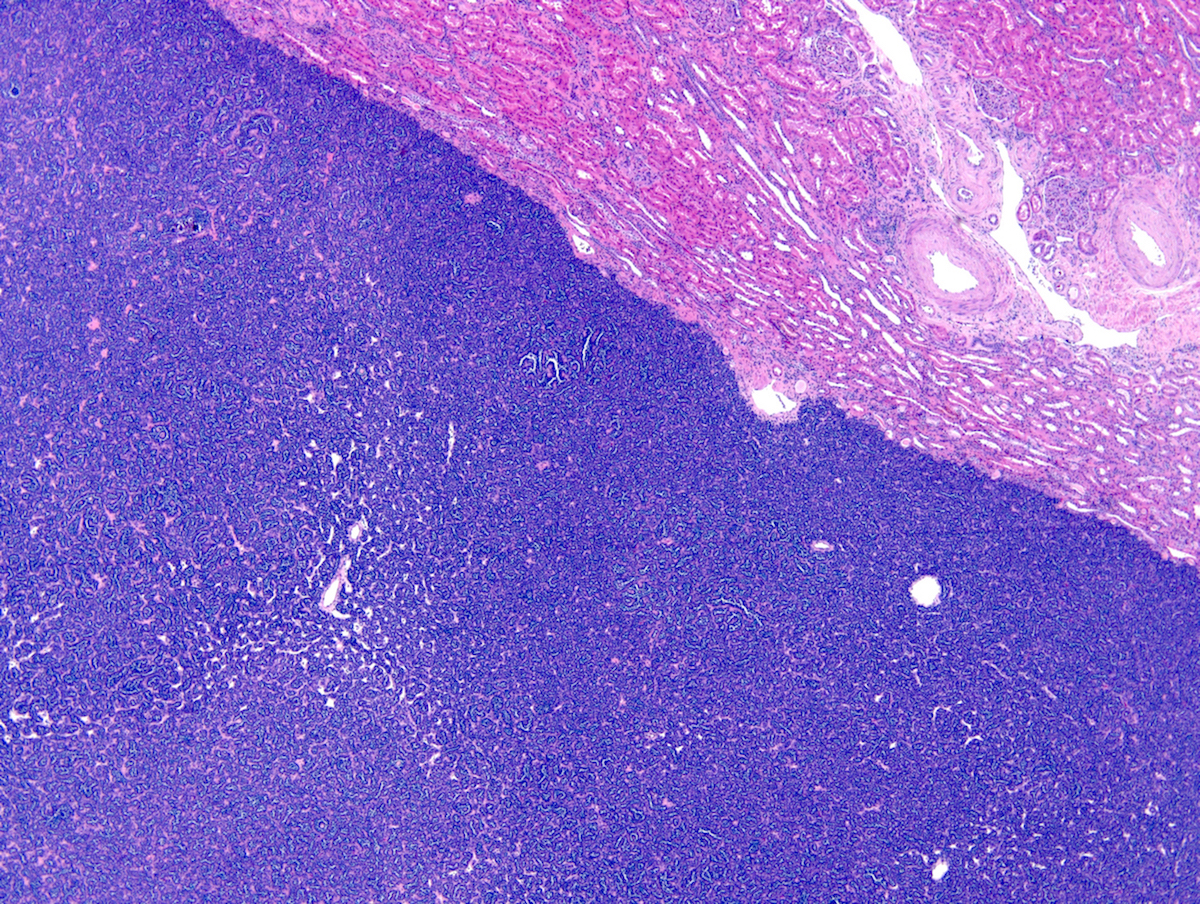
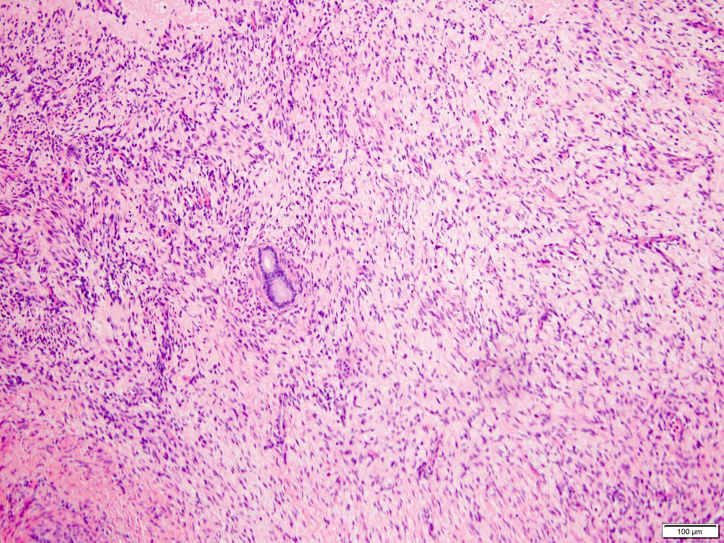
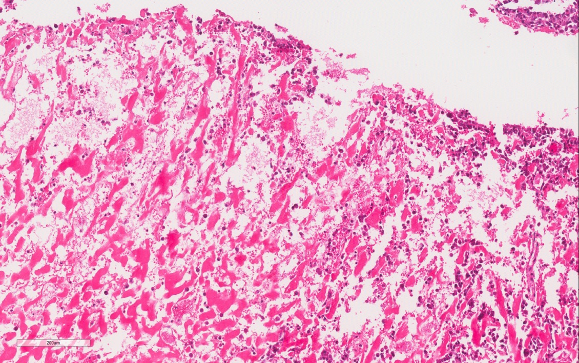
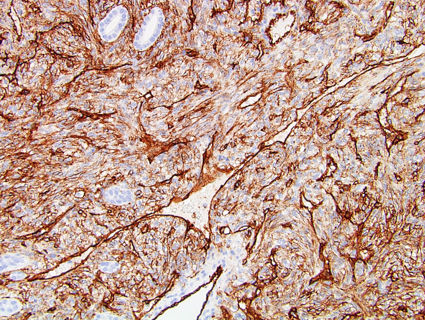
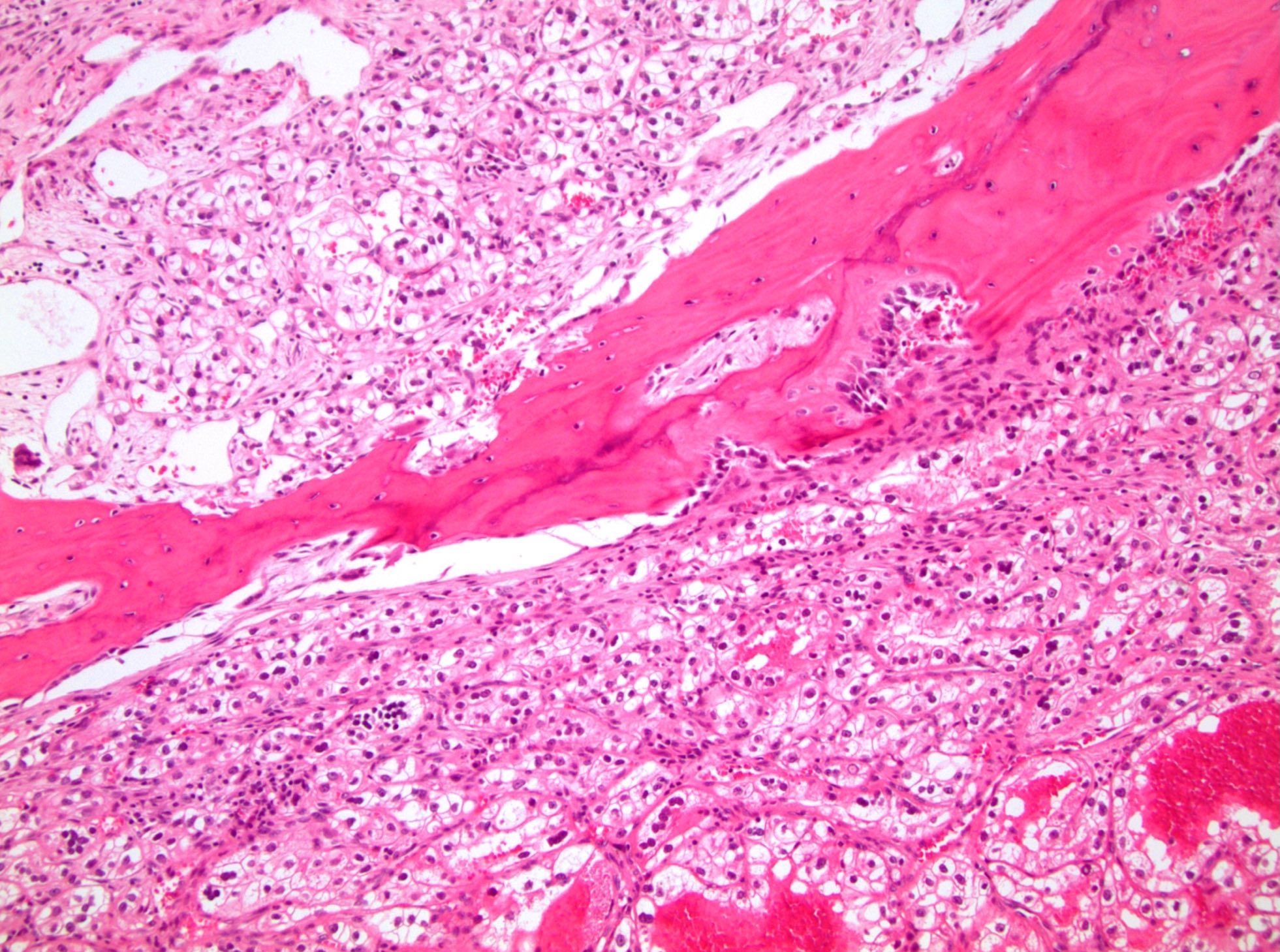
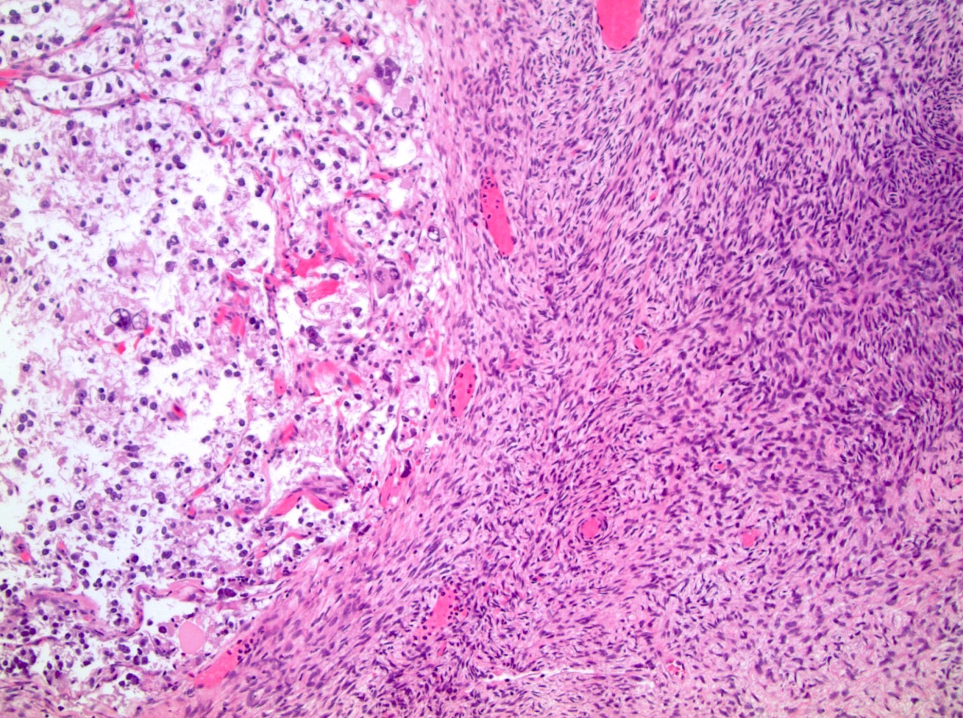
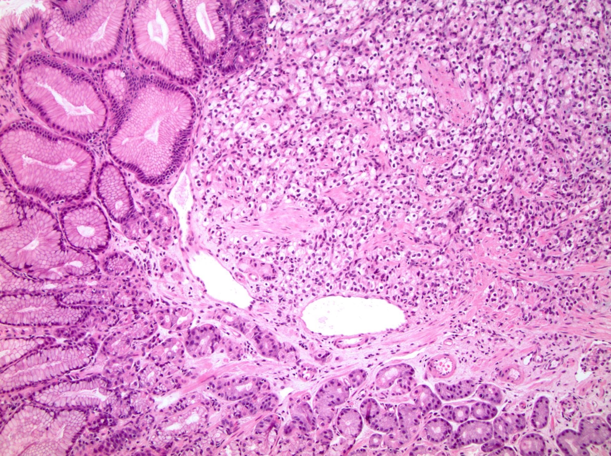
Infiltrating growth with desmoplasia
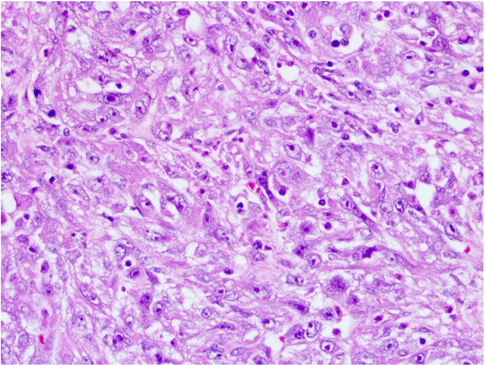
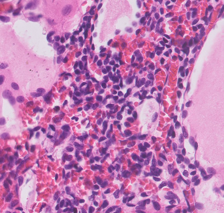
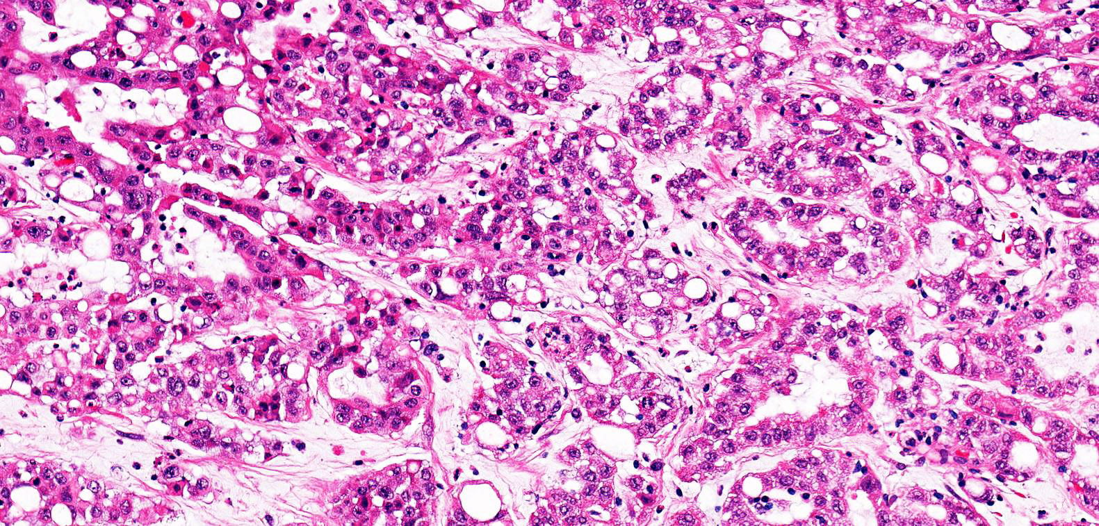
Eosinophilic discohesive pleomorphic cells
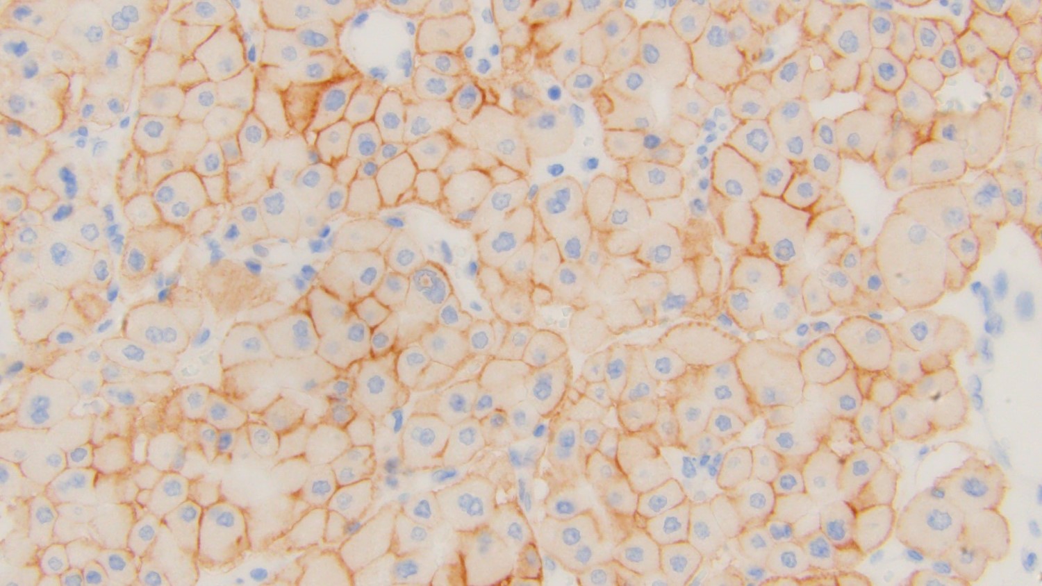
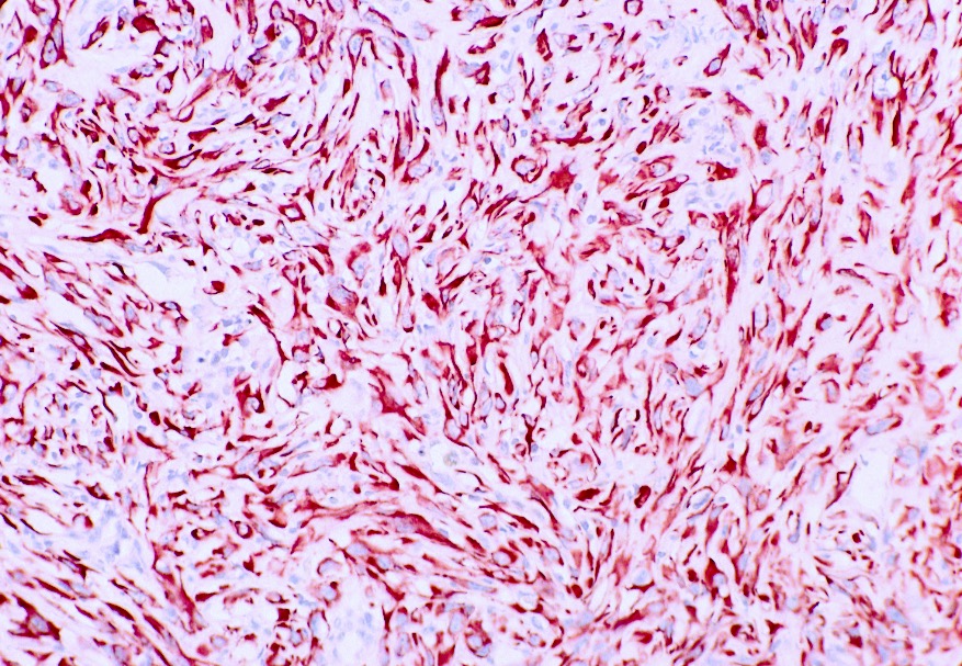
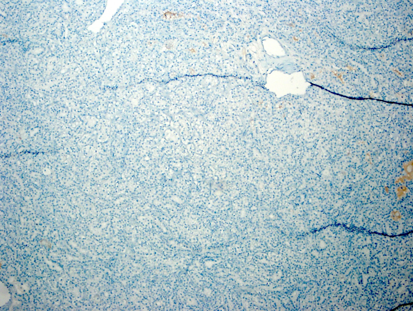
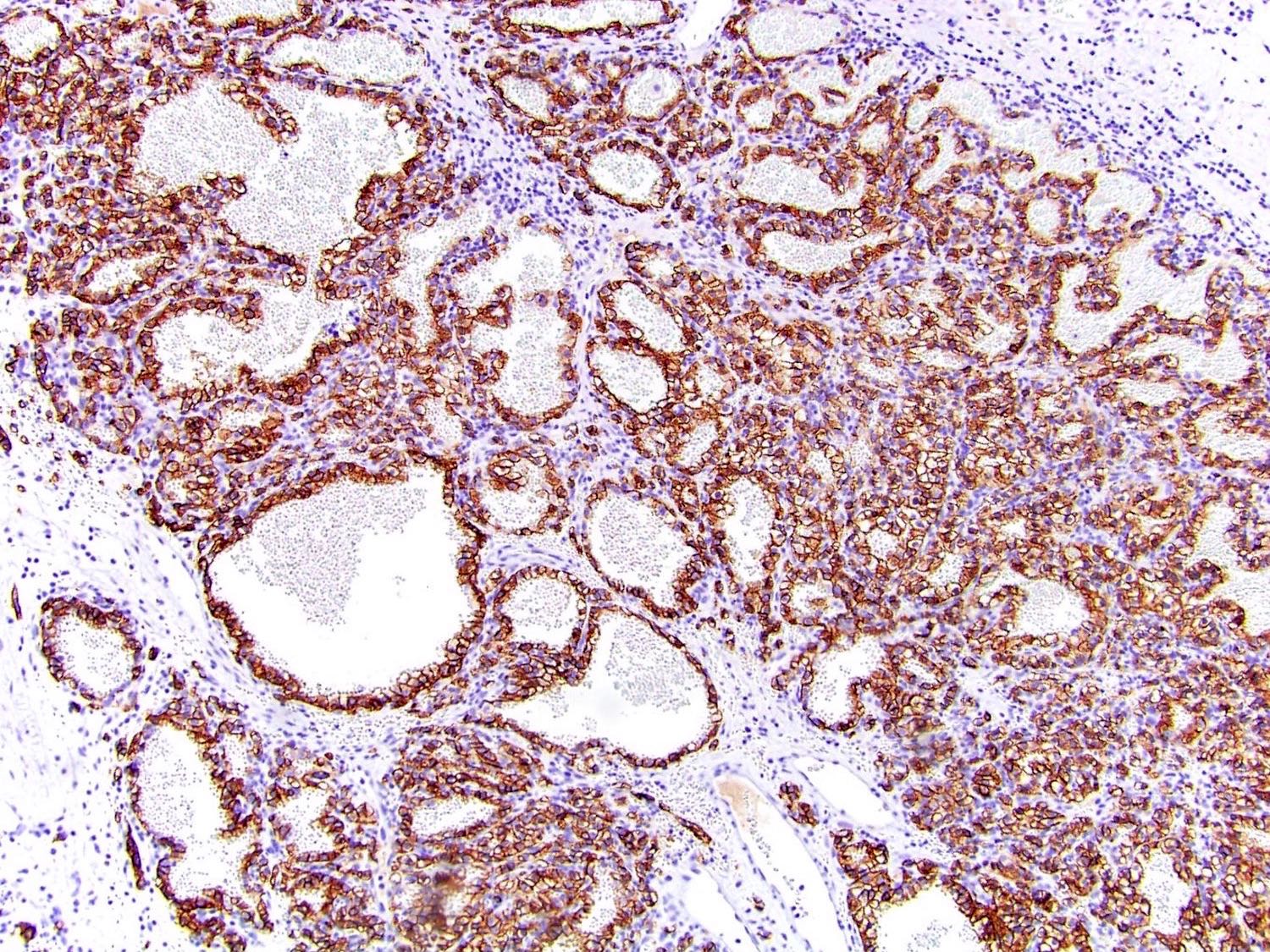
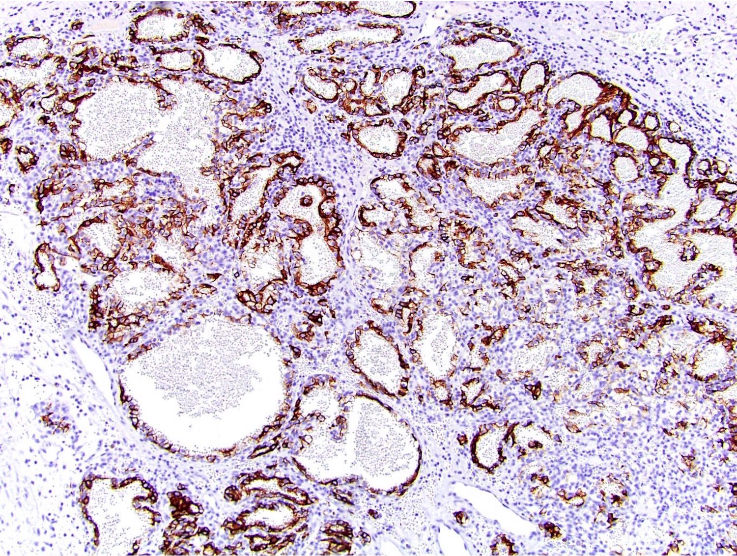
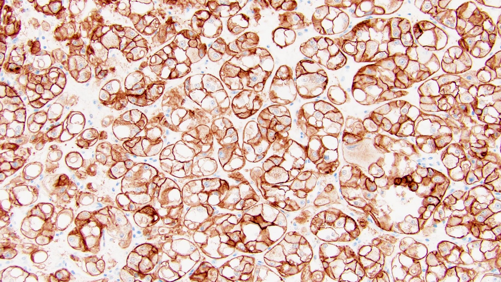
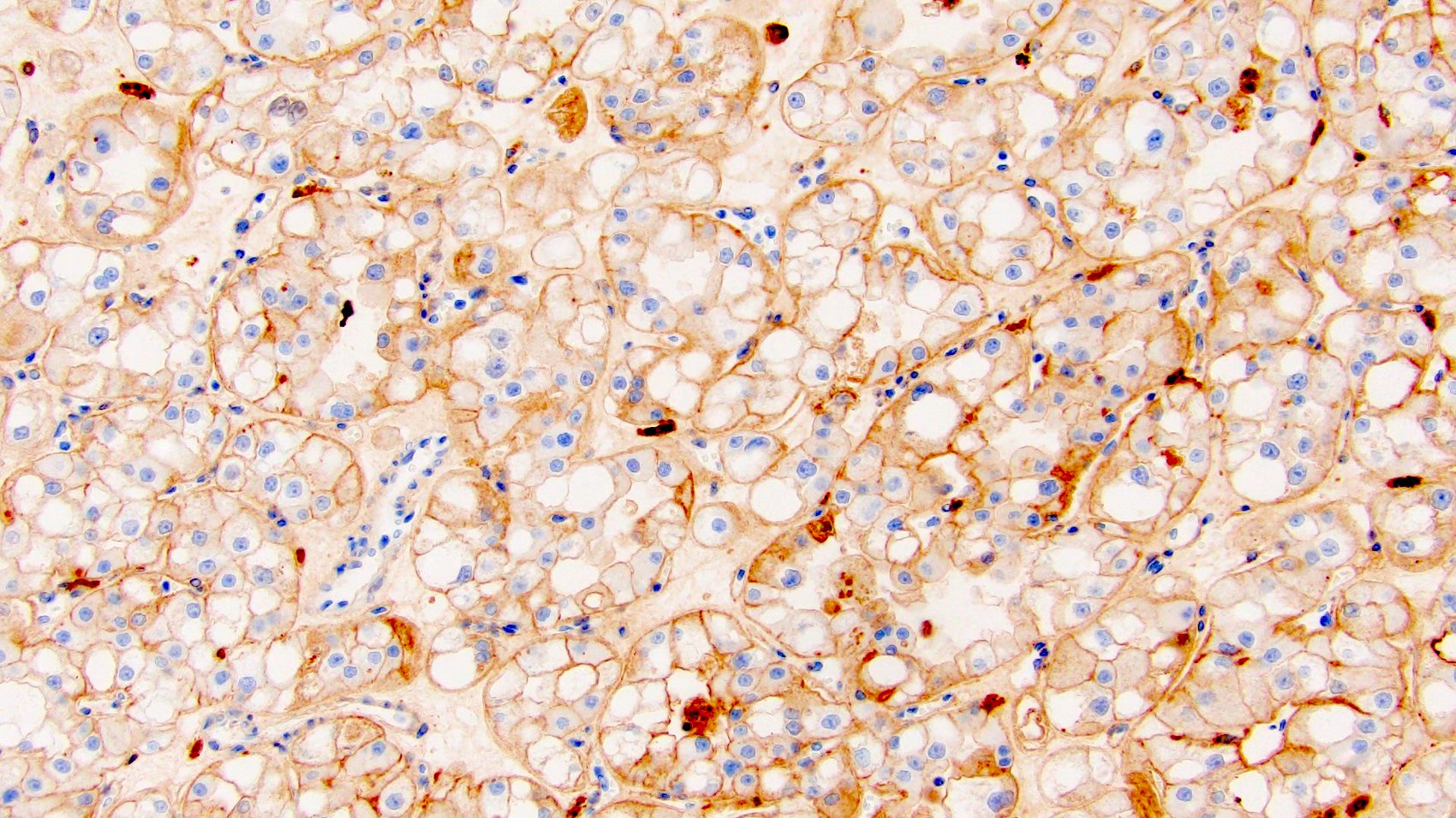
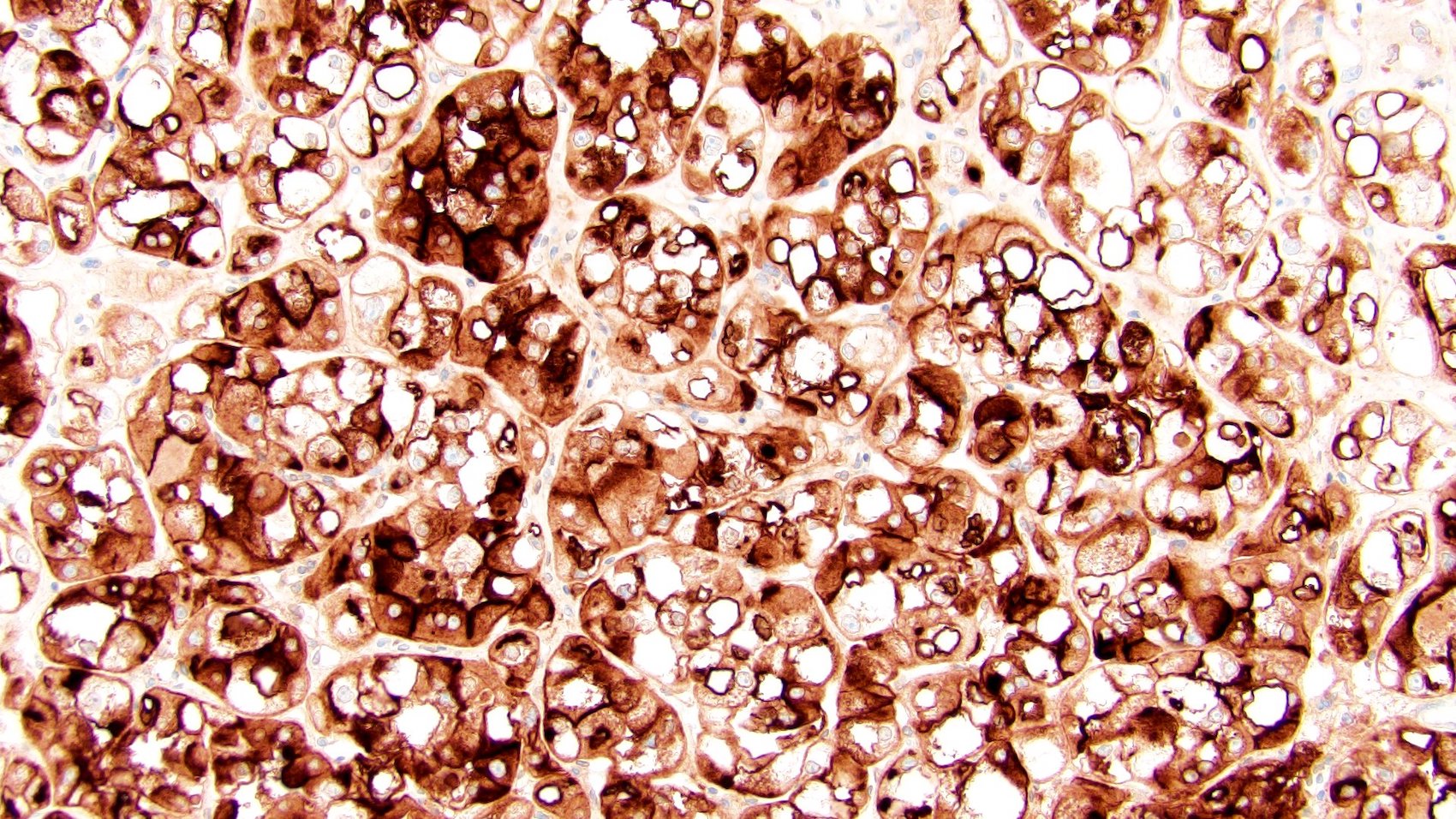
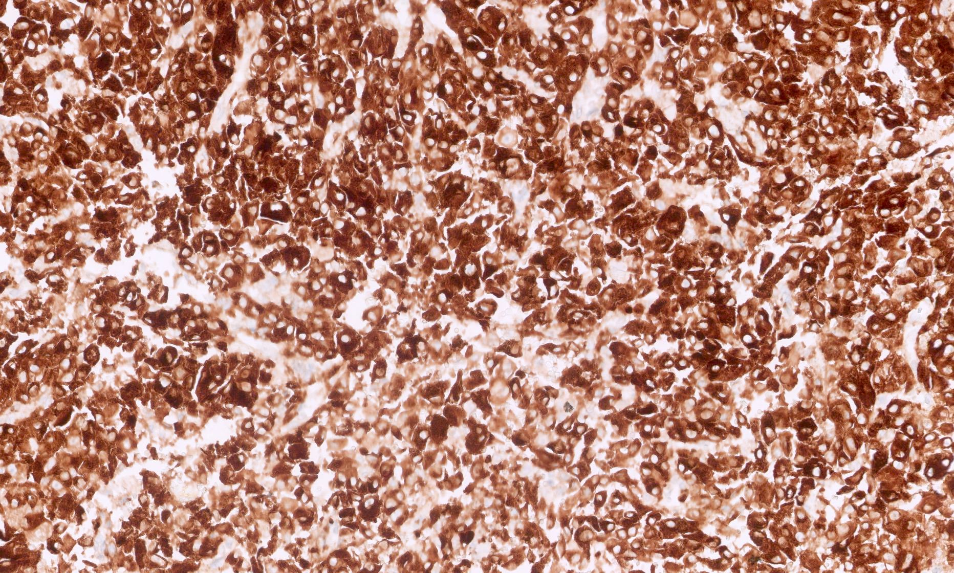
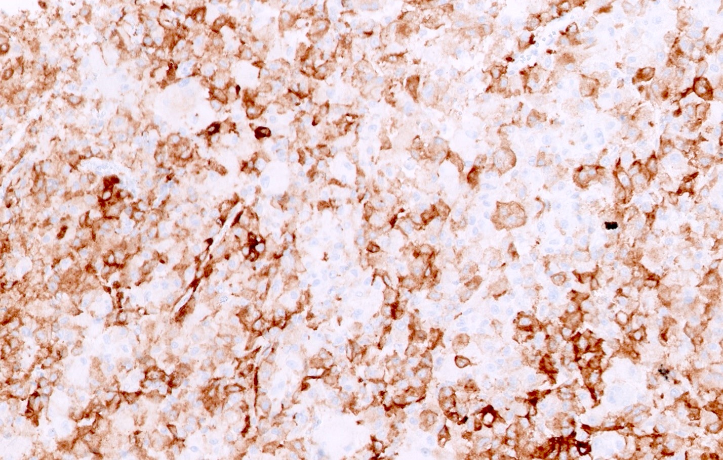
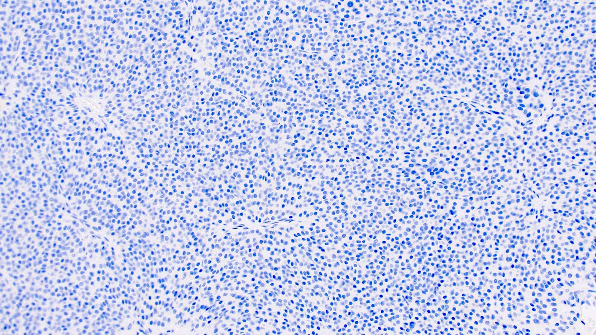
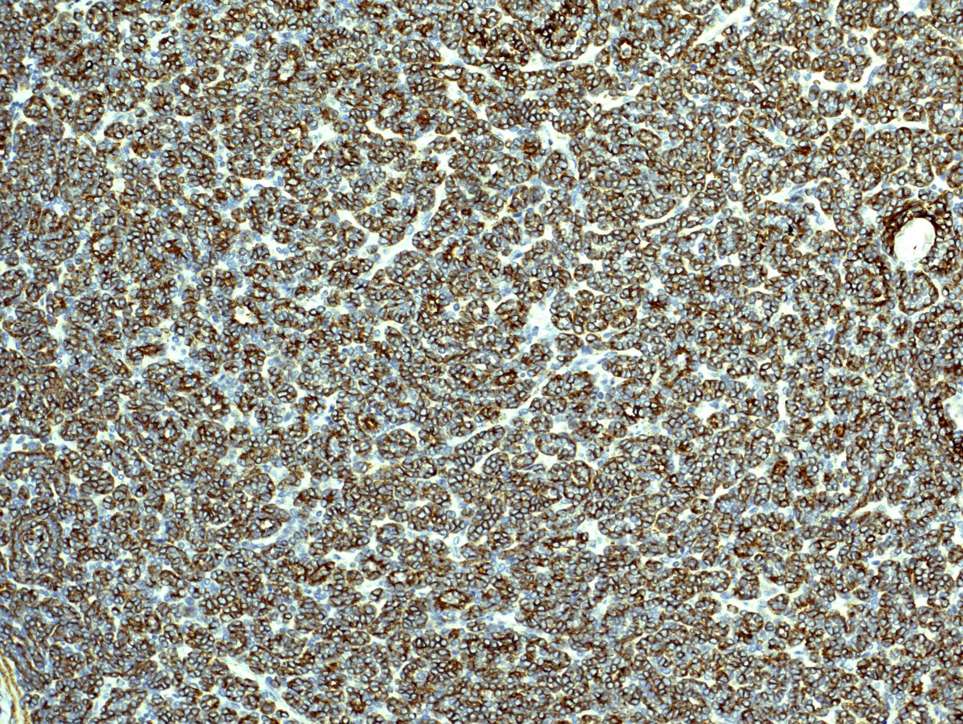
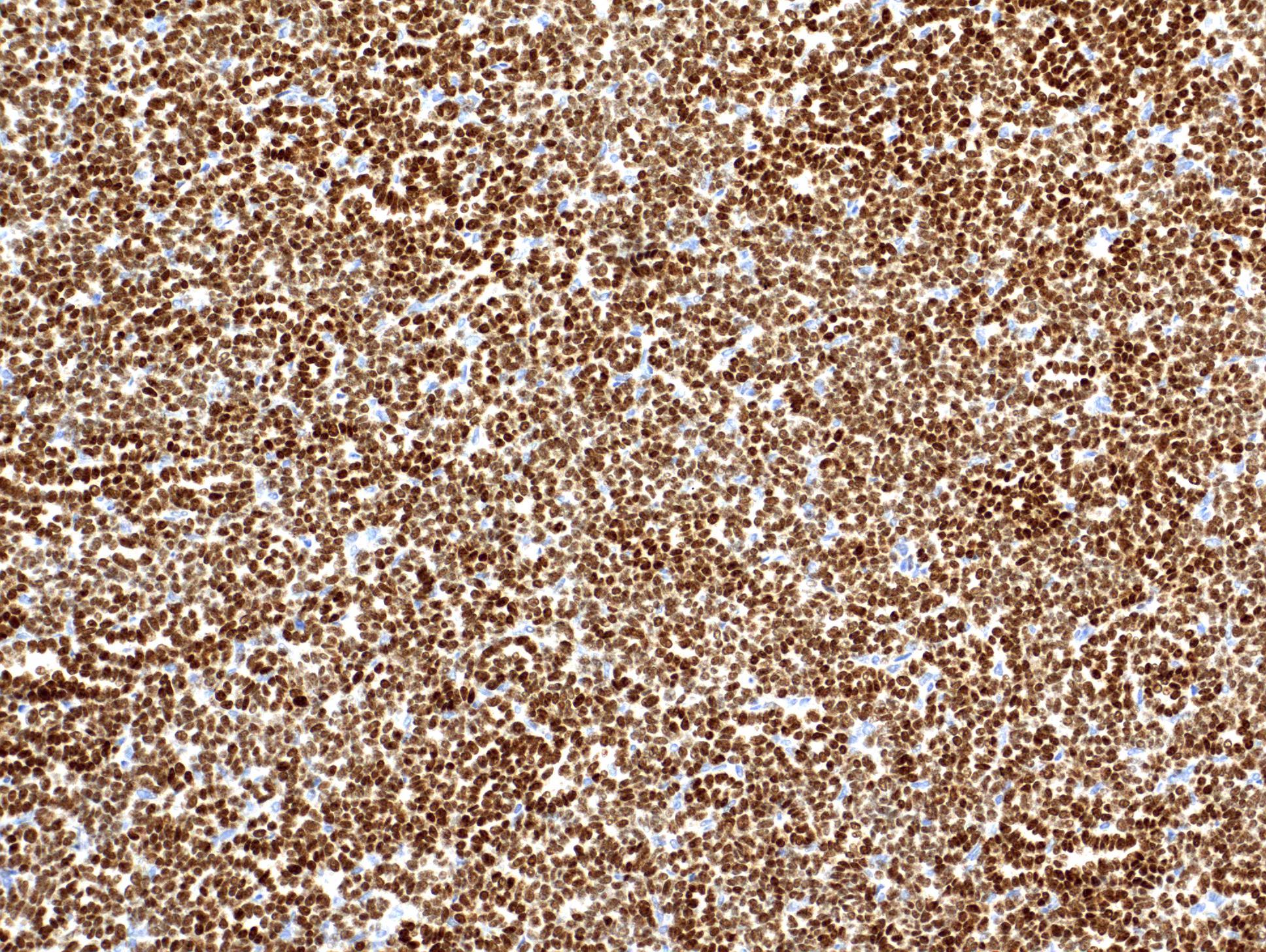
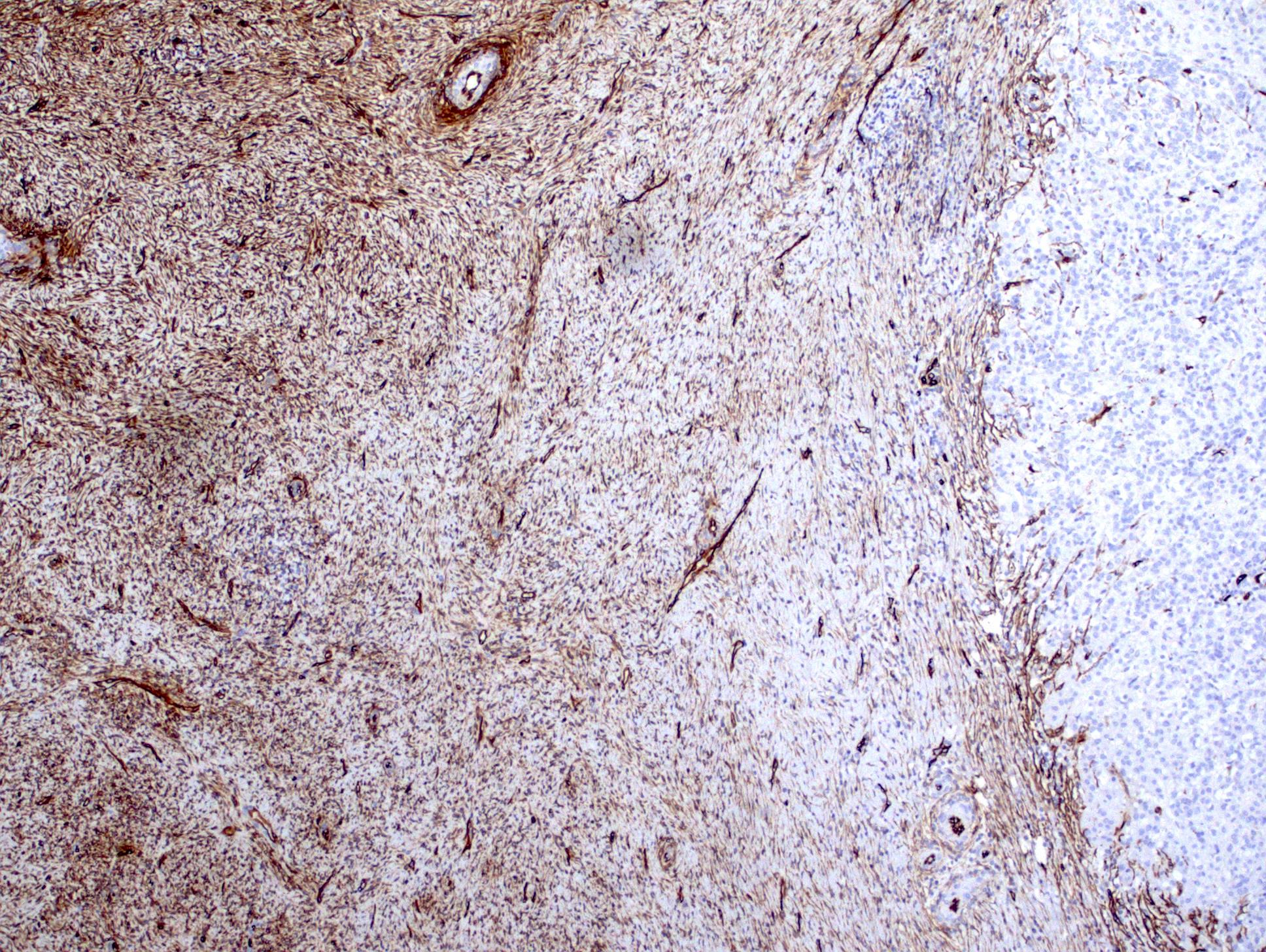
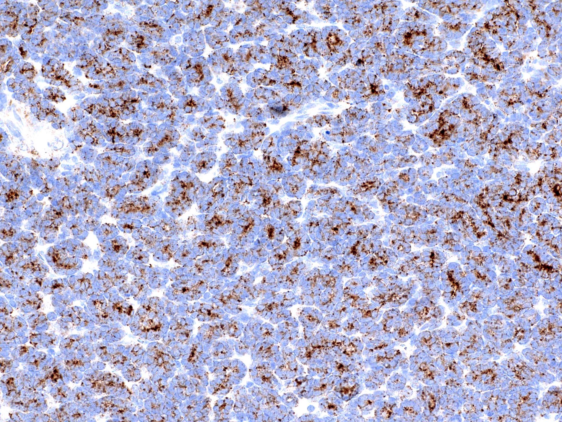
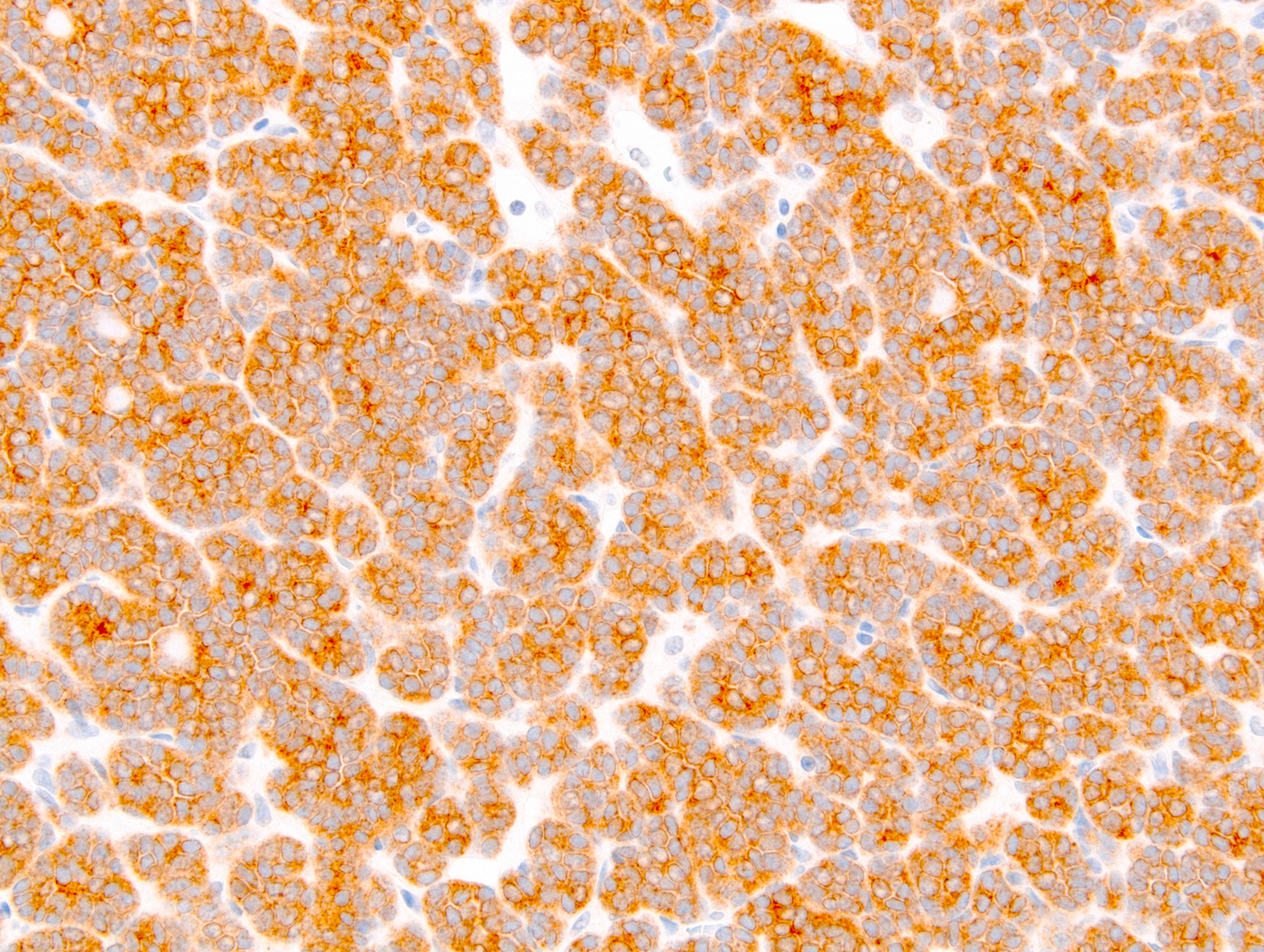
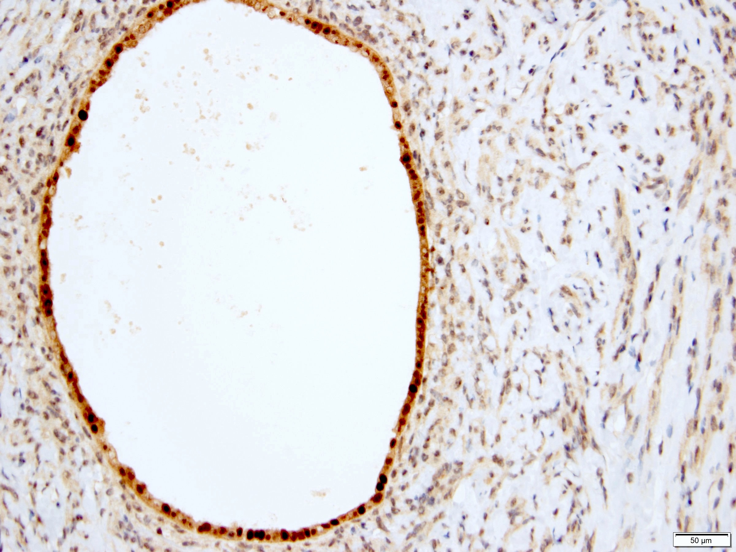
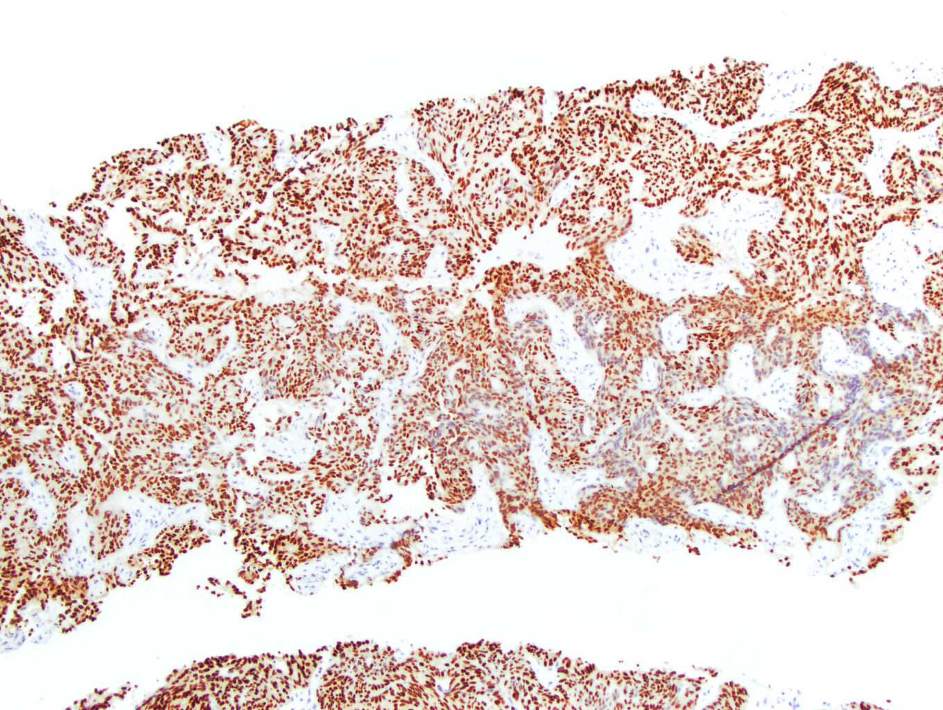
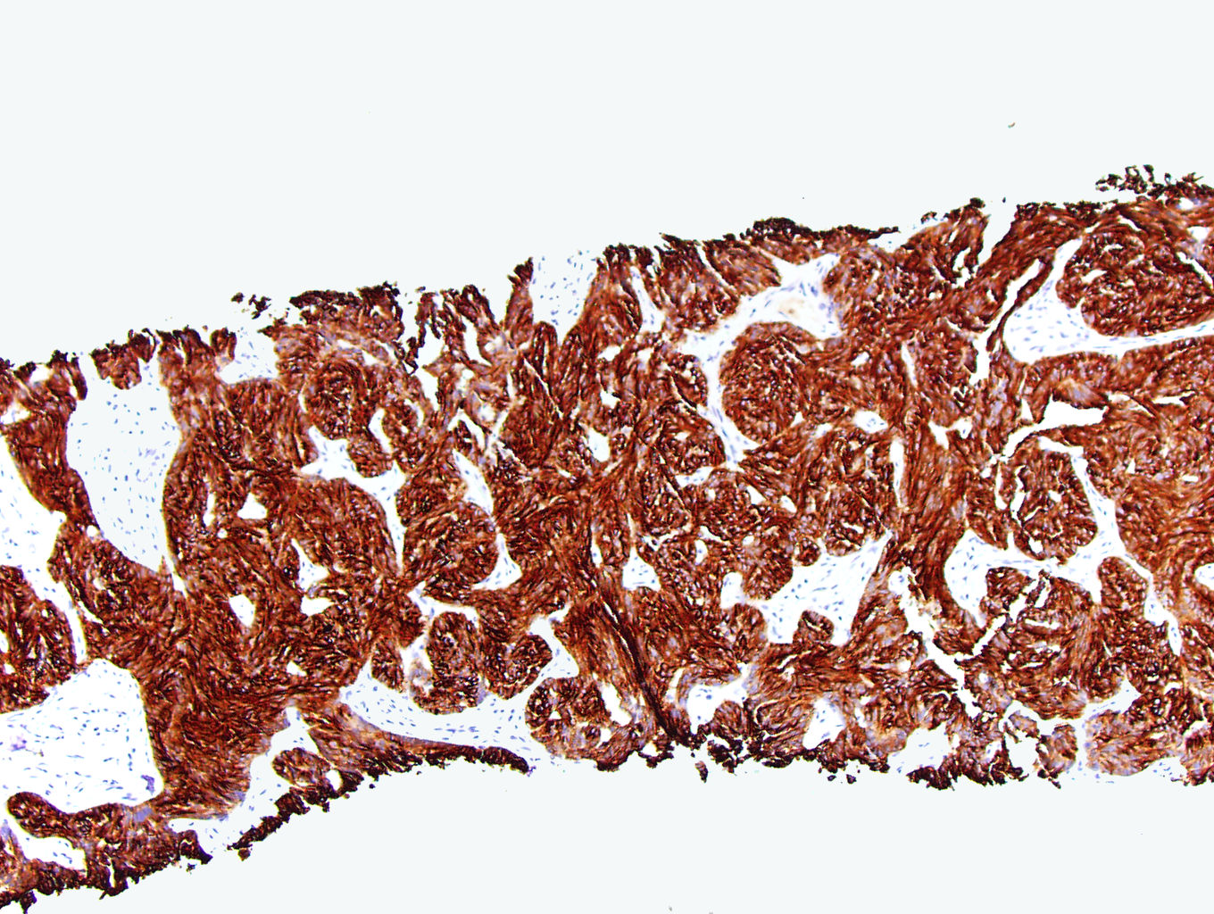
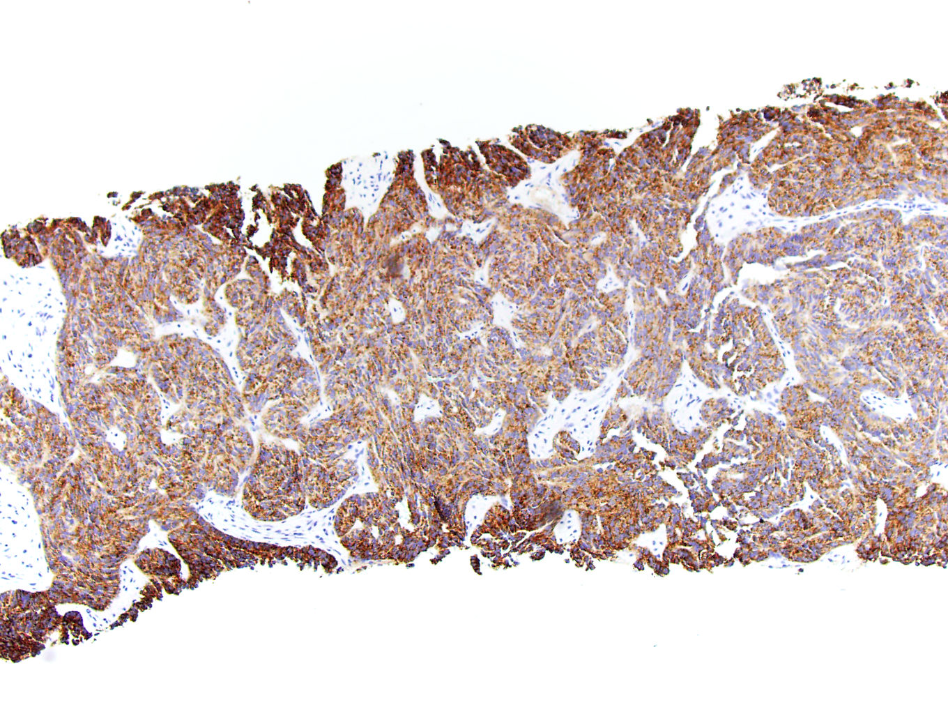
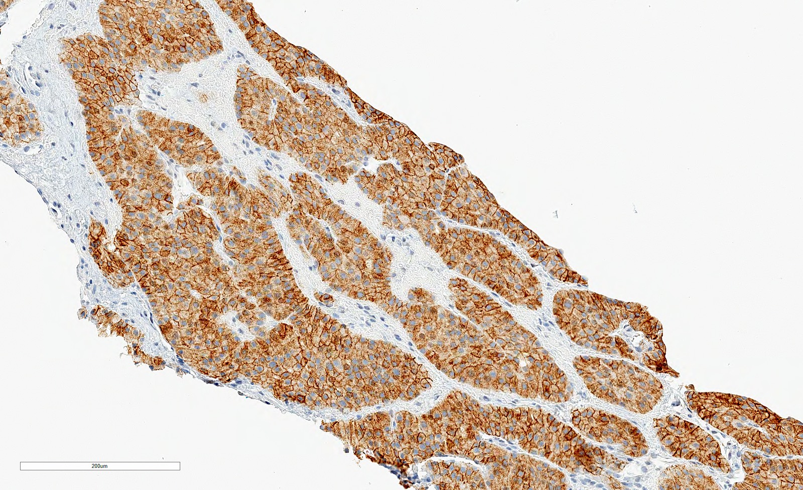
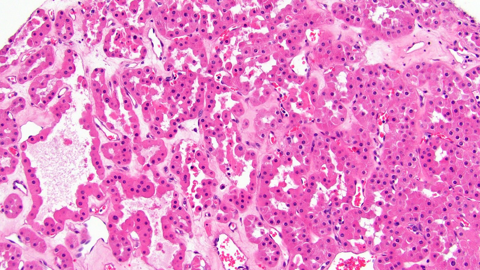
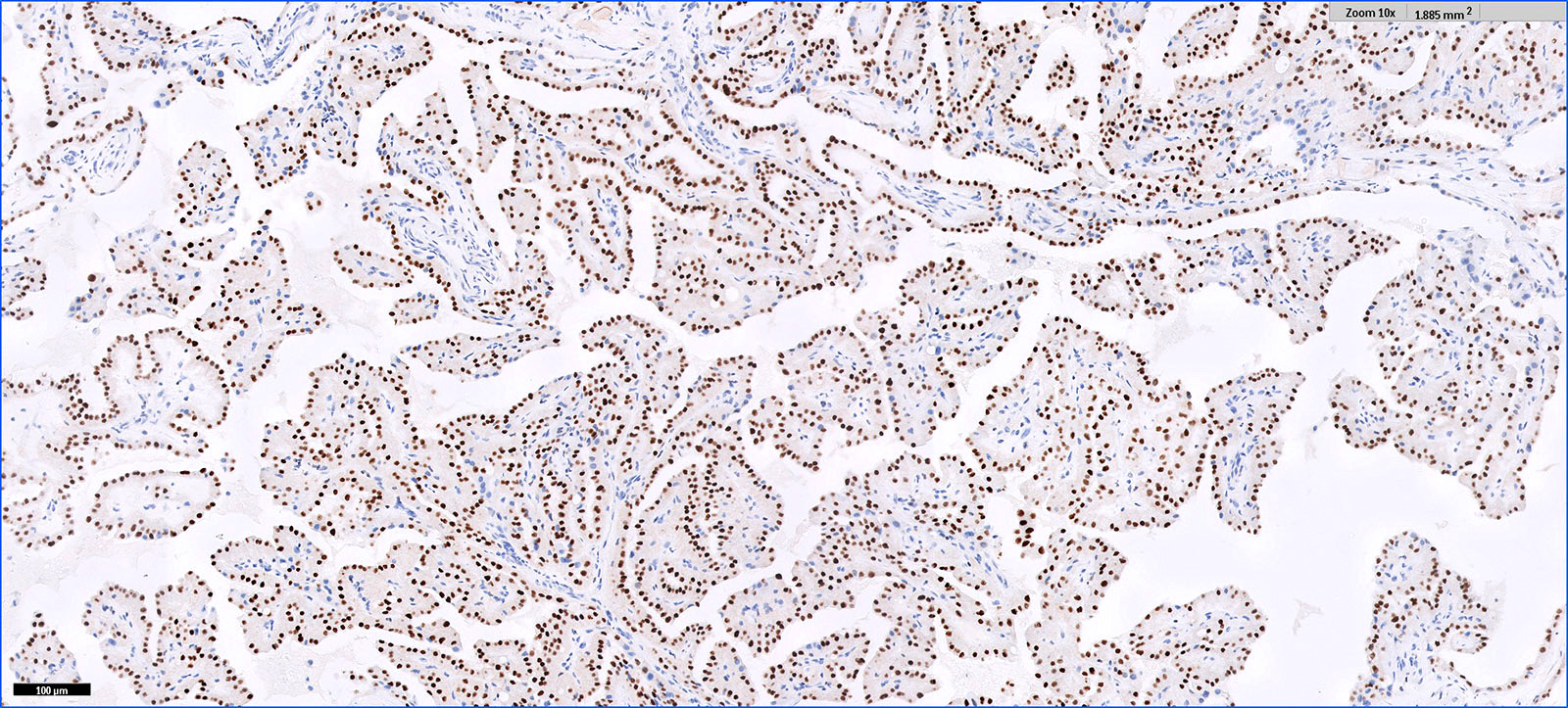
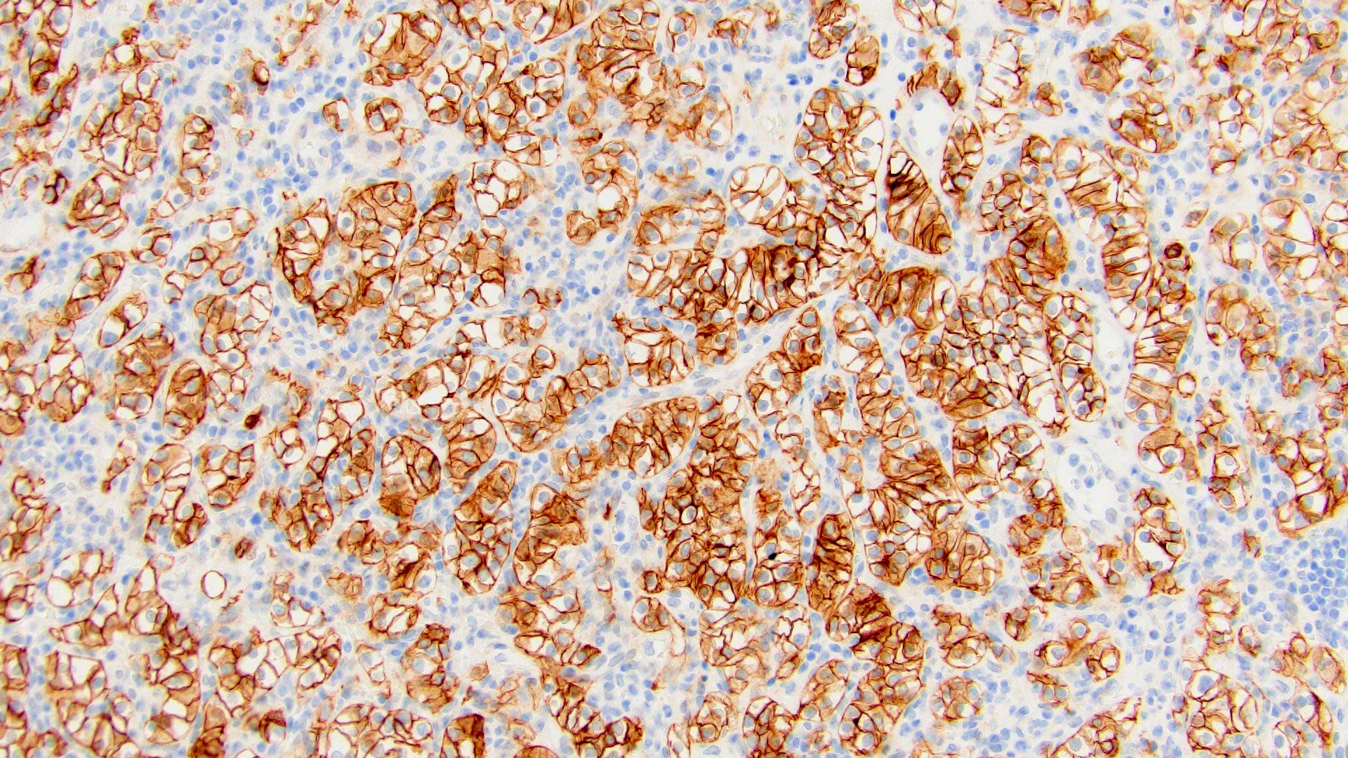
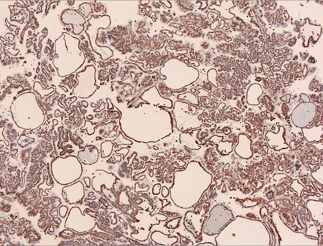
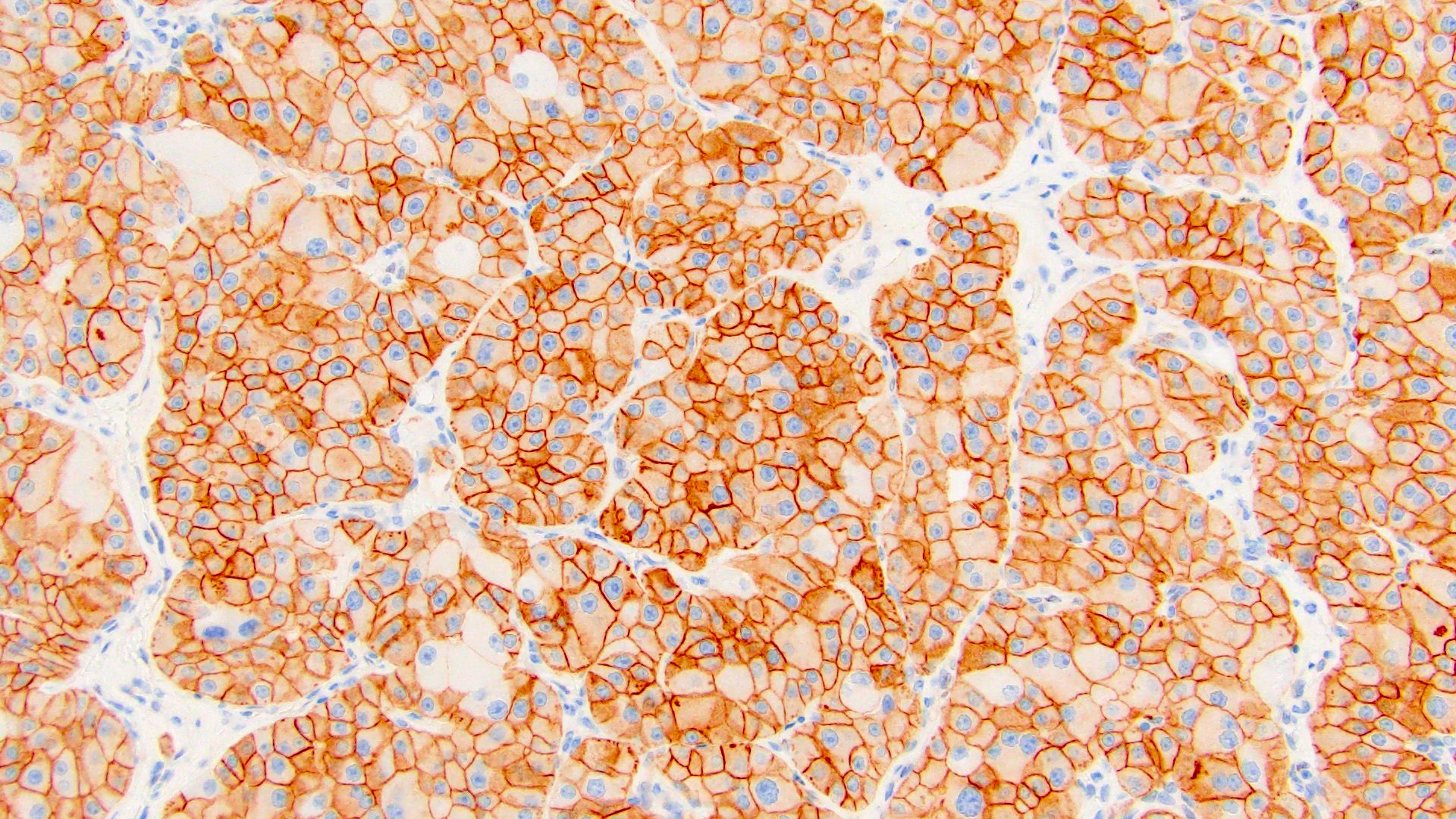
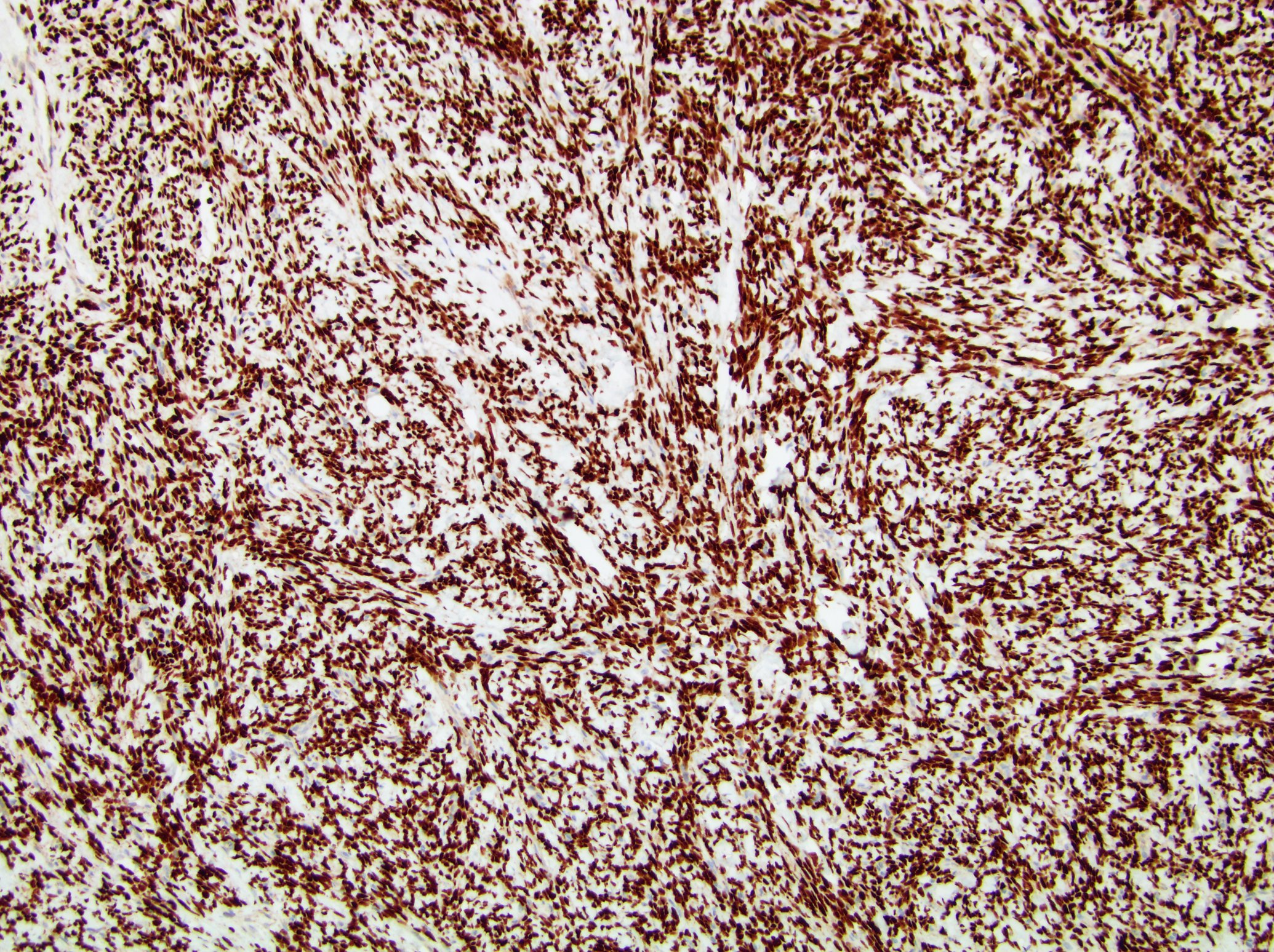
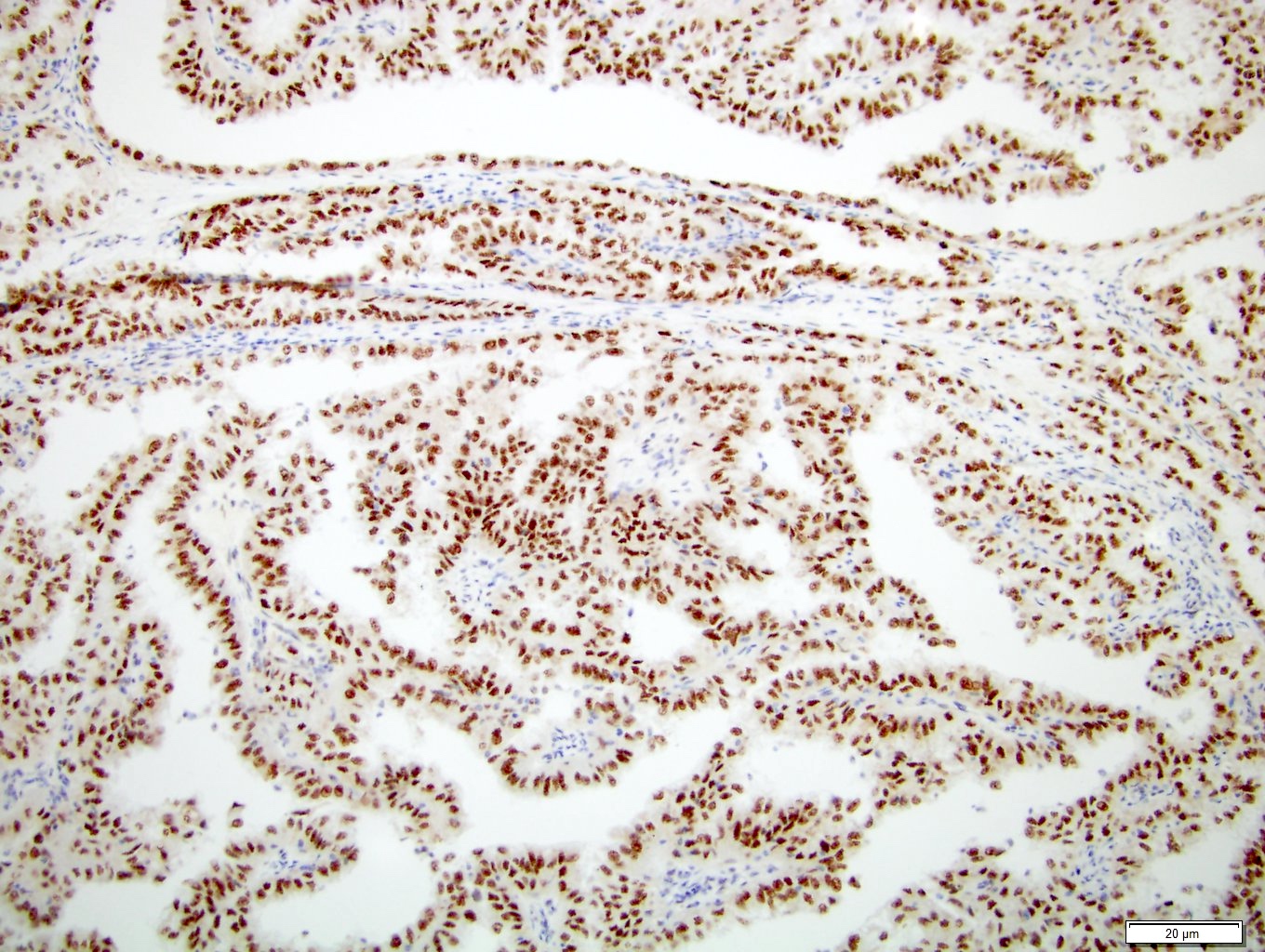
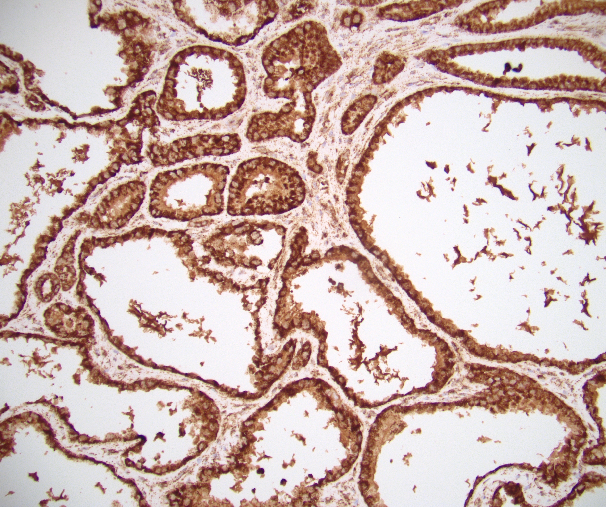
ALK
Images hosted on other servers:
ALK break apart probe with split signals
Karyotype,
metaphase and
interphase FISH
RT-PCR with VCL exon 16 and ALK exon 20
EML4-ALK and
TPM3-ALK
gene fusion points
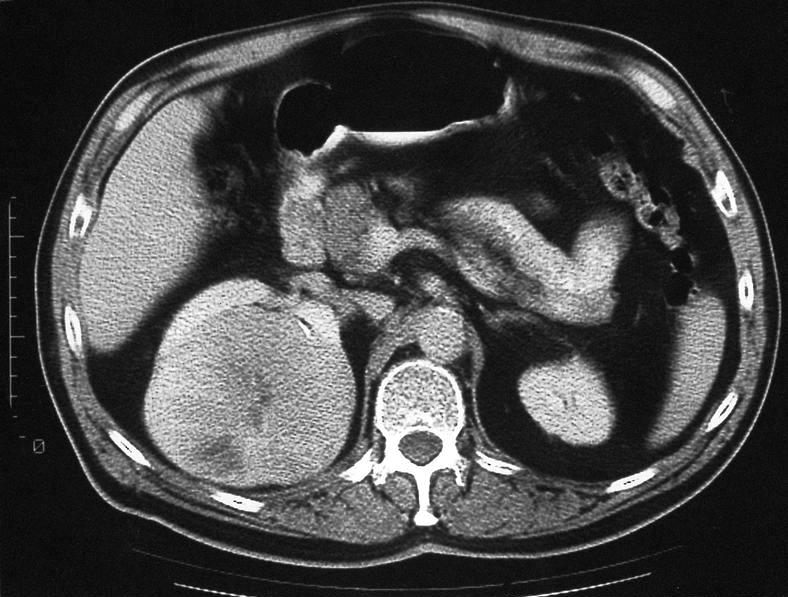
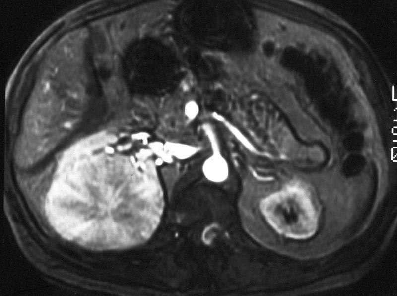
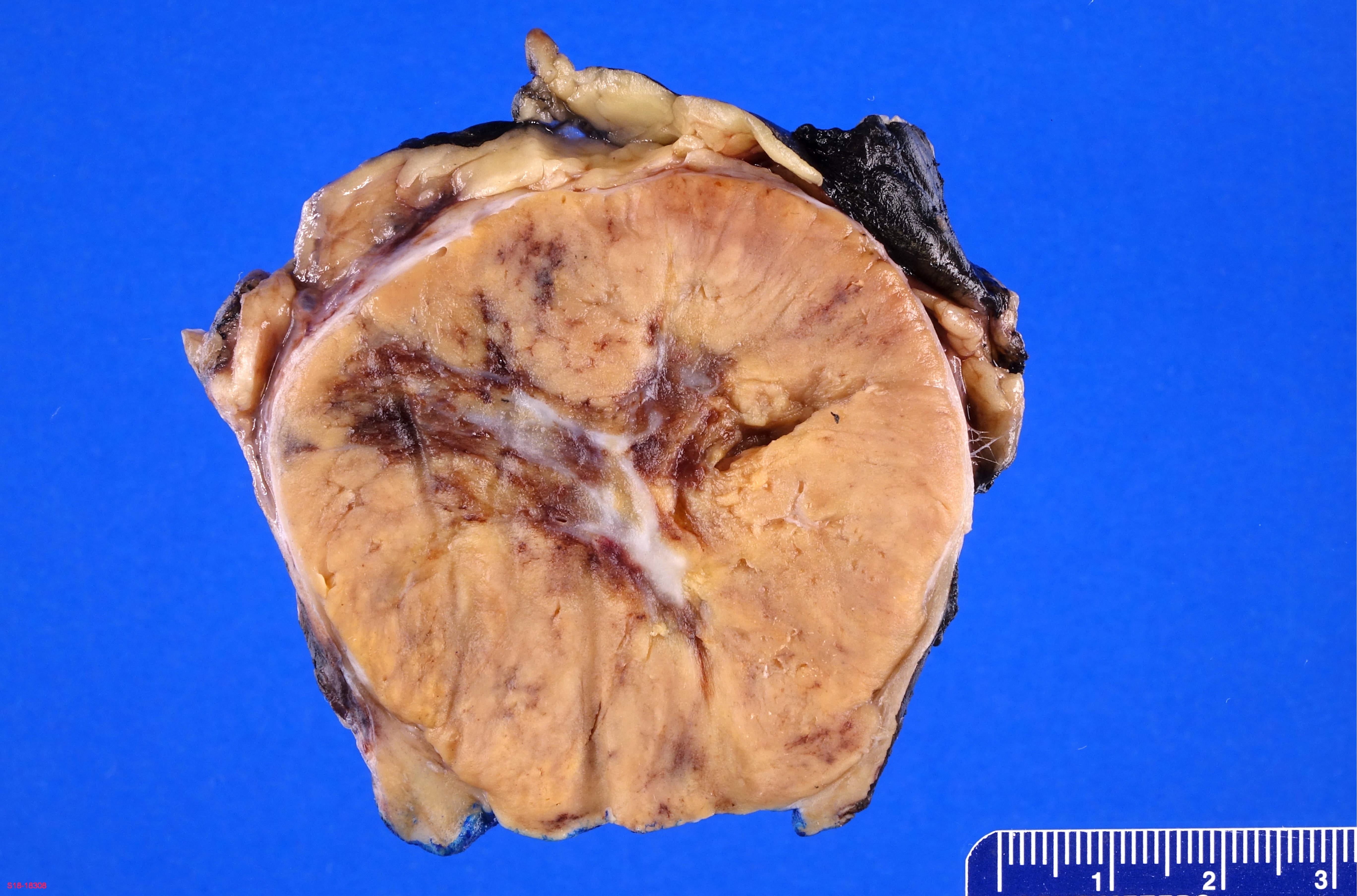
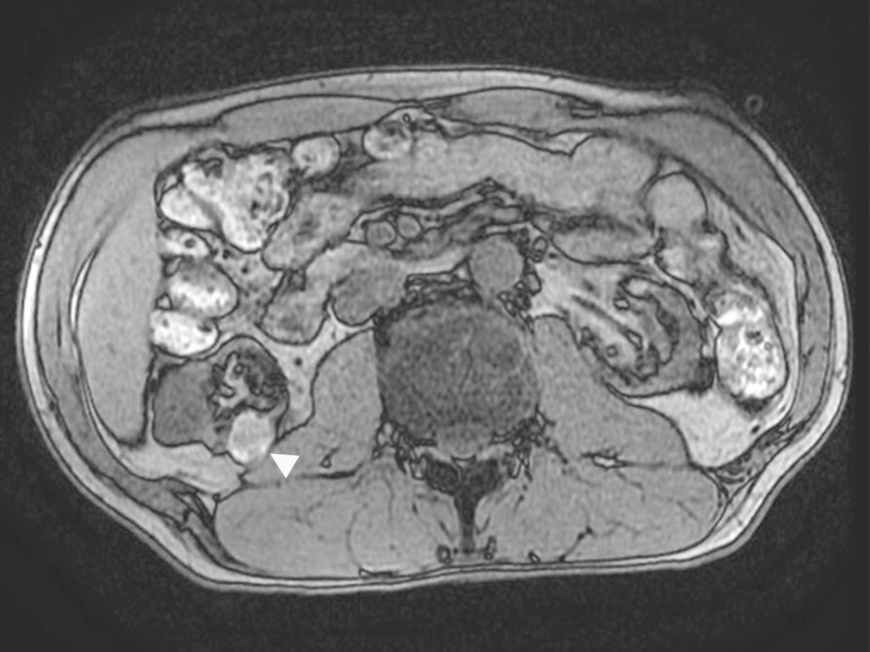
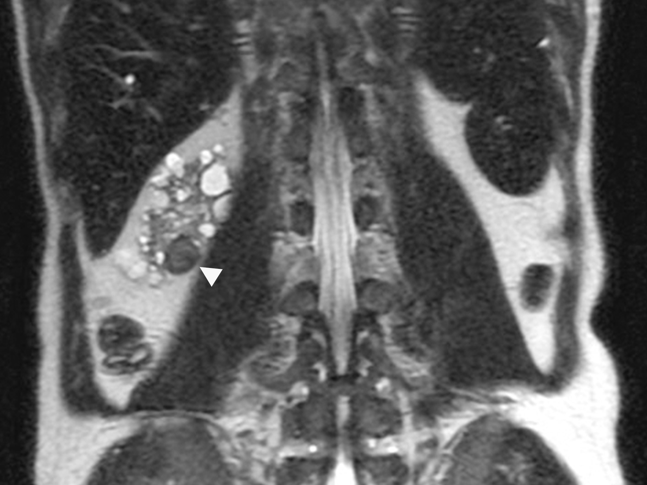
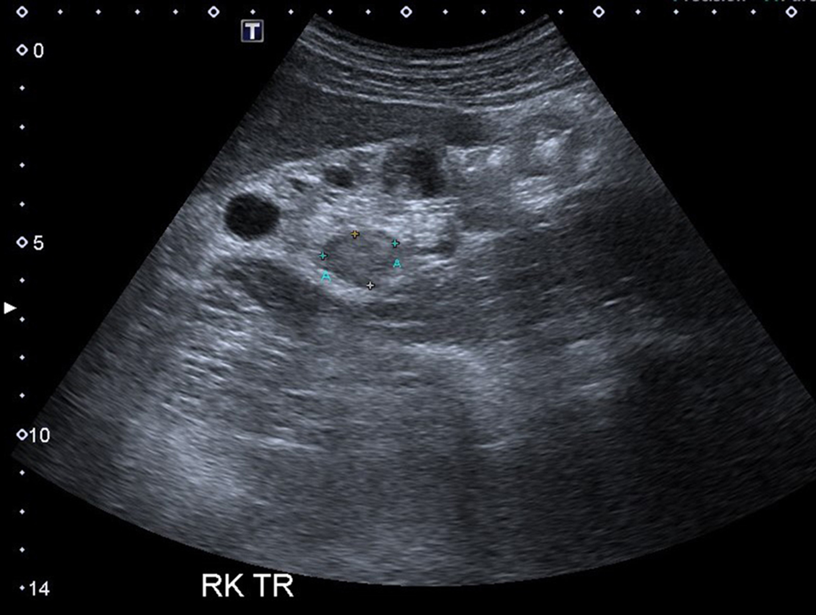
Images hosted on other servers:

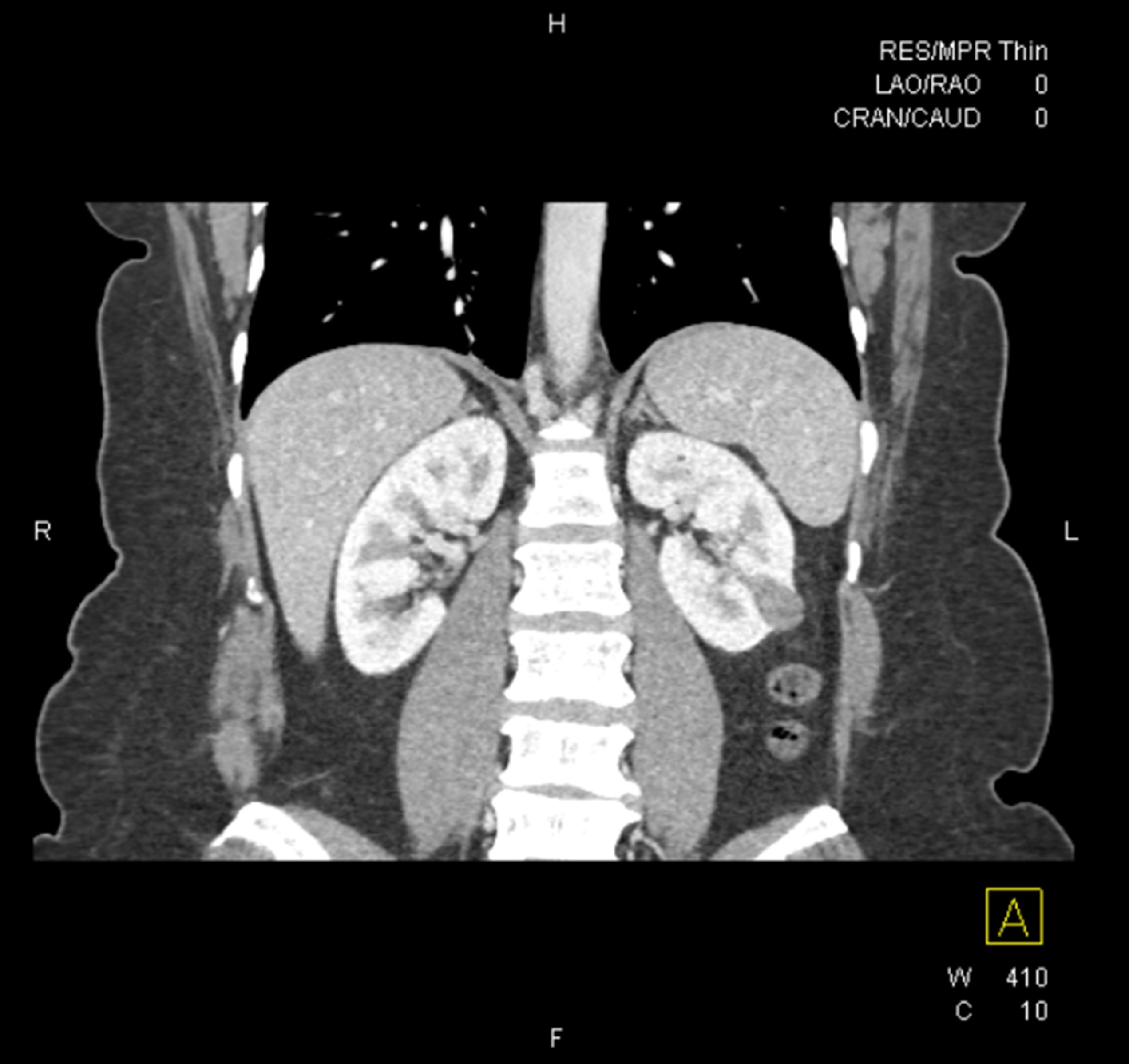
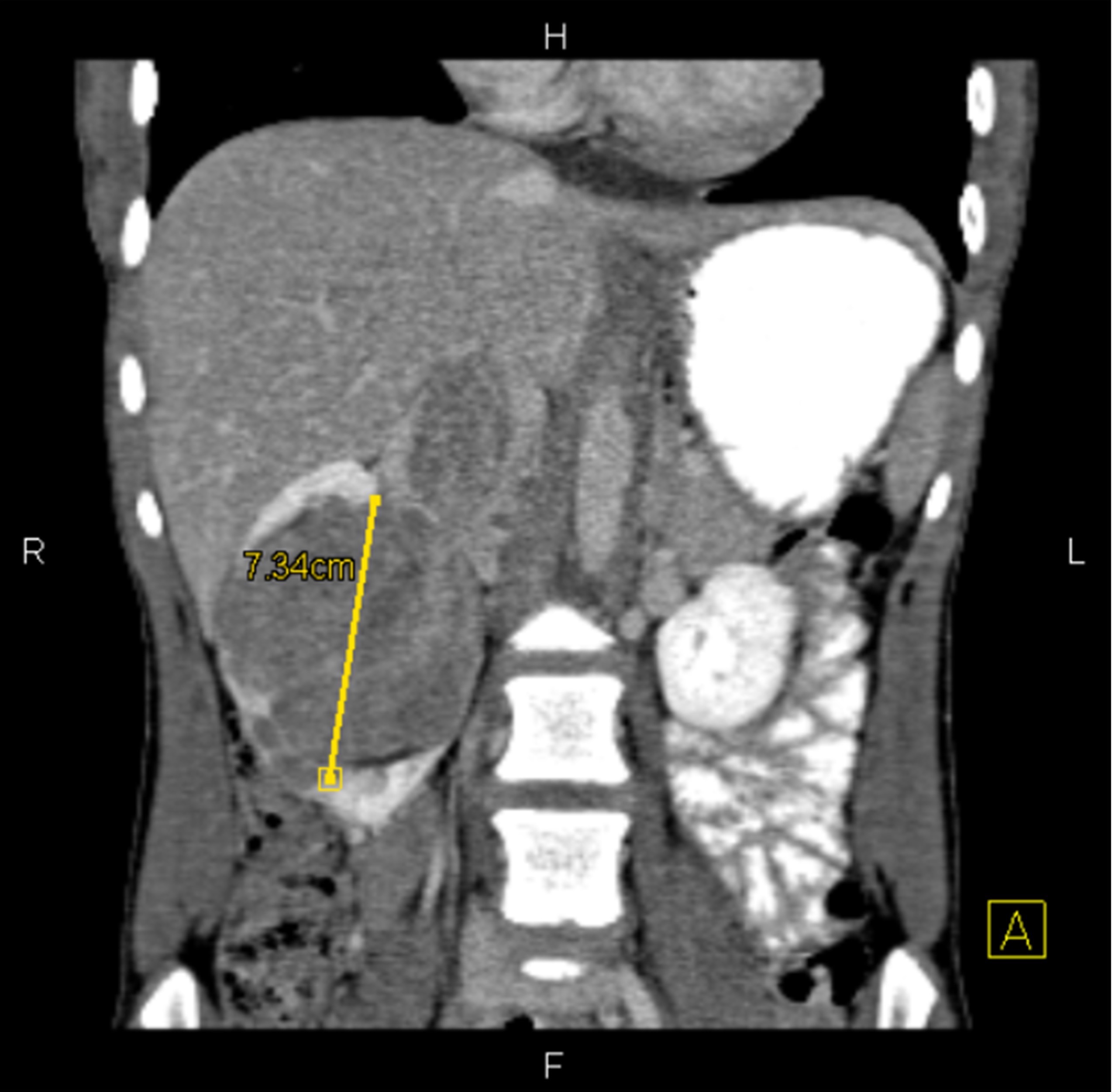
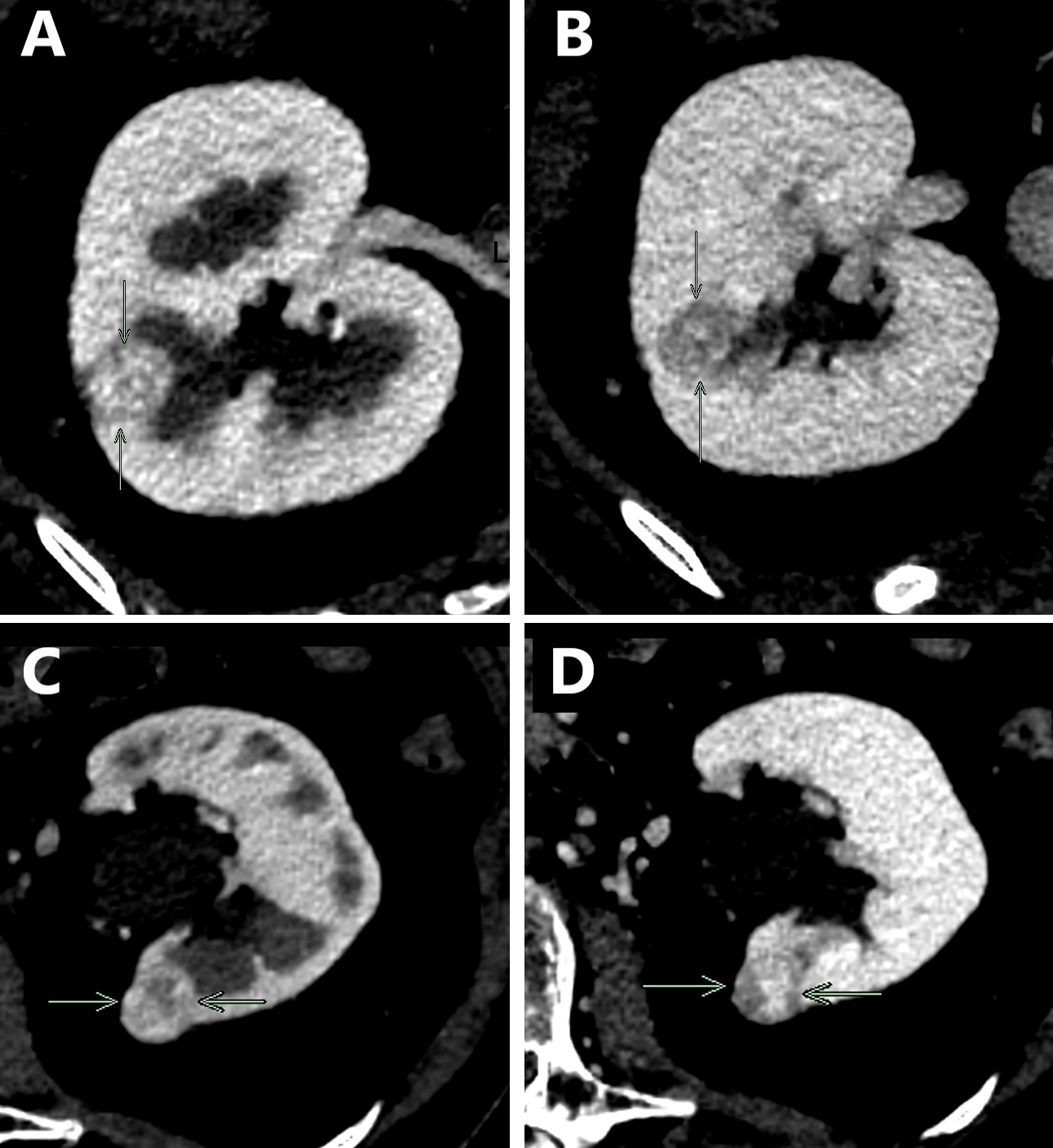
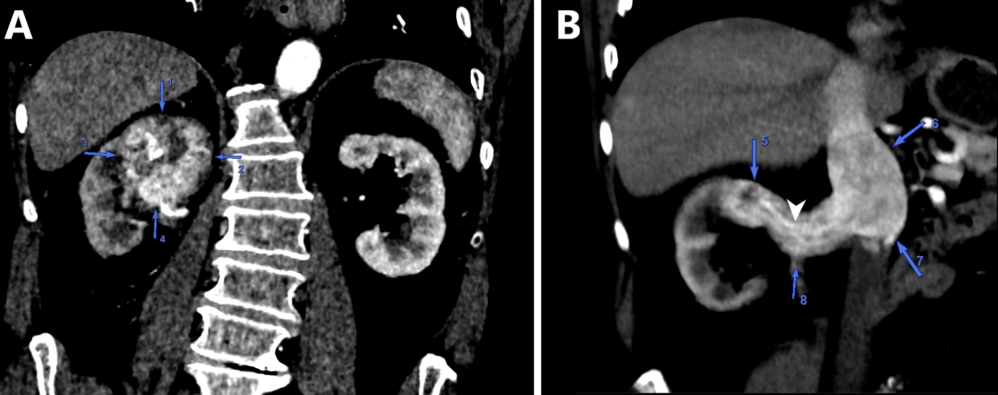
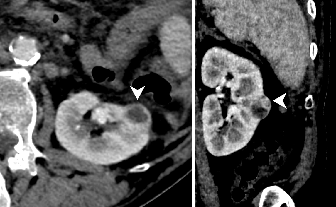
US & CT scan
Contributed by Maria Tretiakova, M.D., Ph.D.
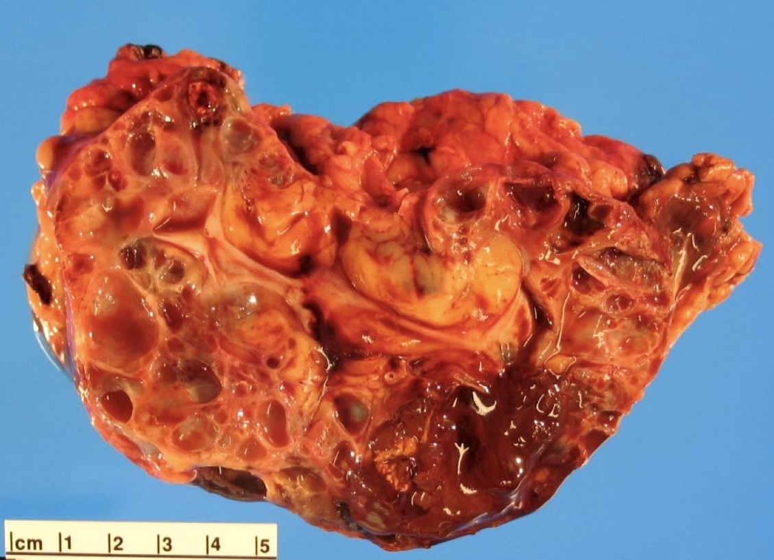
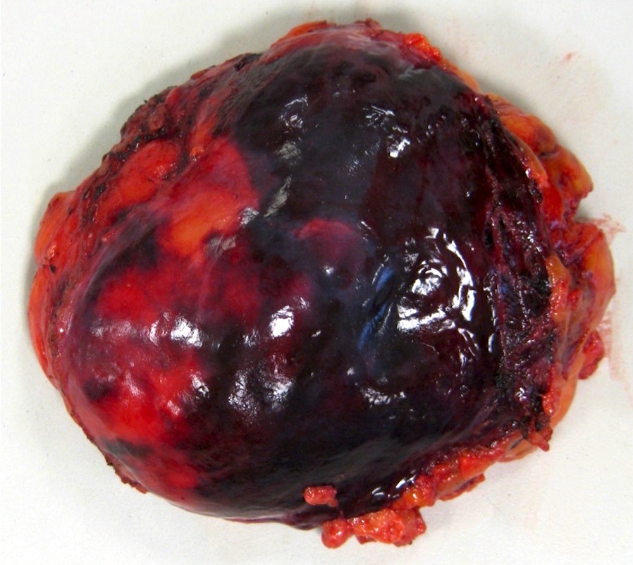
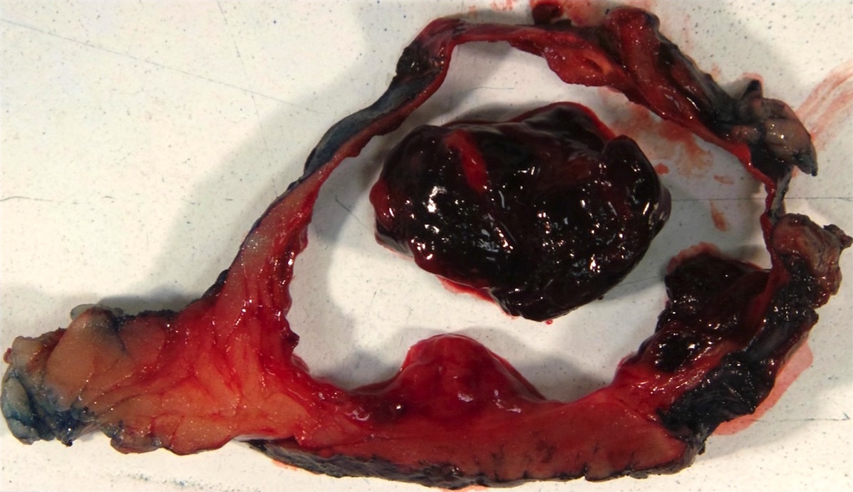
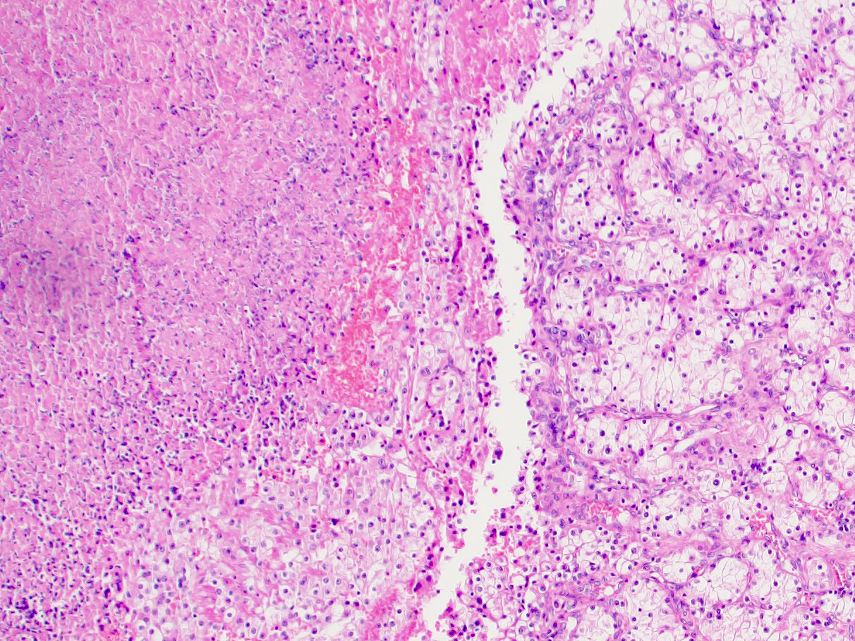
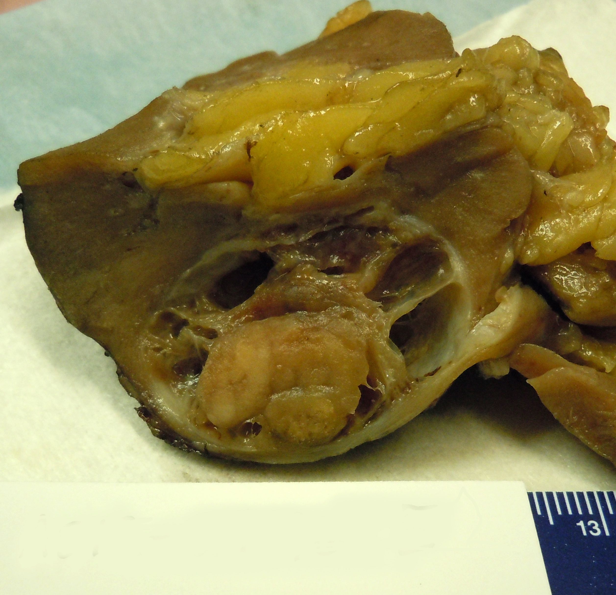
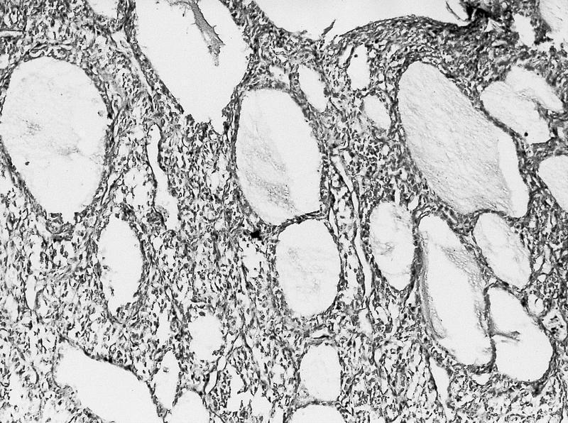
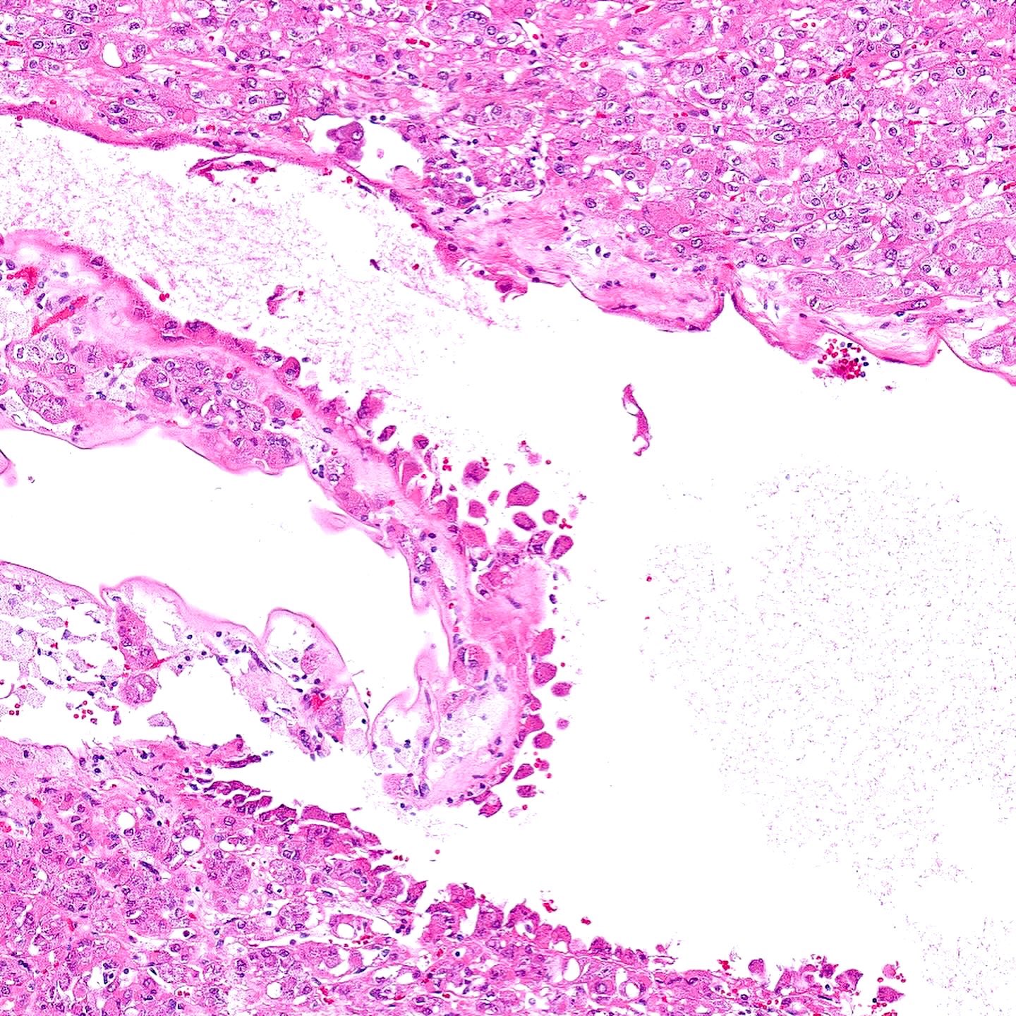
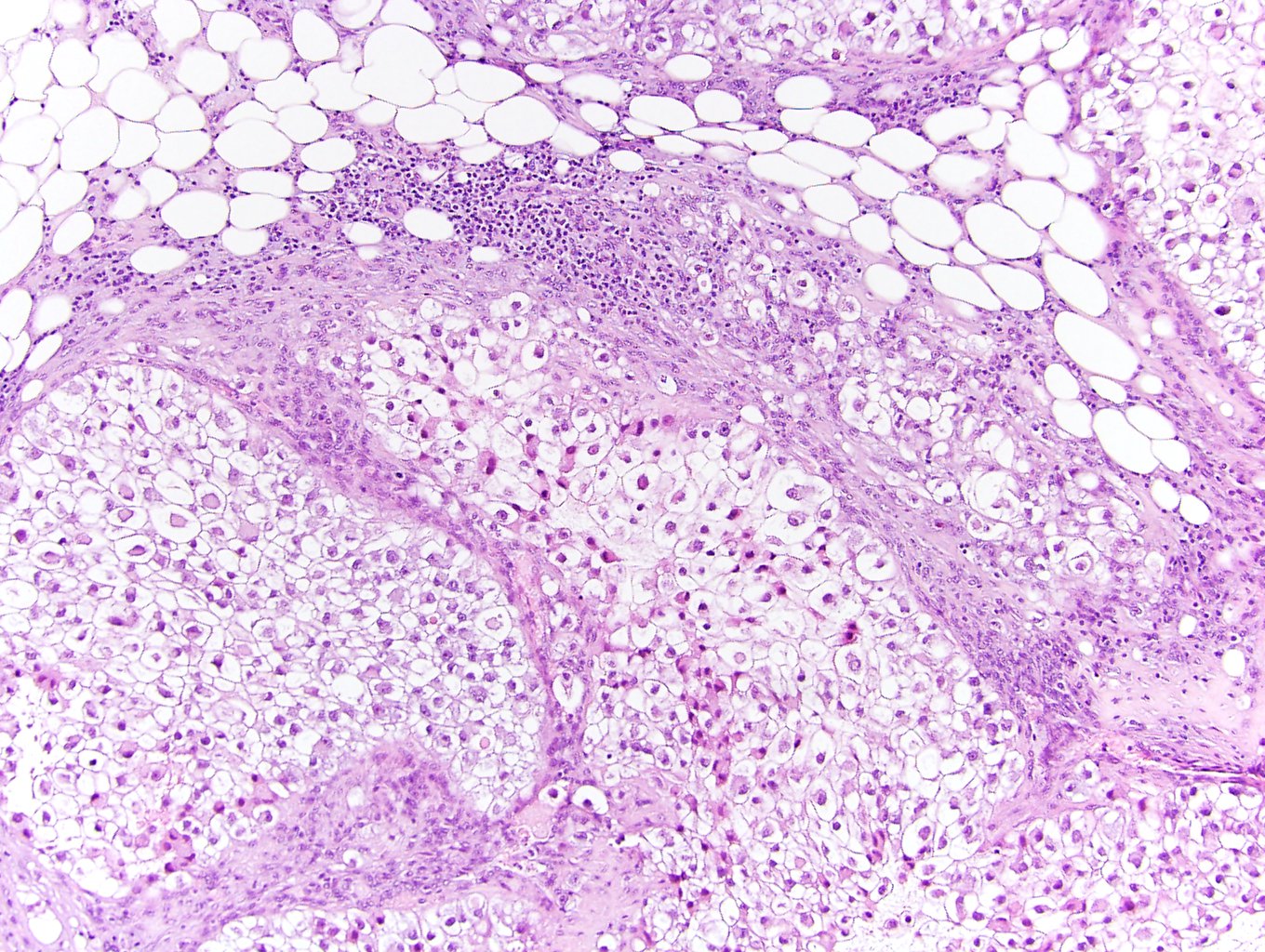
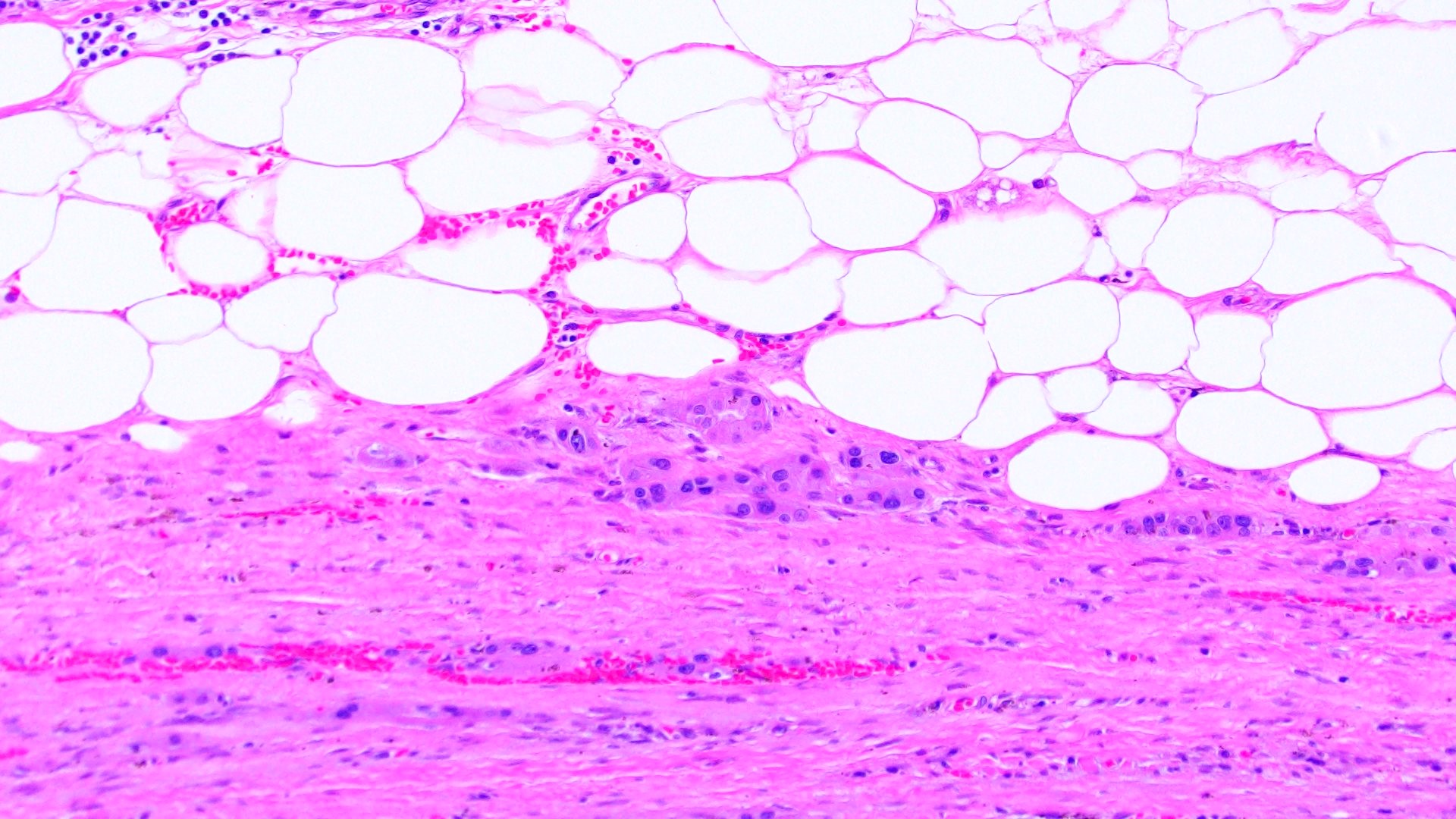
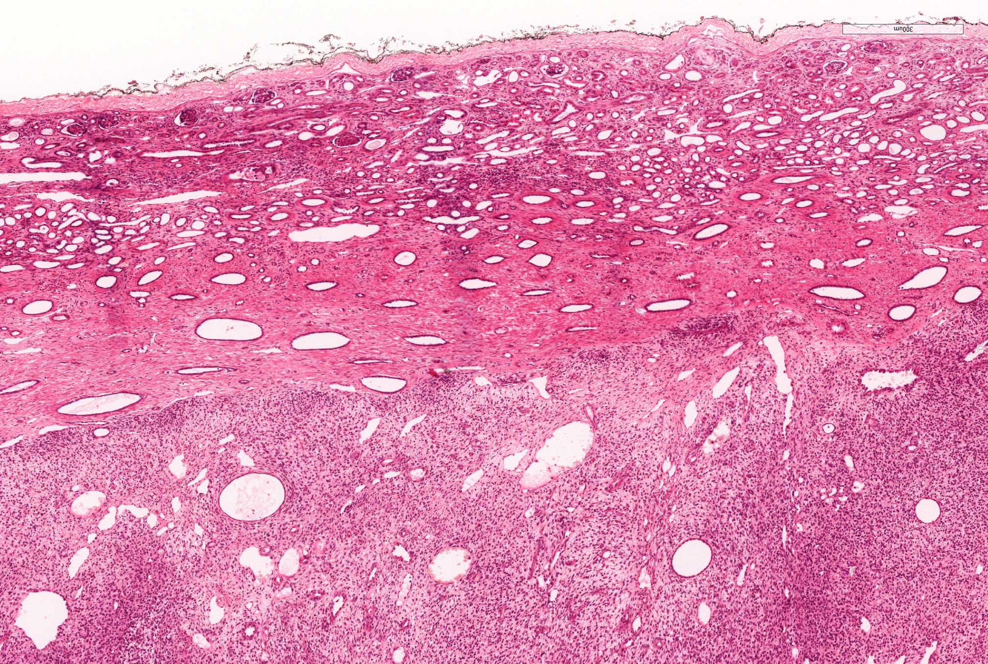
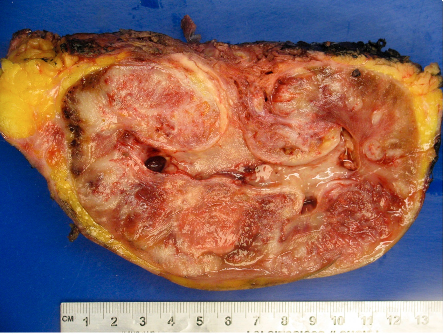
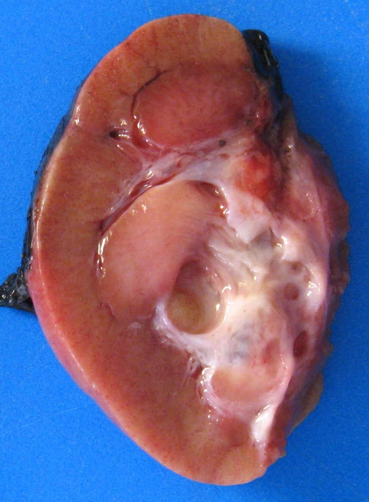
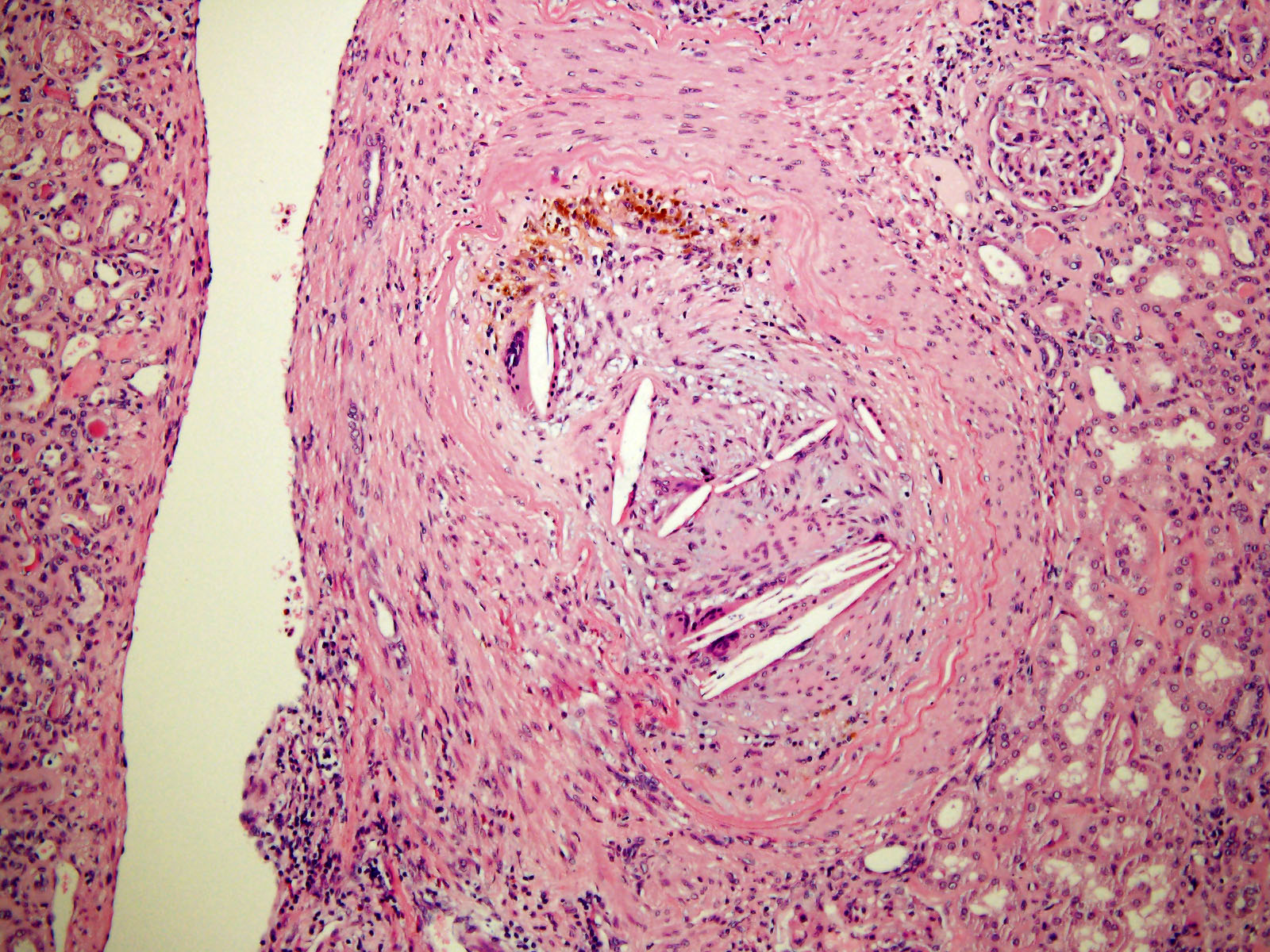
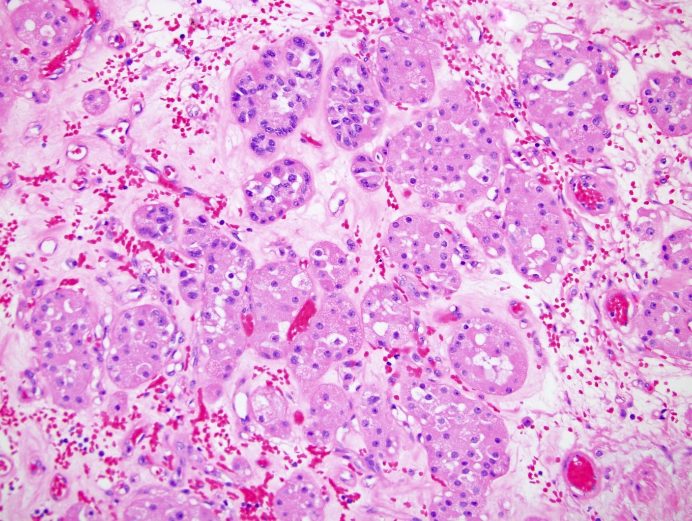
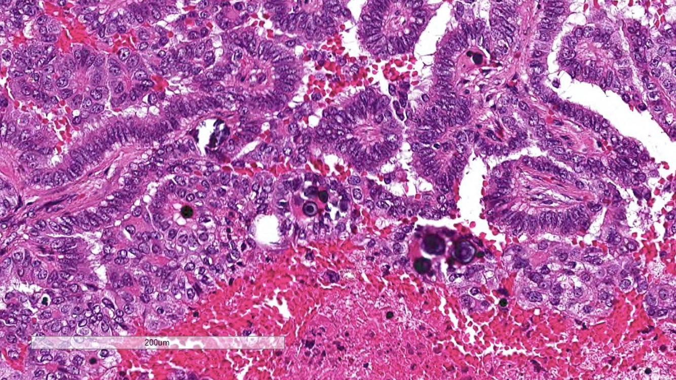
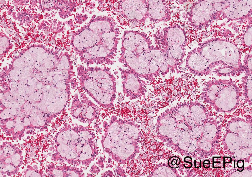
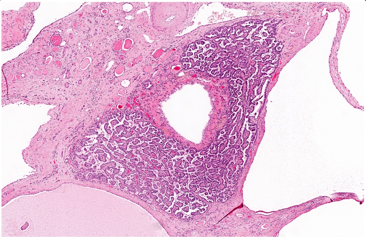
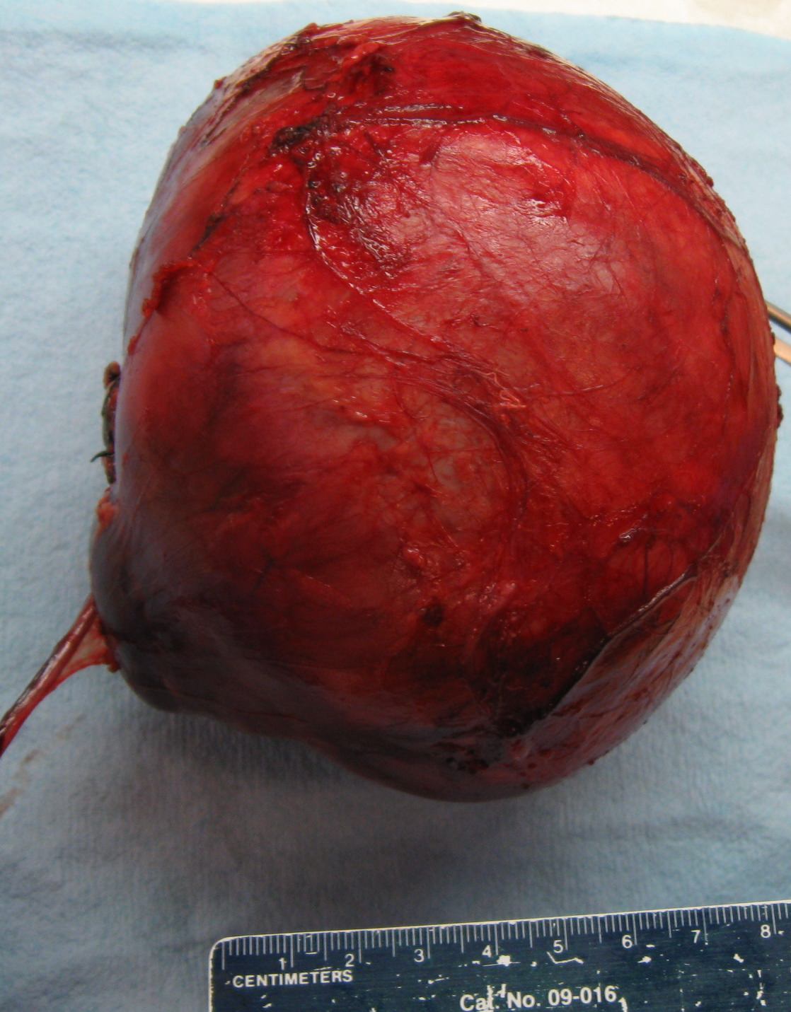
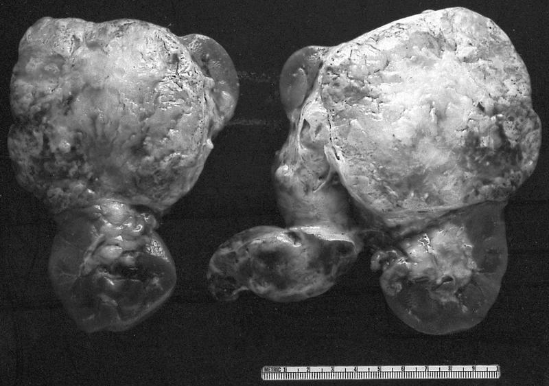
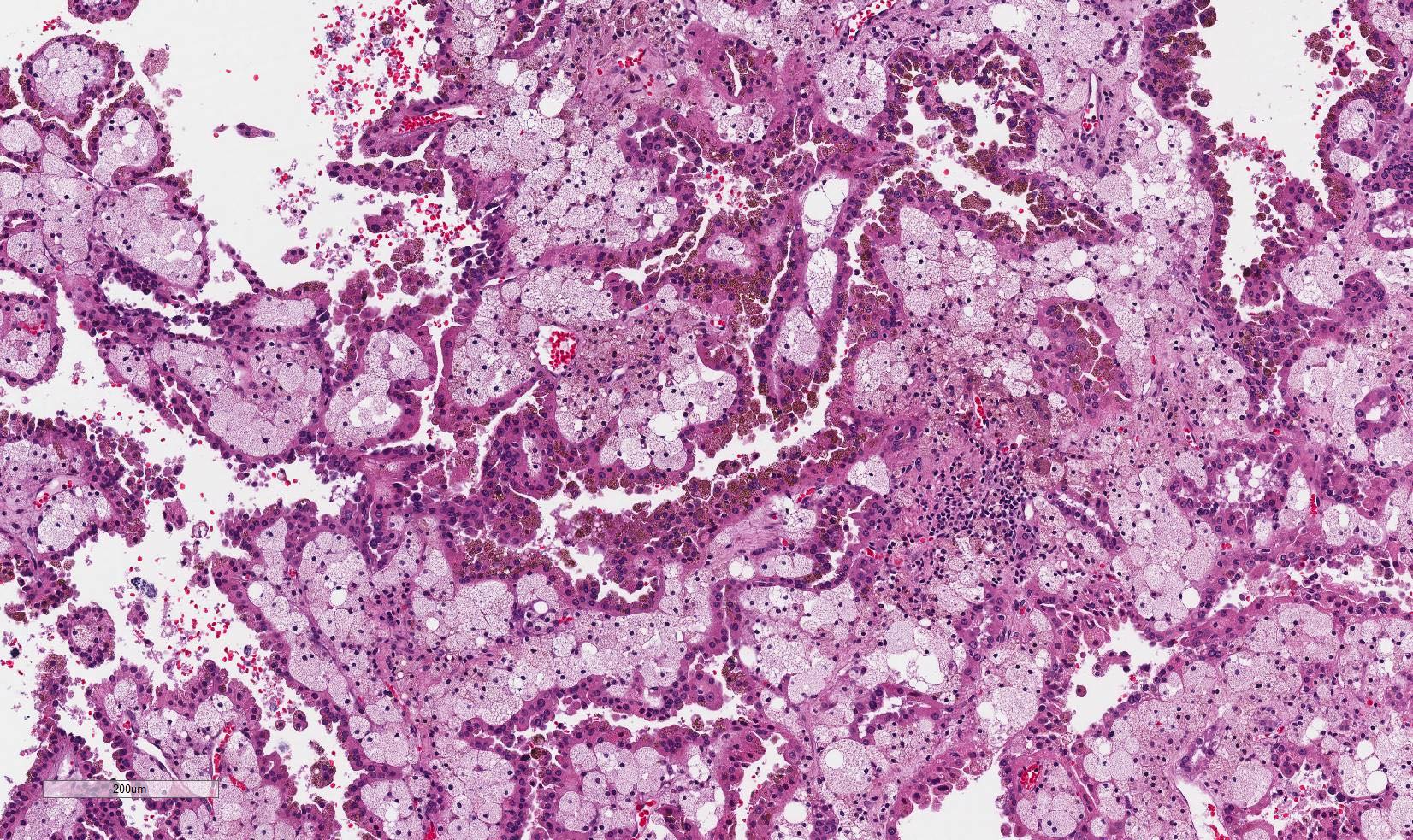
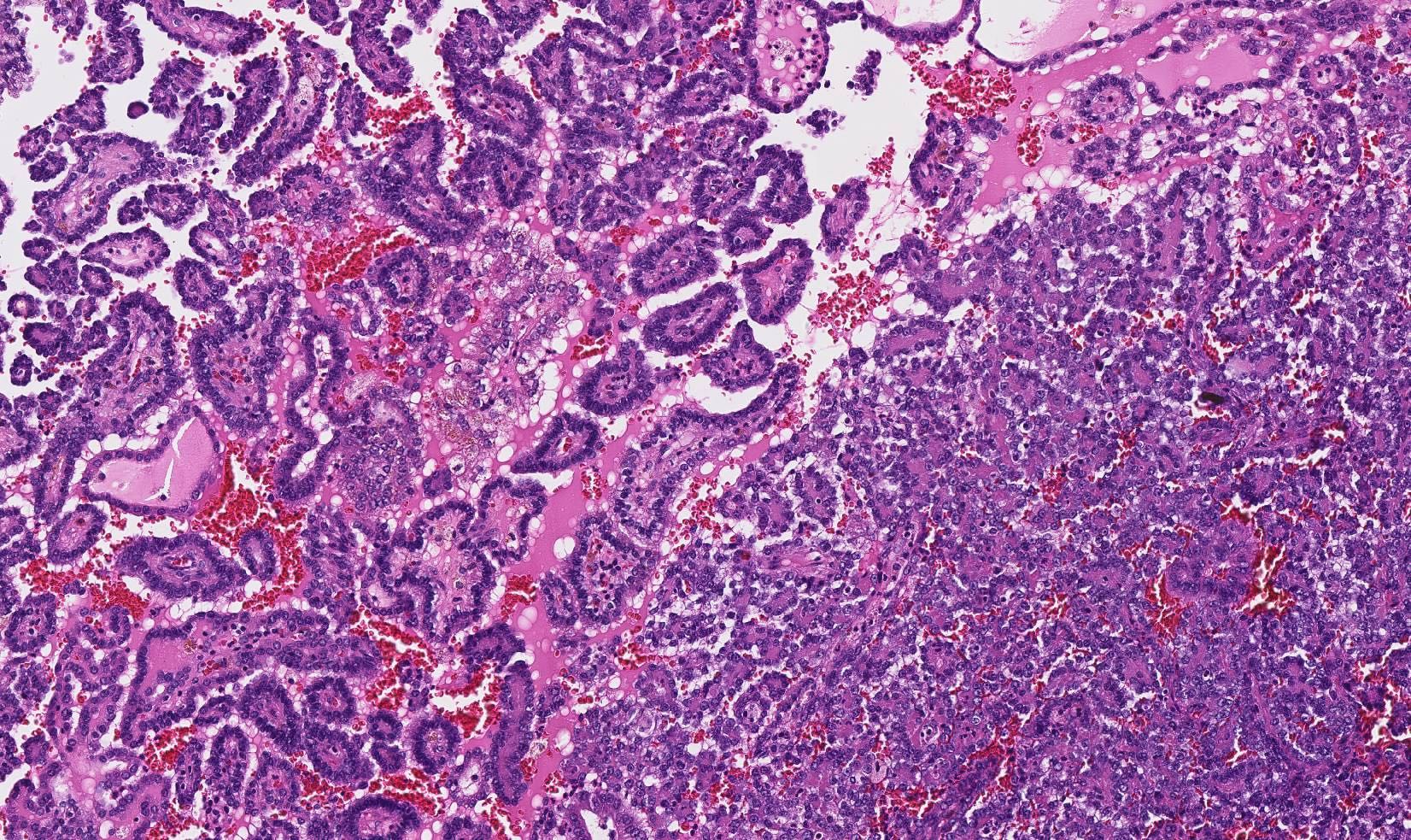
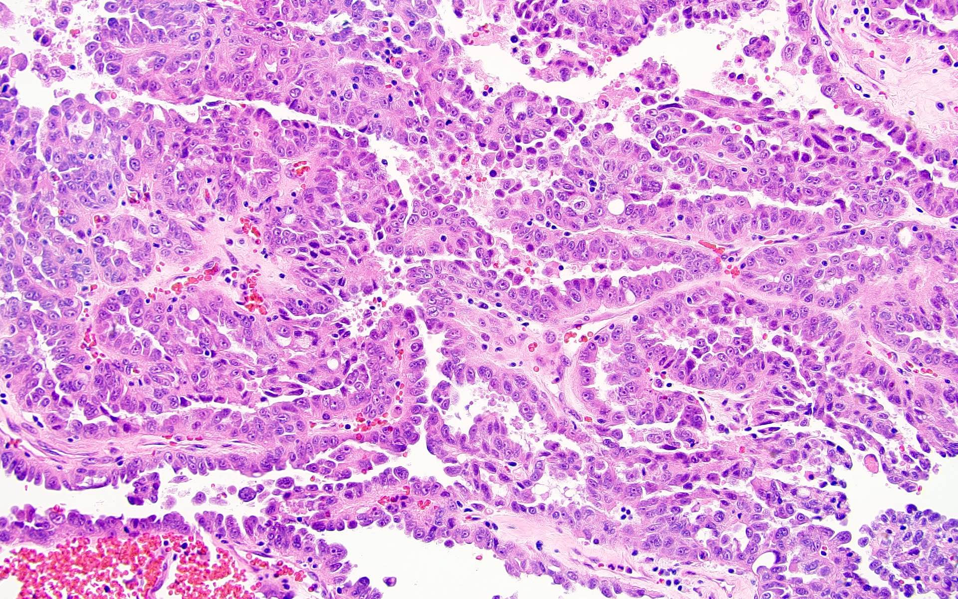
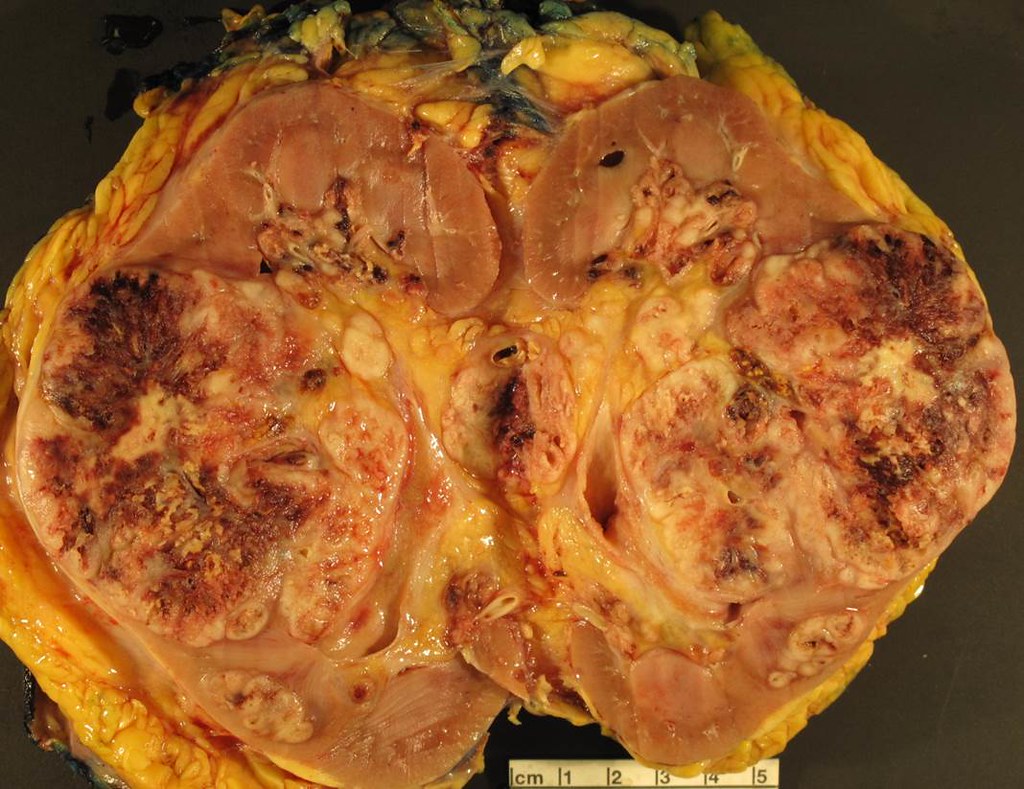
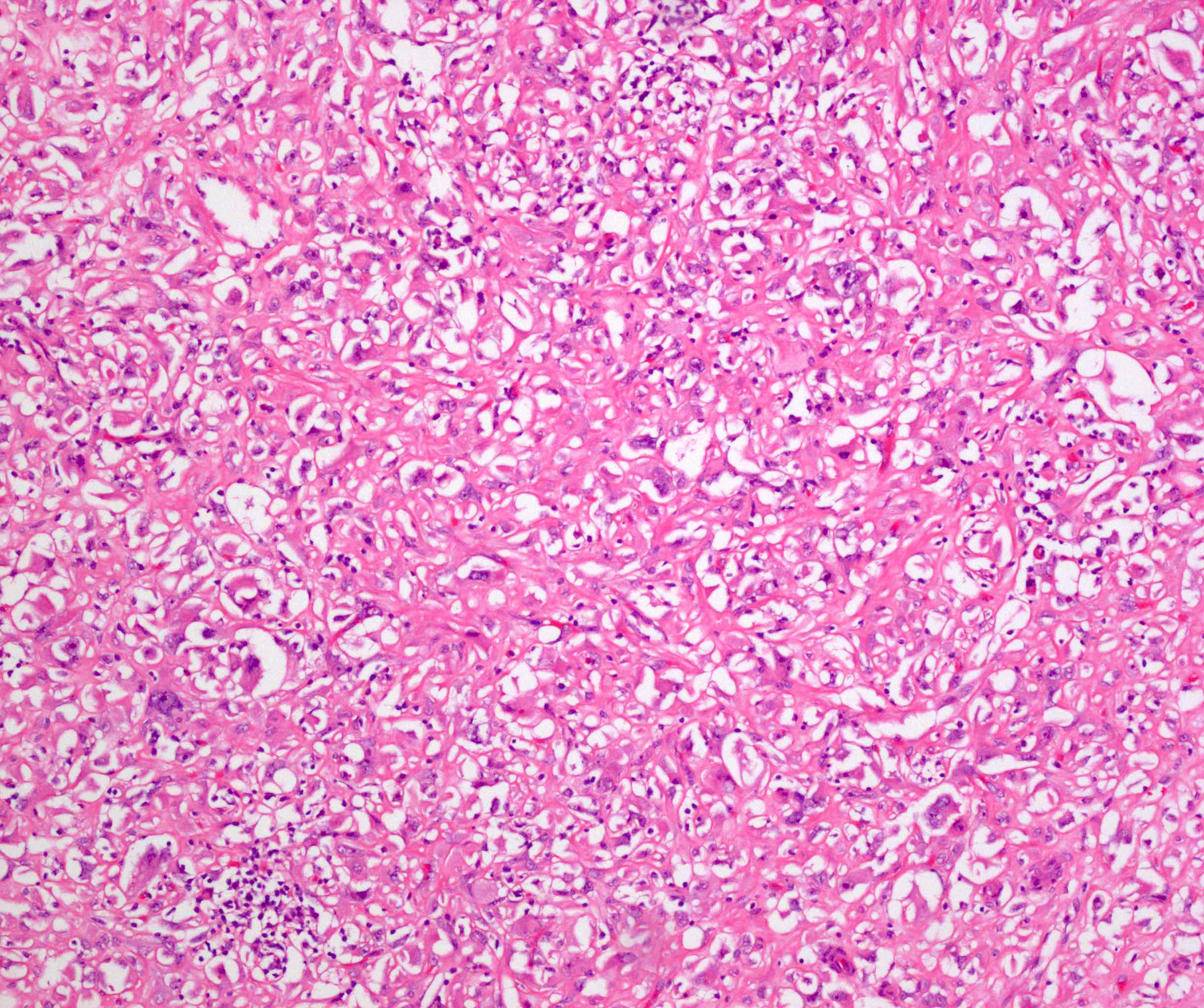
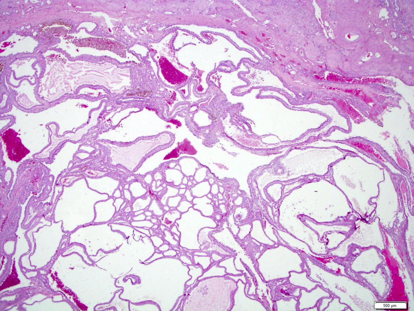
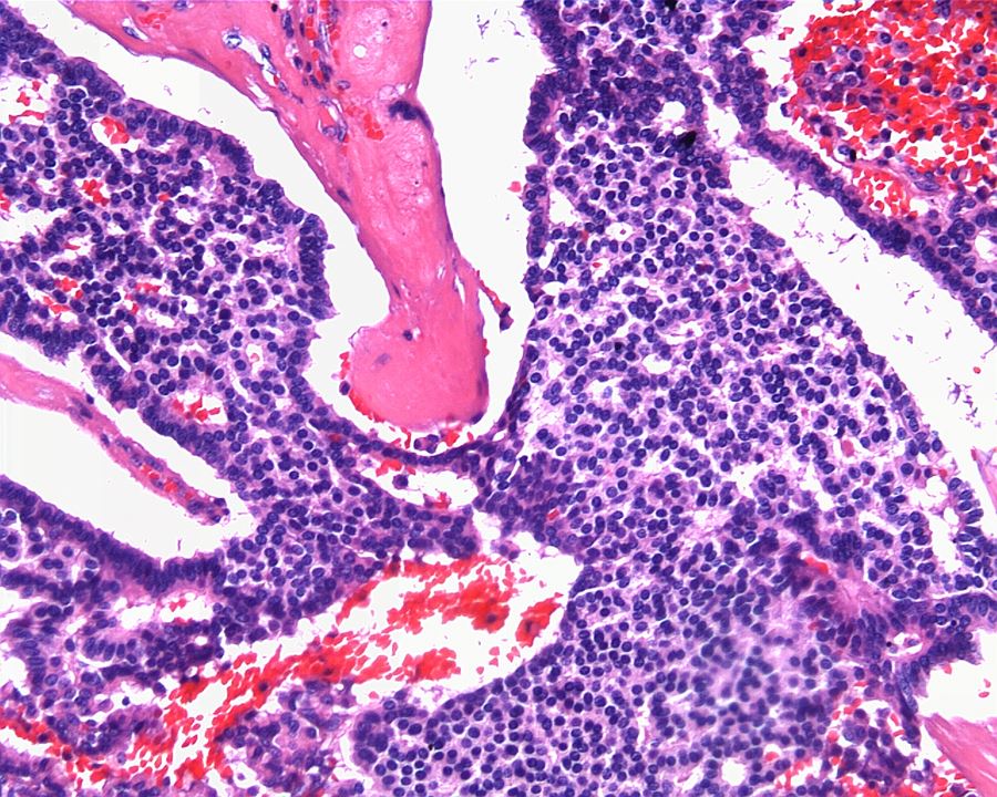
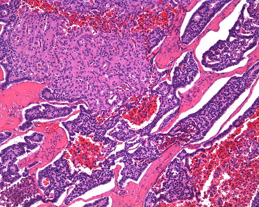
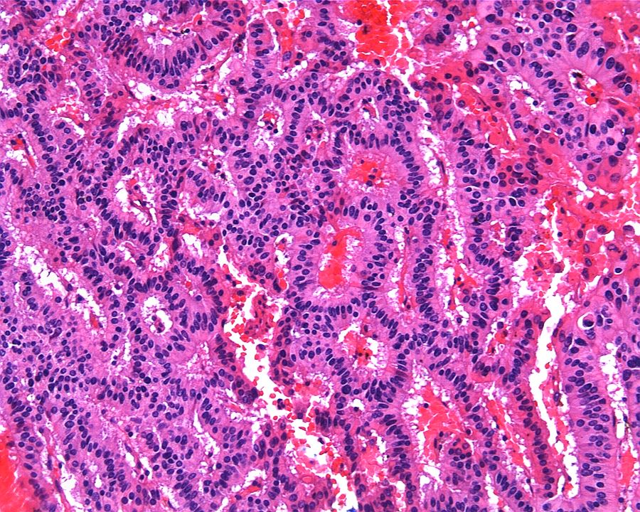
Solid and cystic hemorrhagic neoplasm
Contributed by Nicole Andeen, M.D., Maria Tretiakova, M.D., Ph.D., Gregory T. MacLennan, M.D. and @katcollmd on Twitter
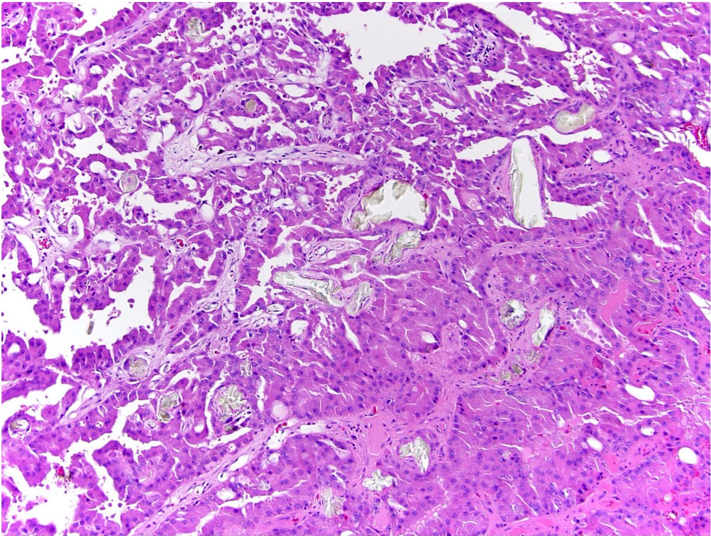

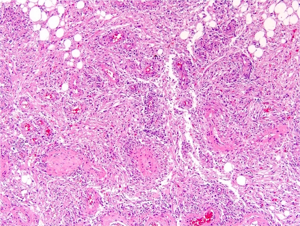
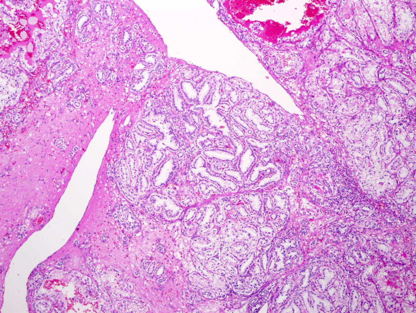
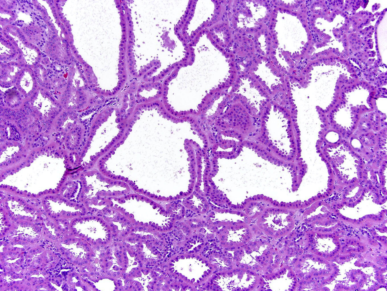
Papillary architecture
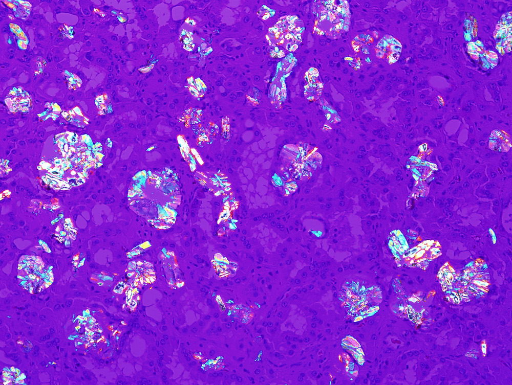
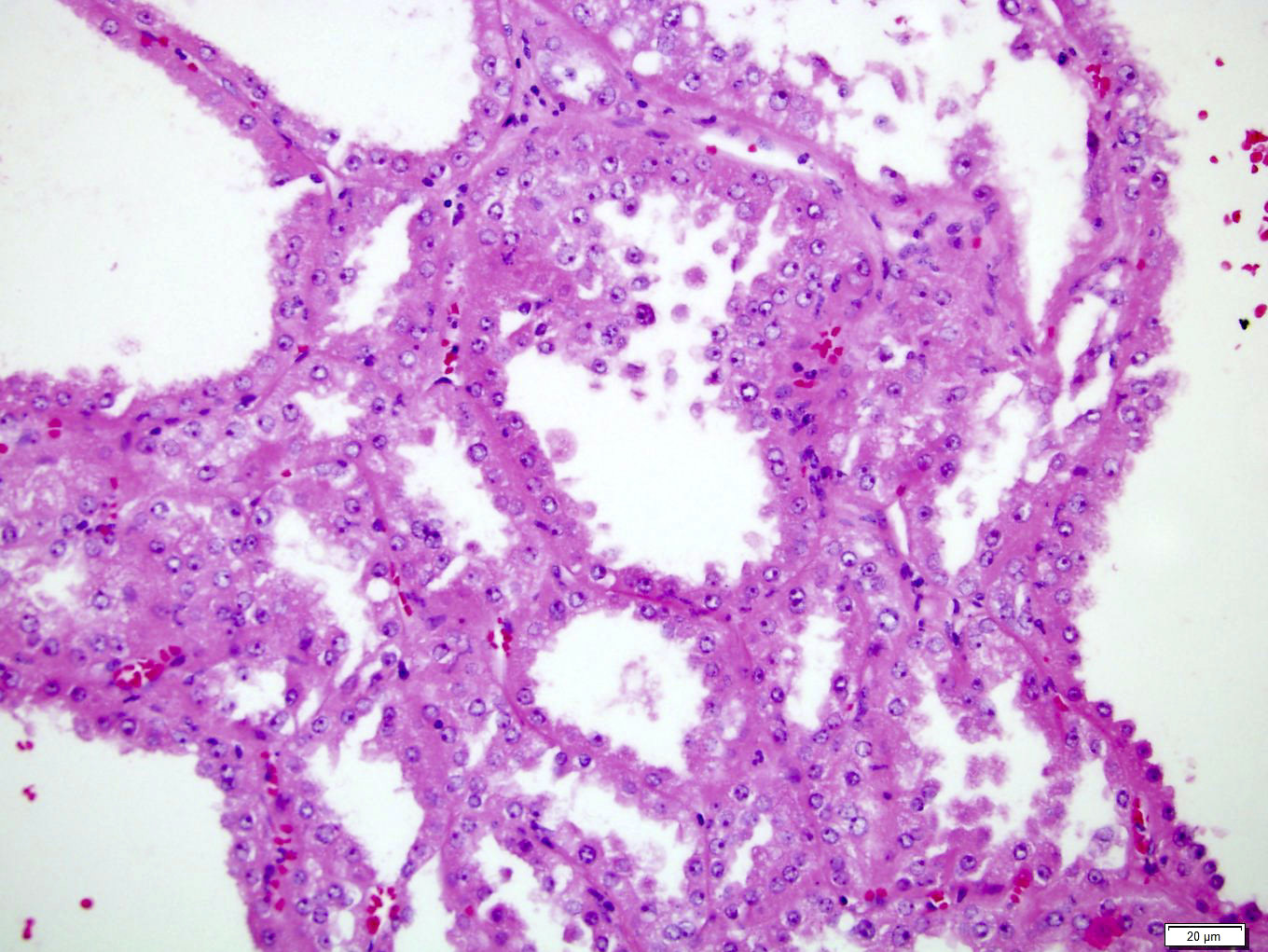
Oxalate crystals

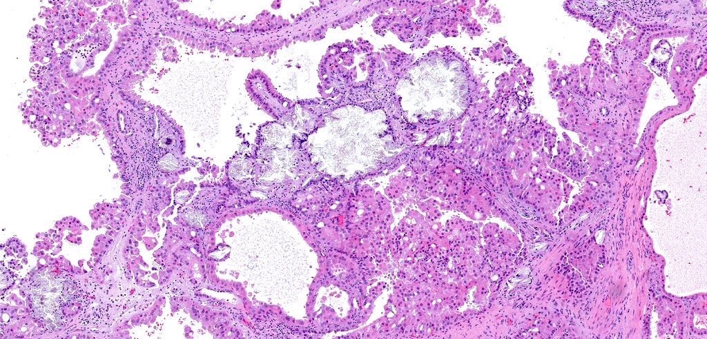


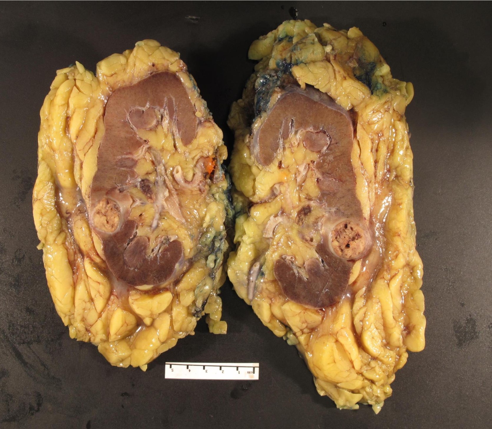
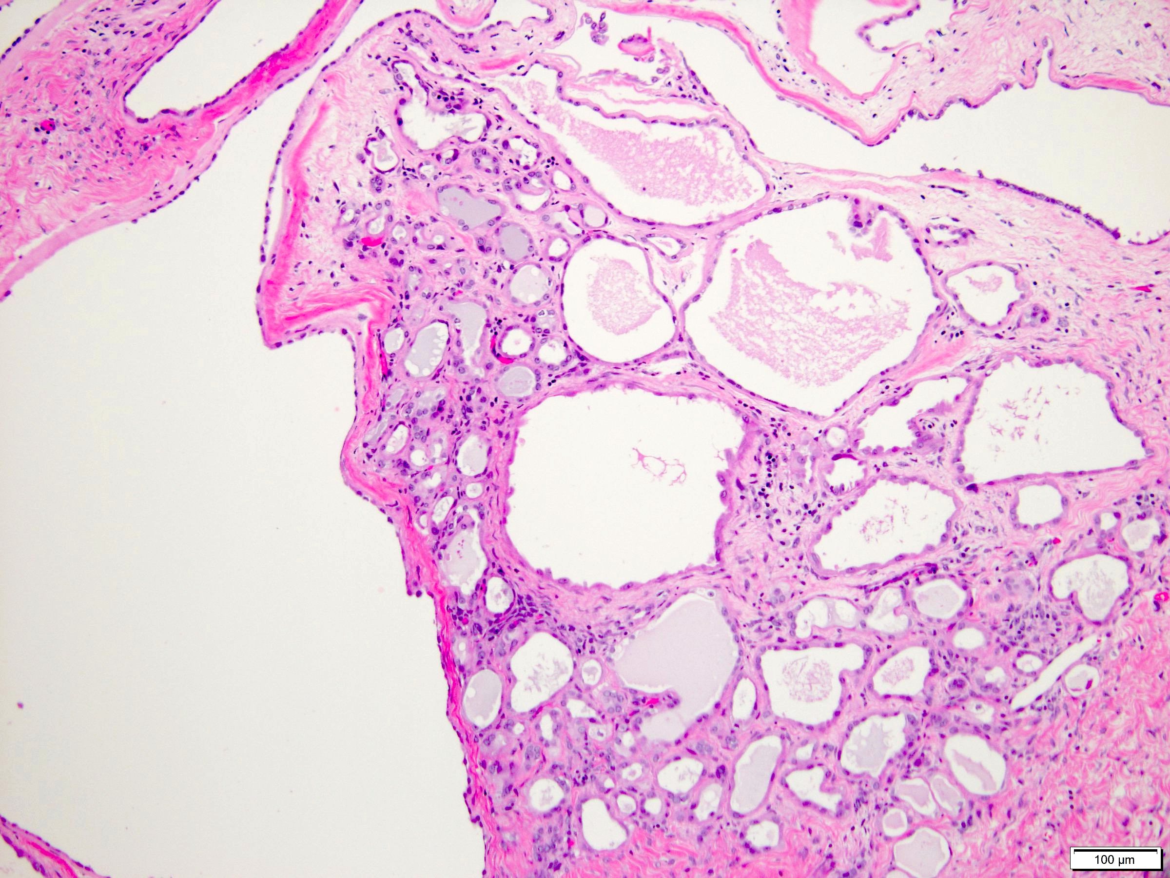
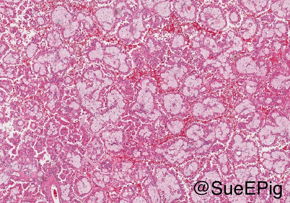
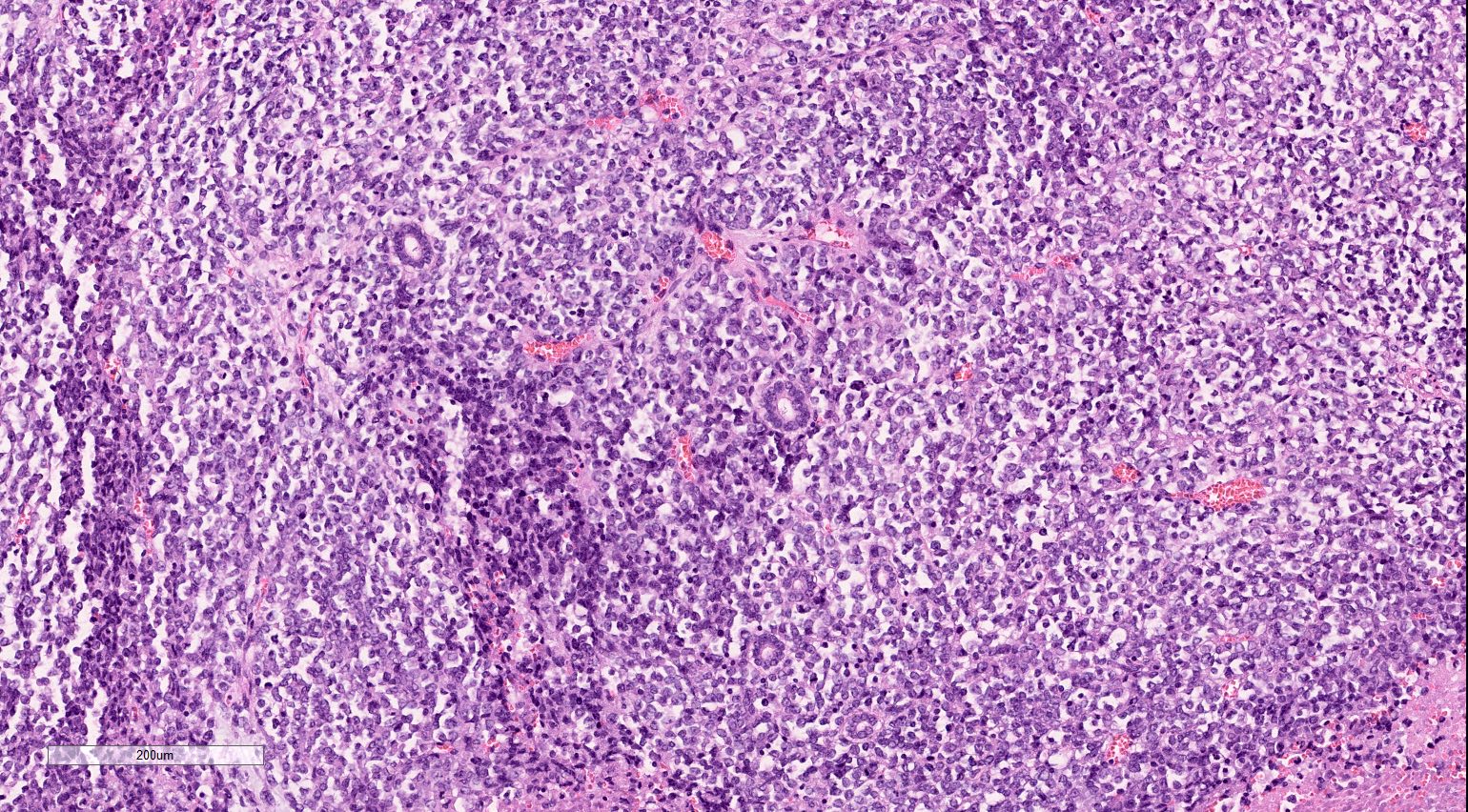
Acquired cystic disease associated renal cell carcinoma
Images hosted on other servers:

Ultrasound

CT
Contributed by Bonnie Choy, M.D.
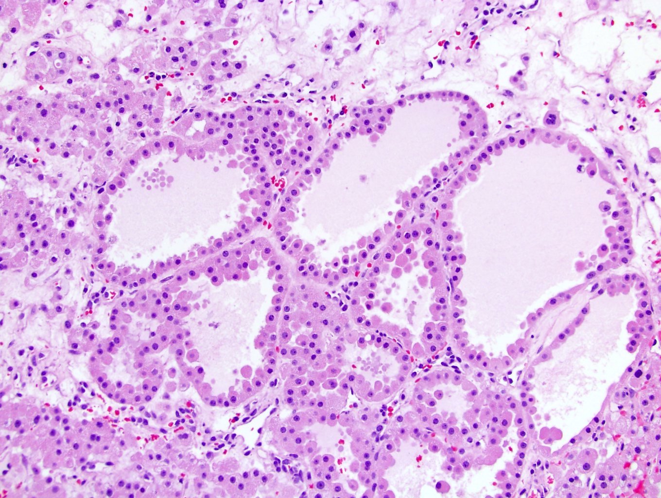
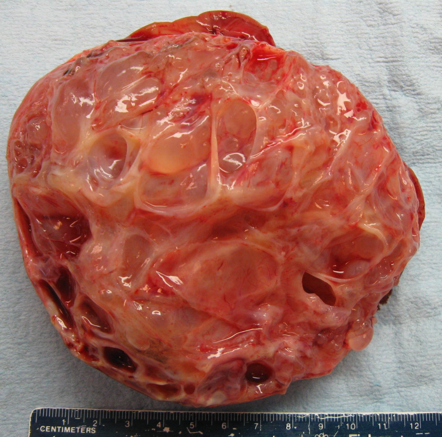
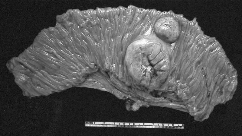
Well circumscribed, multicystic mass
Multilocular cystic mass
Contributed by Bonnie Choy, M.D.
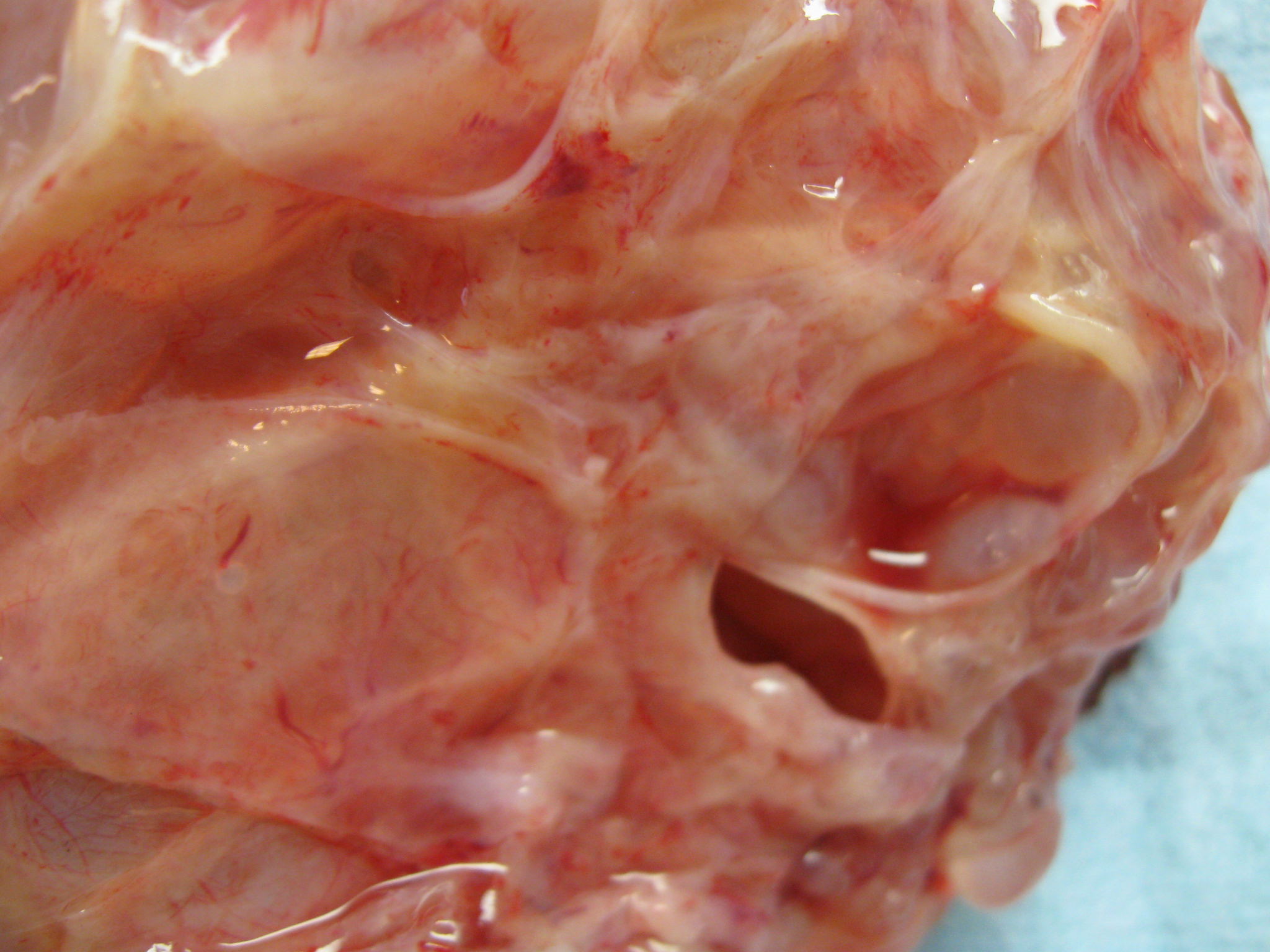
Multicystic tumor
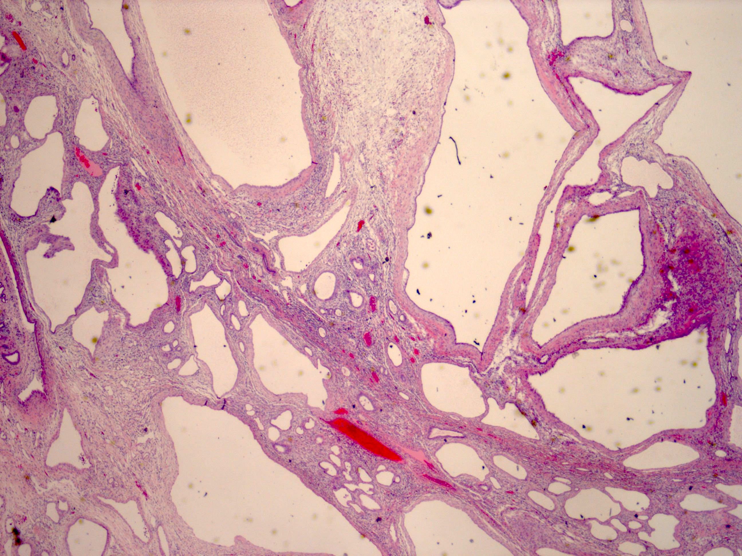
Flat and hobnail epithelium
Hobnail epithelium
Cuboidal epithelium
Stroma with variable cellularity
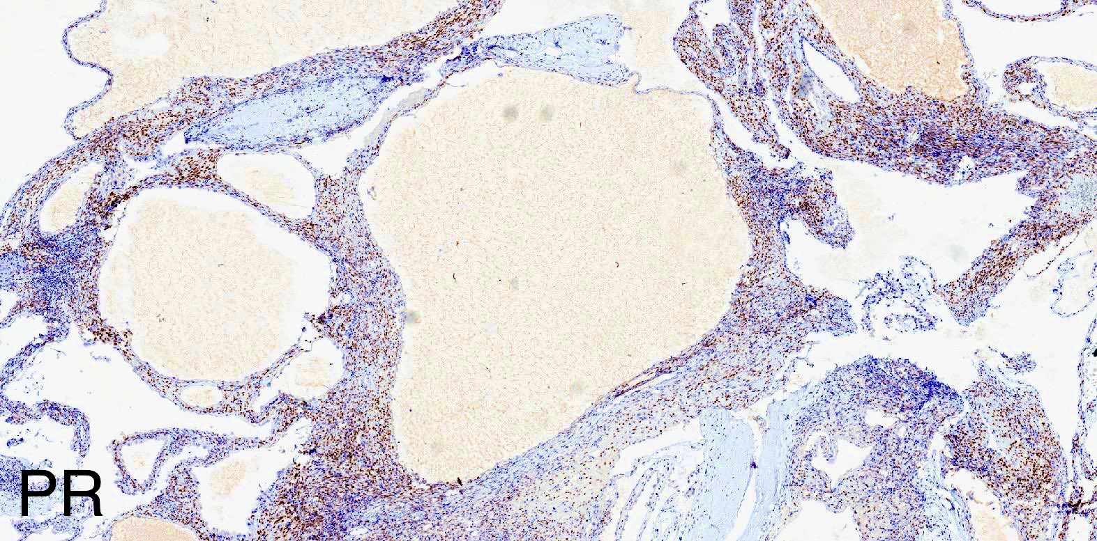
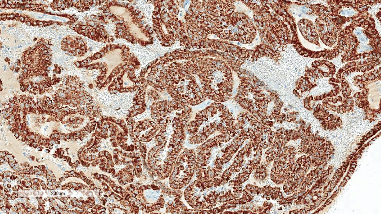
ER
PR
PAX8
Images hosted on other servers:

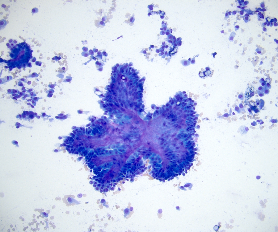
FNA
Contributed by Erdener Özer, M.D., Ph.D.

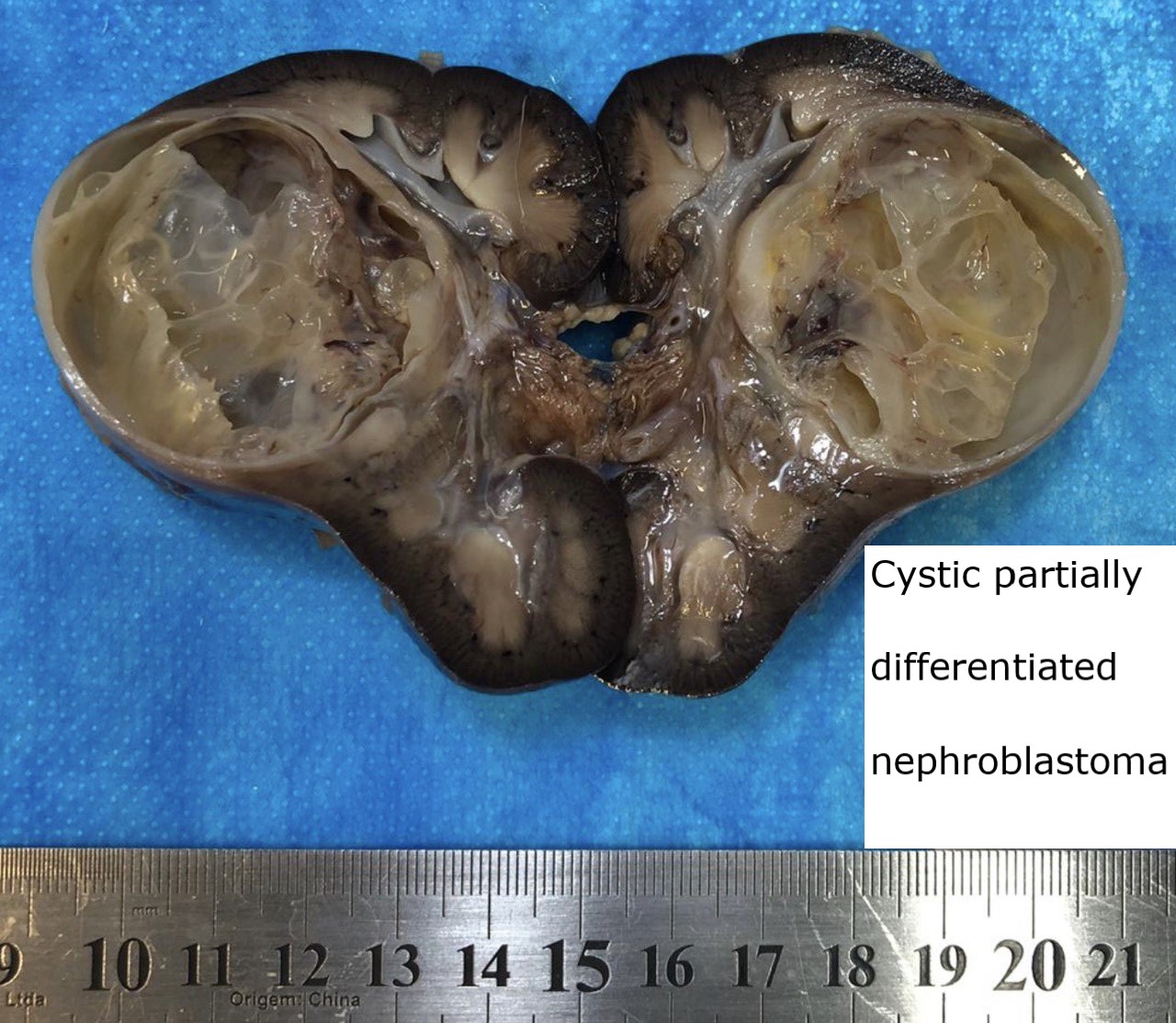
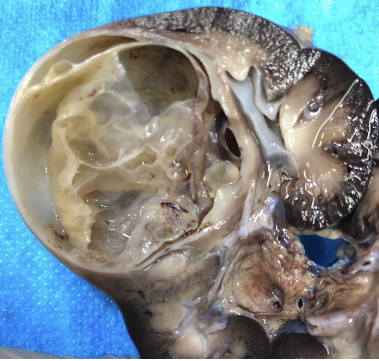
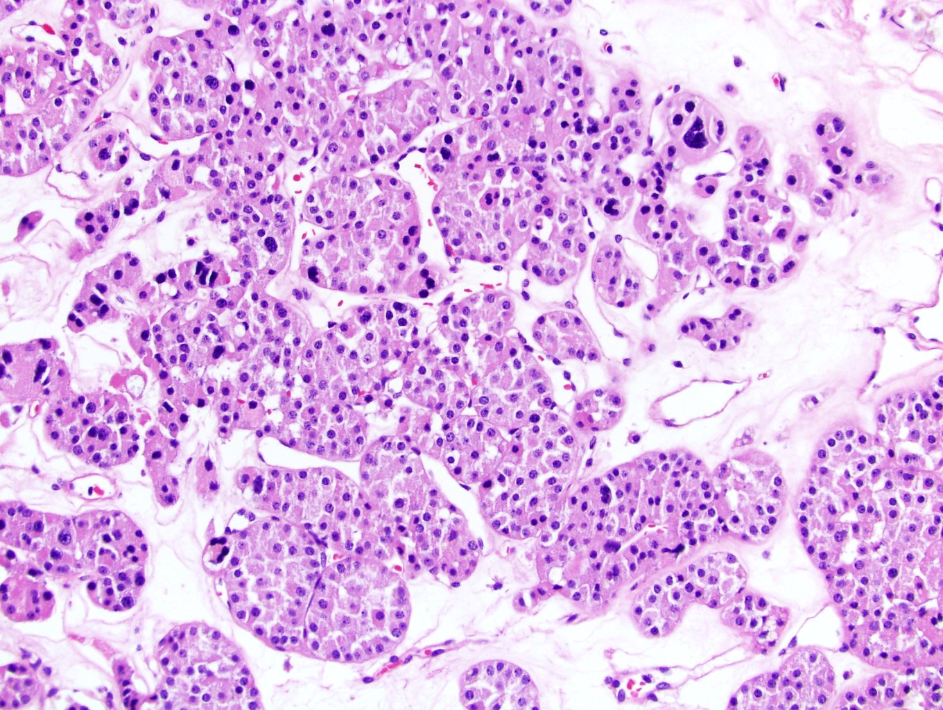
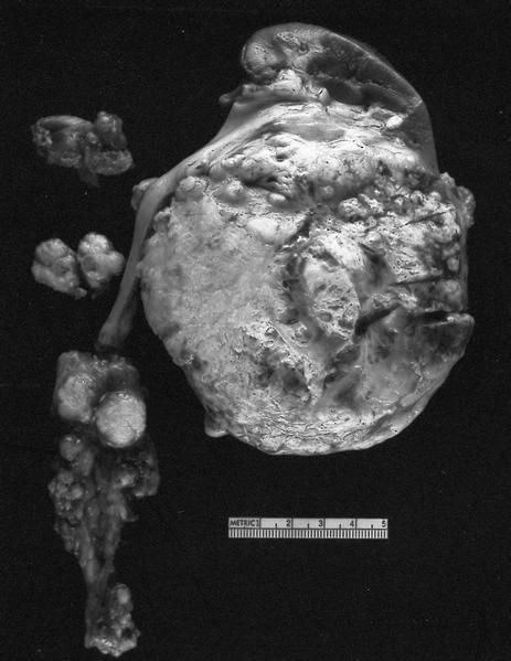
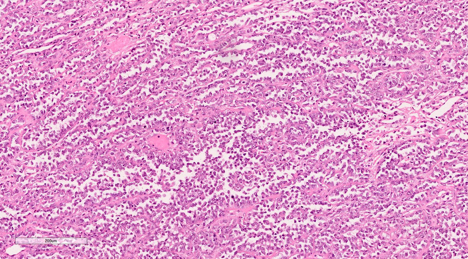
Tumor with solid and cystic parts
Contributed by Maria Laura Galluzzo Mutti, M.D. and Gordan M. Vujanic, M.D., Ph.D.

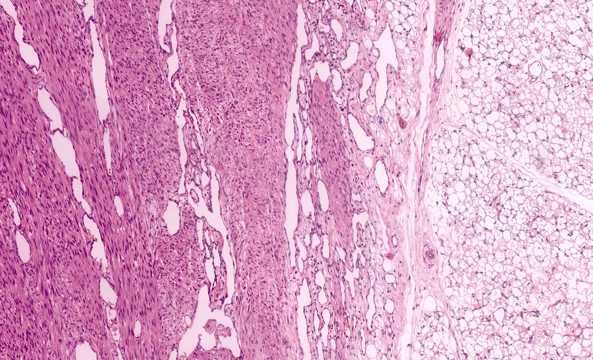
Spindled pattern

Alternating cellularity

Blastema-like area

Diffuse anaplasia

Benign cartilage

Malignant cartilage

Anaplasia

Cystic nephroma-like areas

Cystic pattern
Images hosted on other servers:

CT with renal mass
Contributed by Tatjana Antic, M.D.

Classic variant
Predominant adipose tissue
Fat poor
Hemorrhagic angiomyolipoma
Cystic epithelioid angiomyolipoma with hemorrhage
Contributed by Tatjana Antic, M.D. and Ankur R. Sangoi, M.D. (Case #359)
Classic
Epithelioid
Fat poor

Epithelial cysts (AMLEC)
Sclerosis
SMA
HMB45
Contributed by AFIP and @katcollmd on Twitter
Tan and relatively homogeneous tumor
Chromophobe RCC
Images hosted on other servers:

Yellowish brown tumor with granular cut surface

Nodular tumor with focal hemorrhage and necrosis
Contributed by Maria Tretiakova, M.D., Ph.D., Daniel Anderson, M.D., M.B.A. and @katcollmd on Twitter
Solid sheets and thin septae
Large polygonal and smaller eosinophilic cells
Raisinoid nuclei
Plant-like membranes and binucleation
Plant-like cells
Pale reticular cells
Discohesive
Confluent growth
Chromophobe renal cell carcinoma


Sarcomatoid
Hale colloidal iron
CK7
KIT
Vimentin
Images hosted on other servers:

Two cell populations

Polygonal plant-like cells


Large round to oval cells (Diff-Quik)

Pap stain
AFIP images
Cytoplasmic vesicles of 150 - 300 nm
Paranuclear cytoplasmic vesicles
Images hosted on other servers:

Homogenous enhancement, multifocal calcifications
Contributed by Timothy Isaac Miller, M.D., M.A. and Maria Tretiakova, M.D., Ph.D.
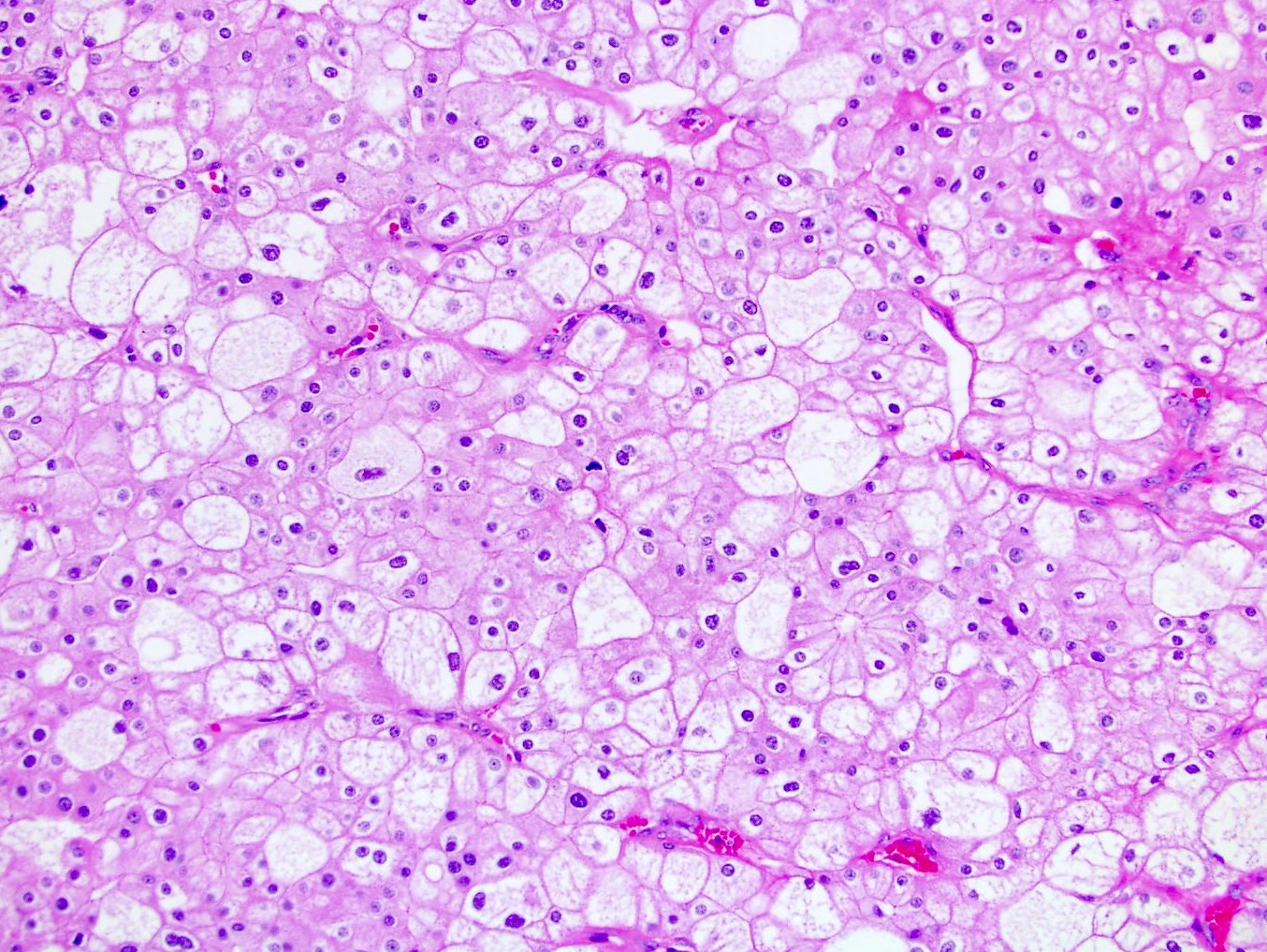
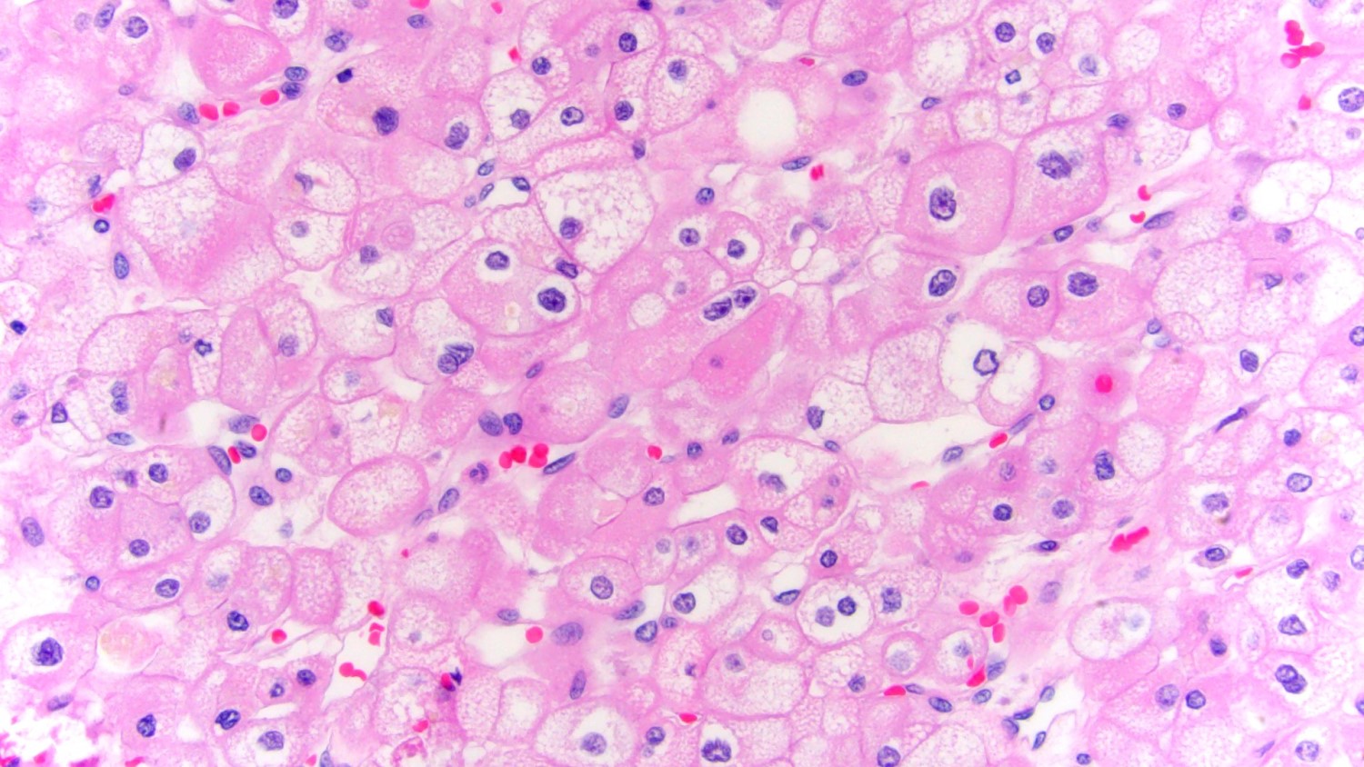
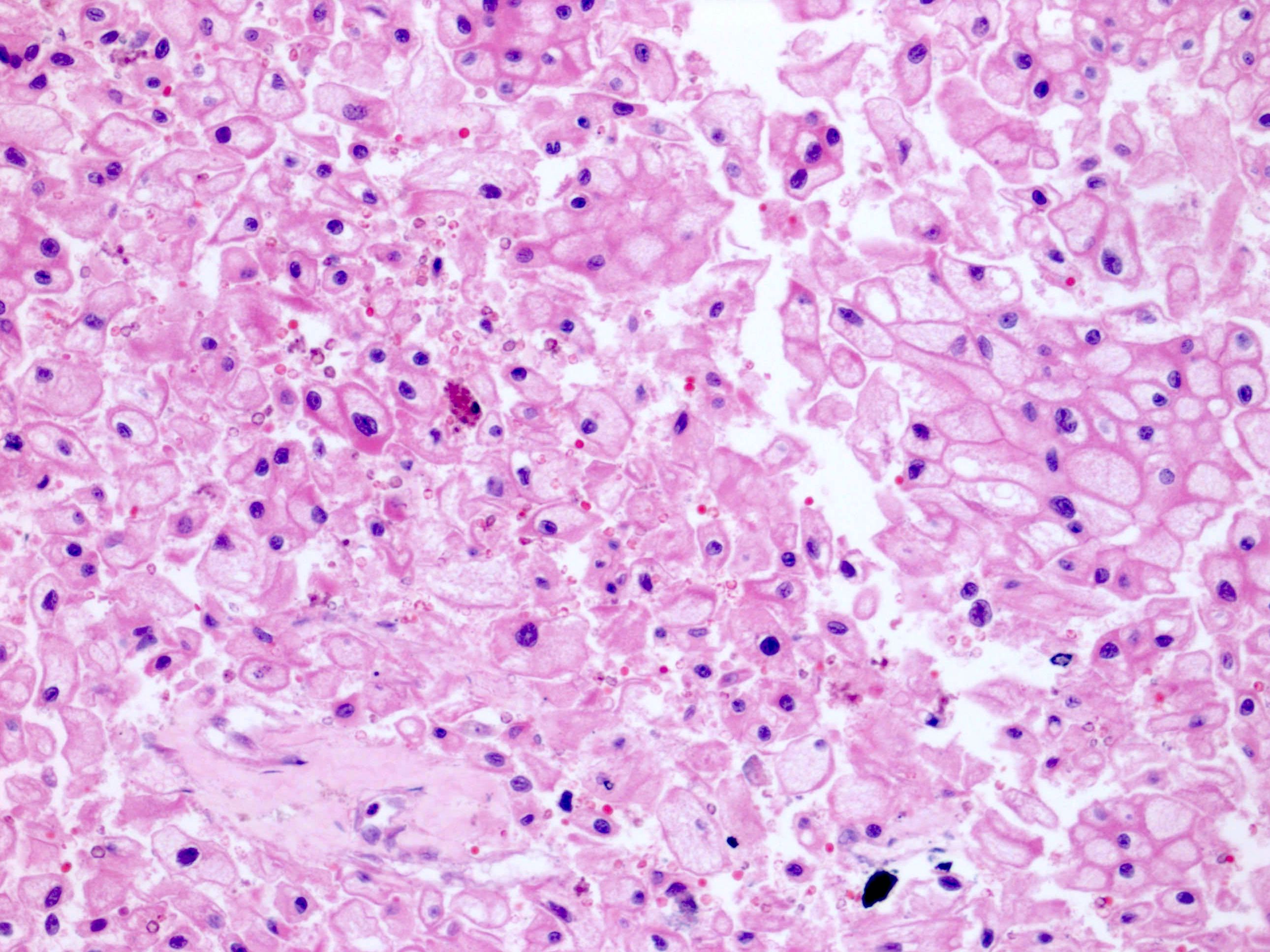
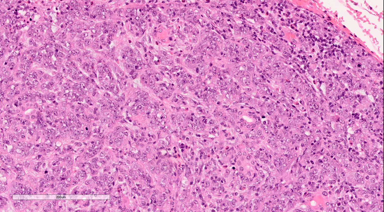
Nephrectomy with ChRCC eosinophilic variant
Contributed by Timothy Isaac Miller, M.D., M.A. and Maria Tretiakova, M.D., Ph.D.

Mild cytological atypia
Prominent perinuclear clearing
Alveolar architecture
Focal classic cells
Increased cytological atypia
Extension to perinephric fat
Distinguishing eosinophilic chromophobe RCC from oncocytoma
Images hosted on other servers:


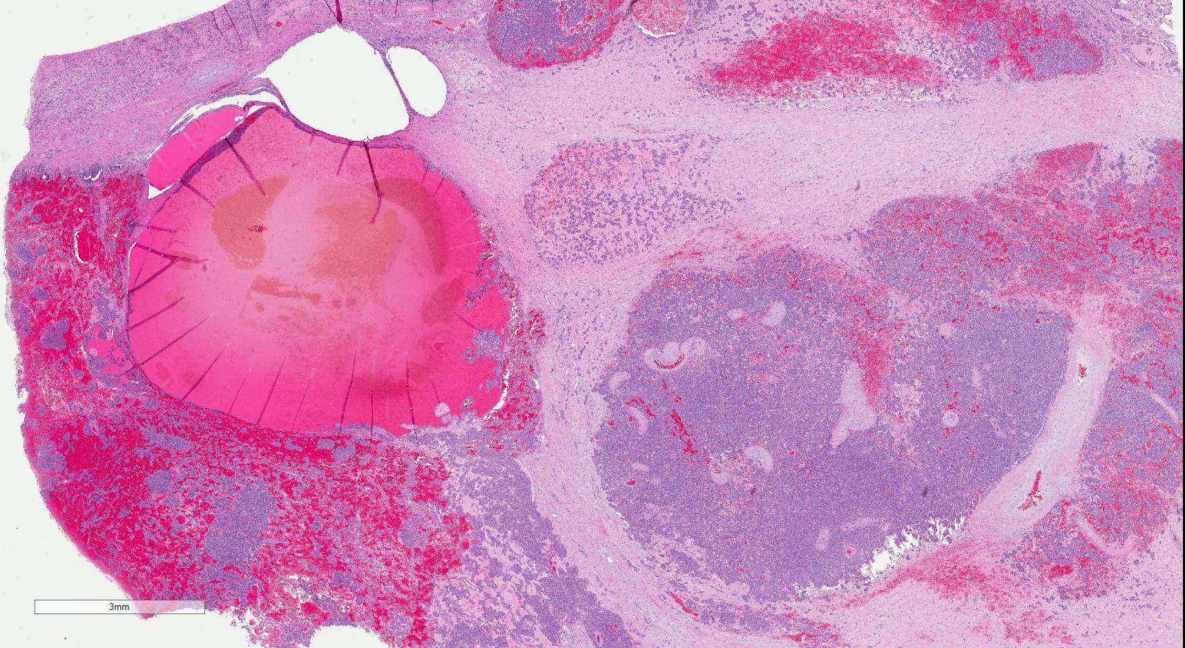
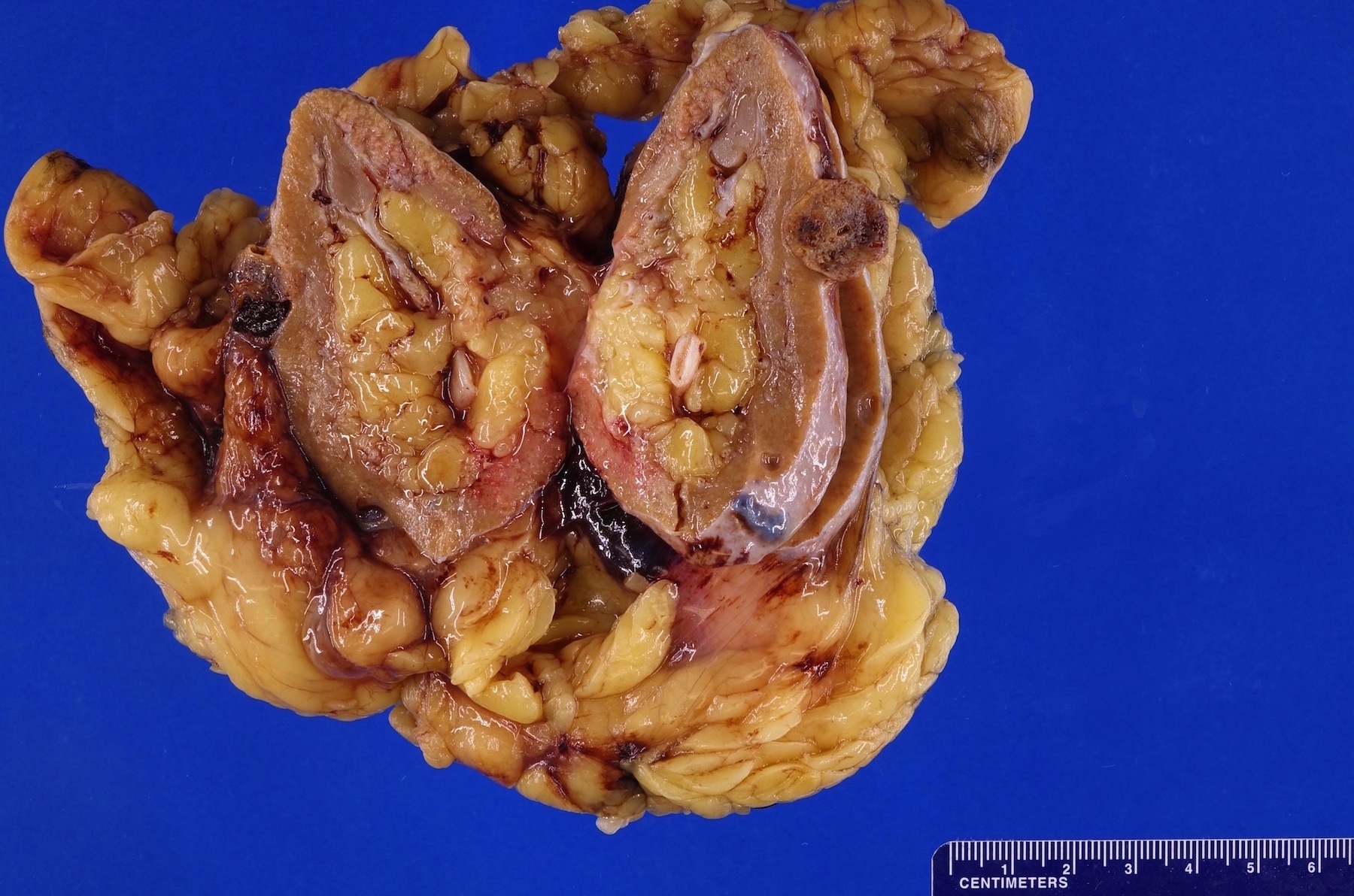
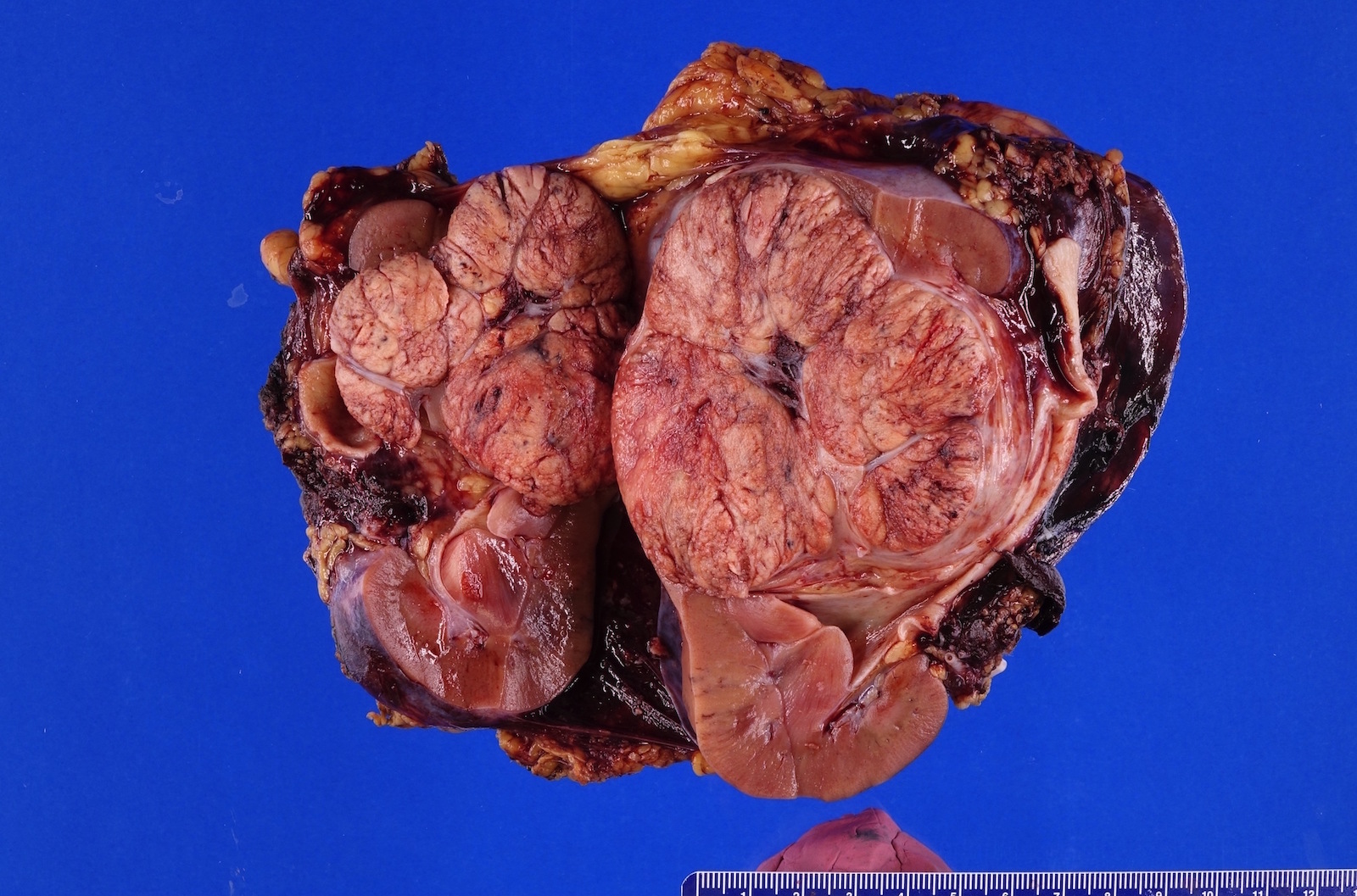
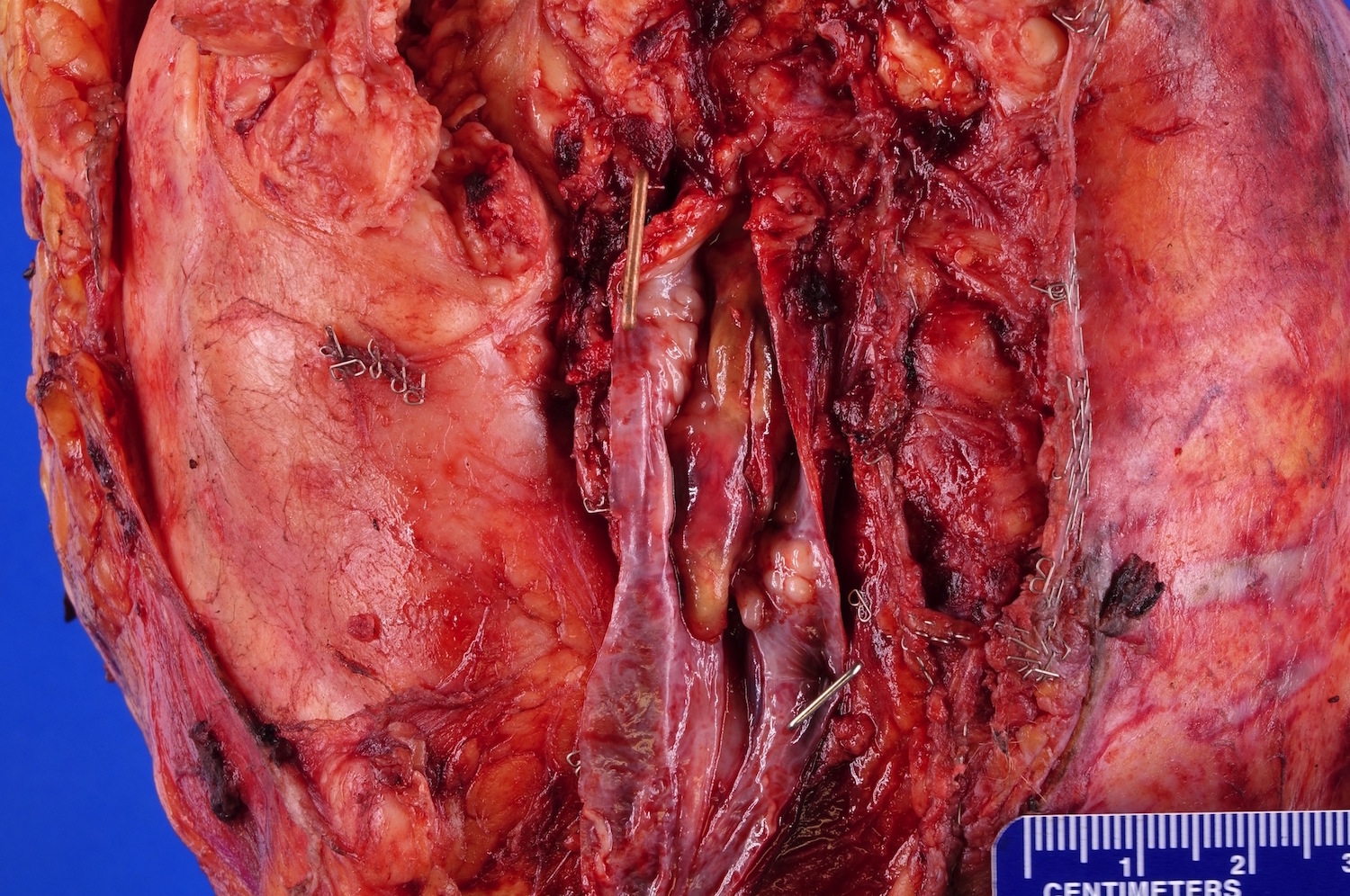
Left sided mass (CT scan)
Contributed by Gregory T. MacLennan, M.D. and Behtash Nezami, M.D.

Involving Gerota fascia (pT4)
Confined T1b
Multifocal
In renal vein
Invading sinus fat and pelvicalyceal system
Variegated cut surface
Cystic changes
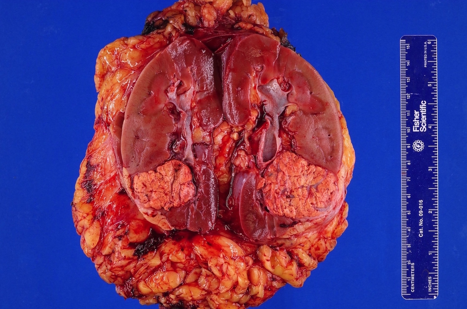
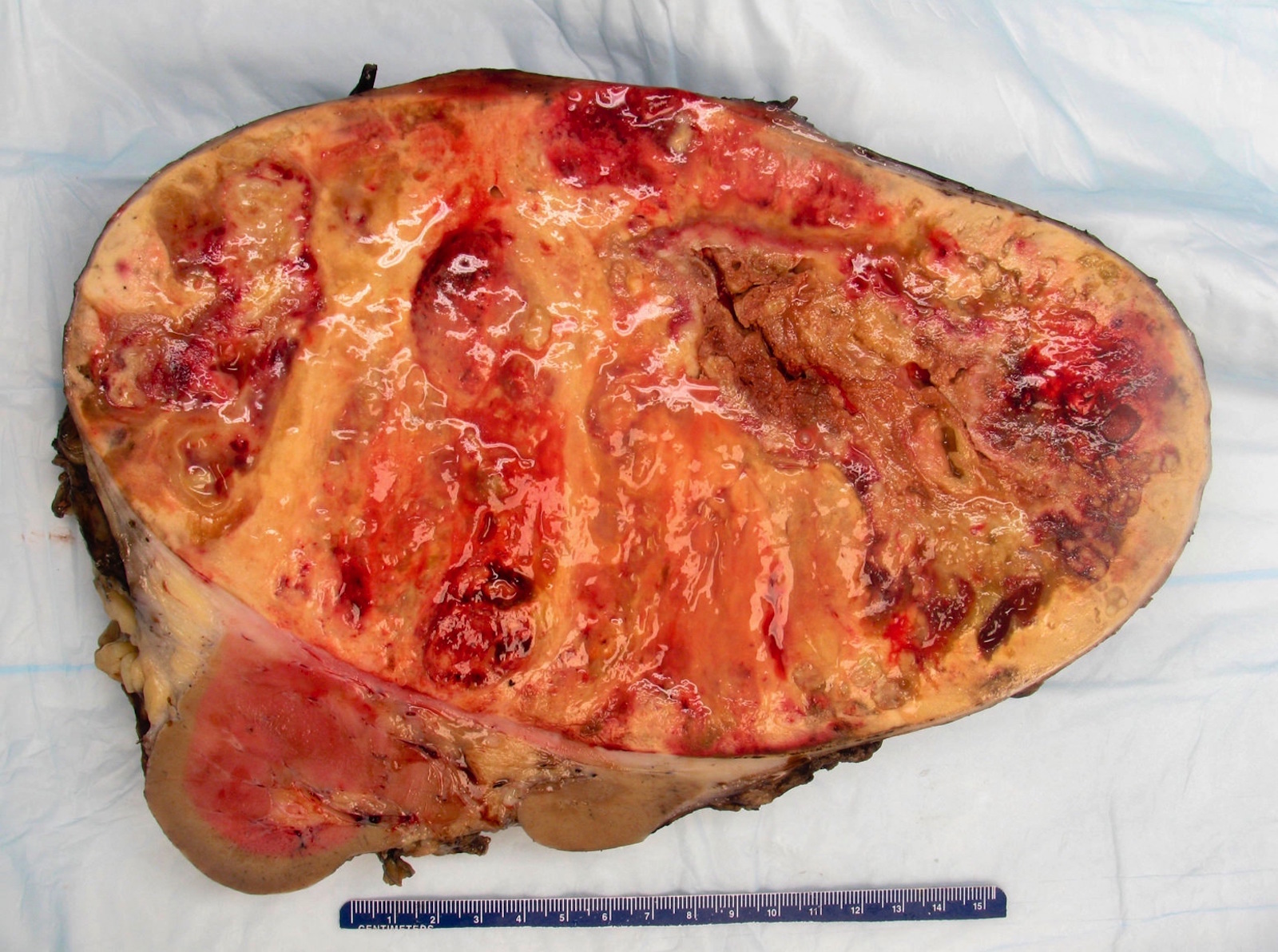
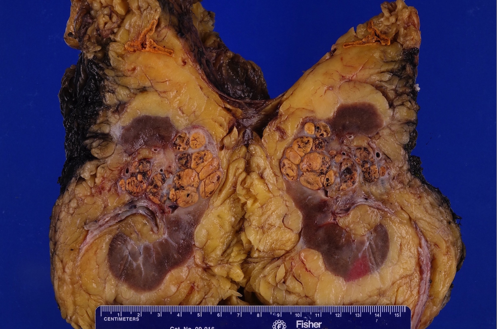
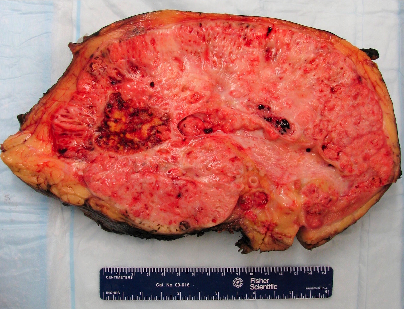
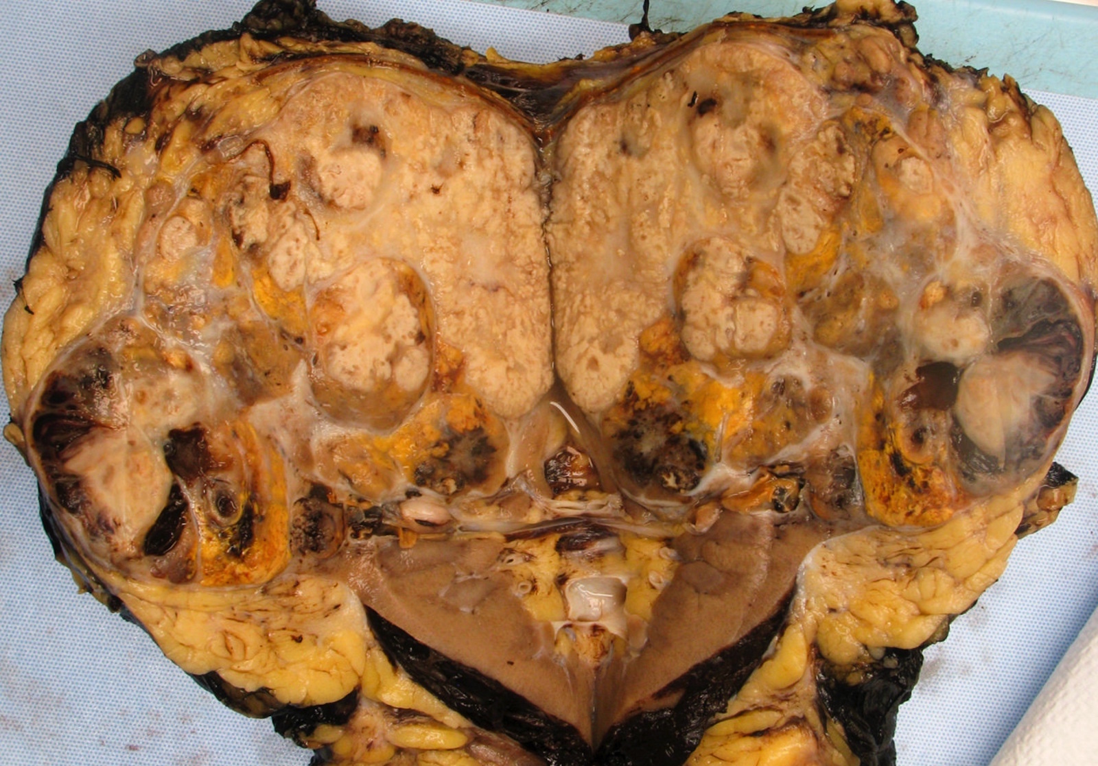
Contributed by Gregory T. MacLennan, M.D., Behtash Nezami, M.D., Nicole K. Andeen, M.D. and Maria Tretiakova, M.D., Ph.D.
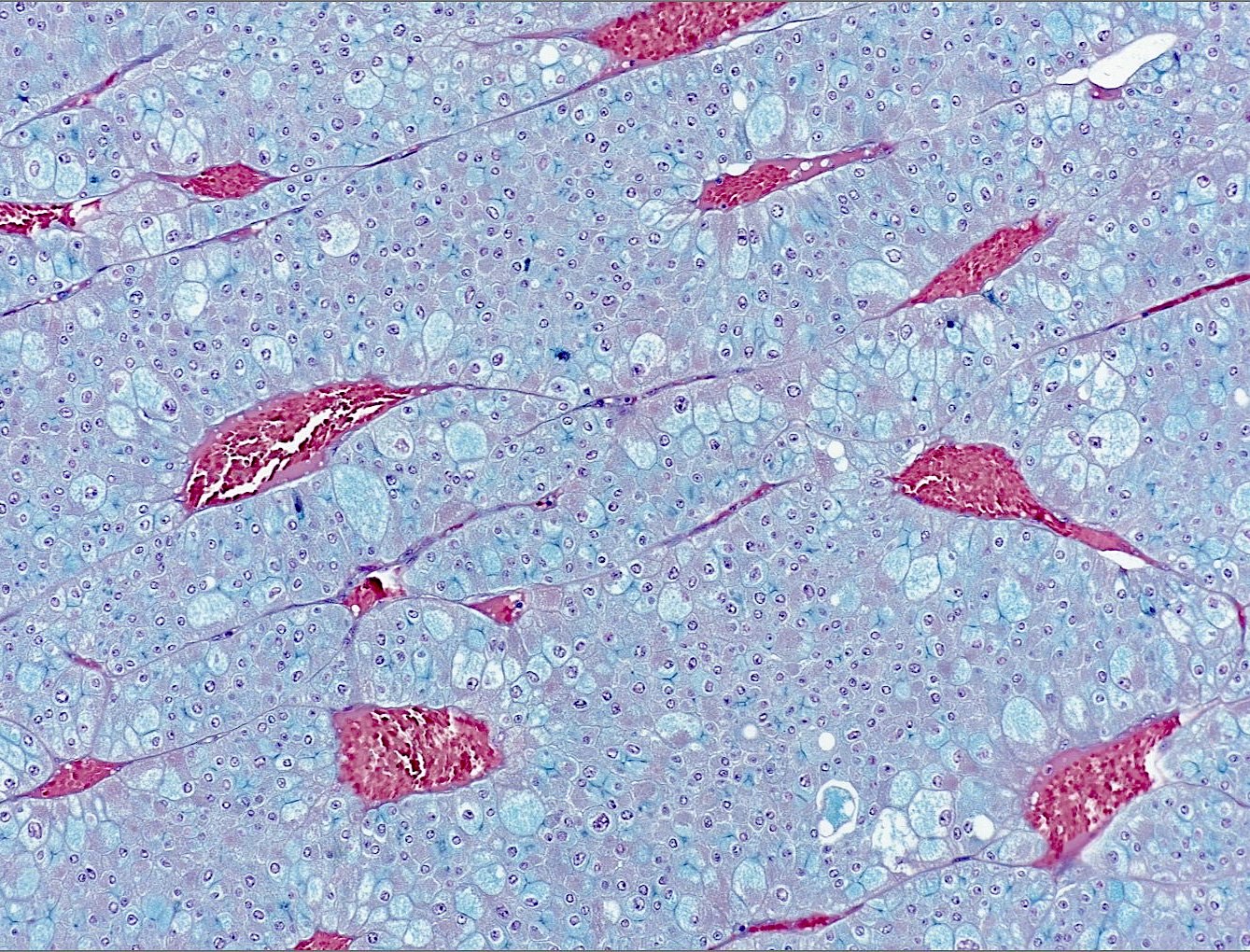
Sinusoidal vasculature
Micro and macrocysts
With renal capsule invasion

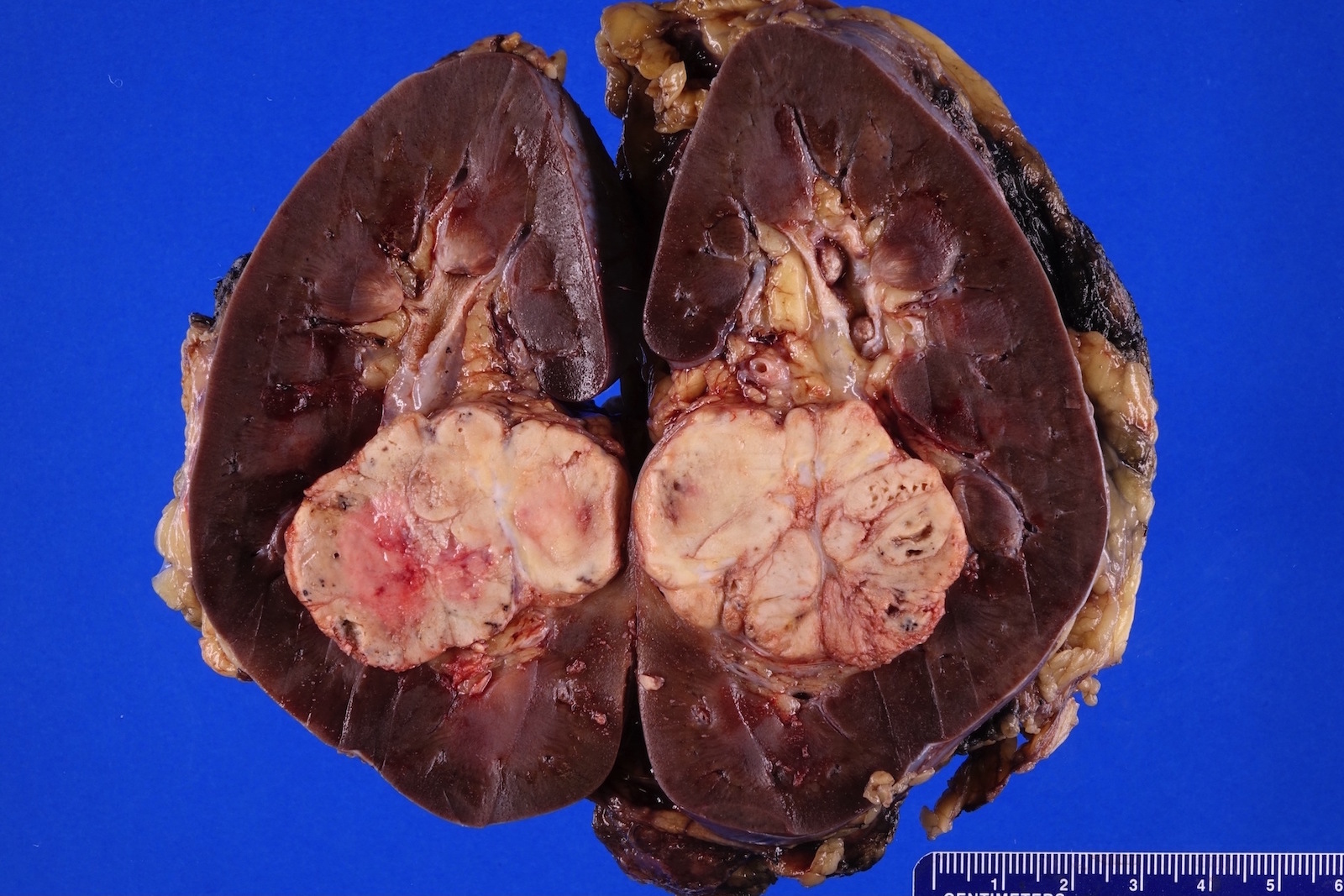
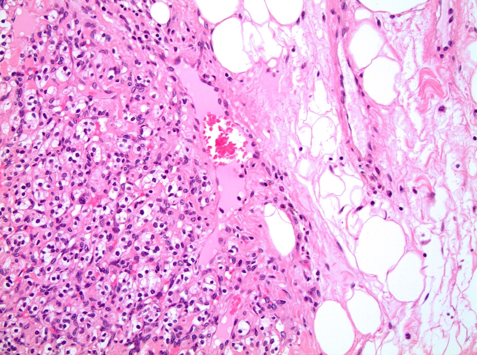
ISUP grade 1
ISUP grade 2
ISUP grade 3
ISUP grade 4
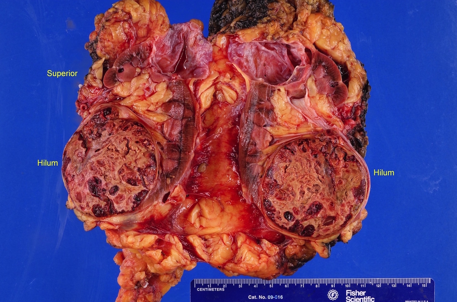
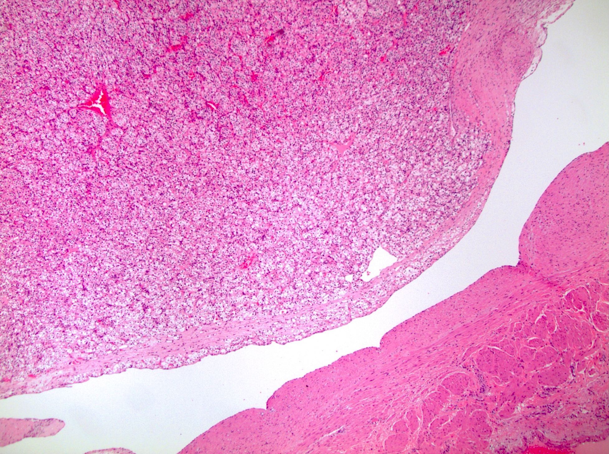
In renal vein
In perirenal fat
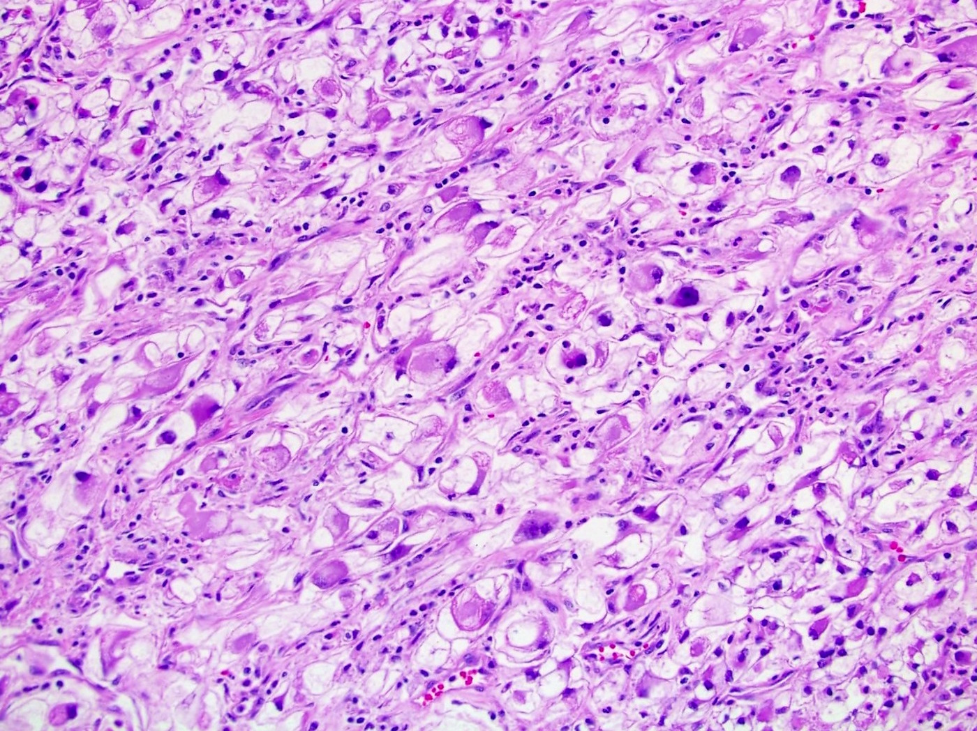
Sarcomatoid
Sarcomatoid
Rhabdoid differentiation

Rhabdoid and sarcomatoid areas
Necrosis
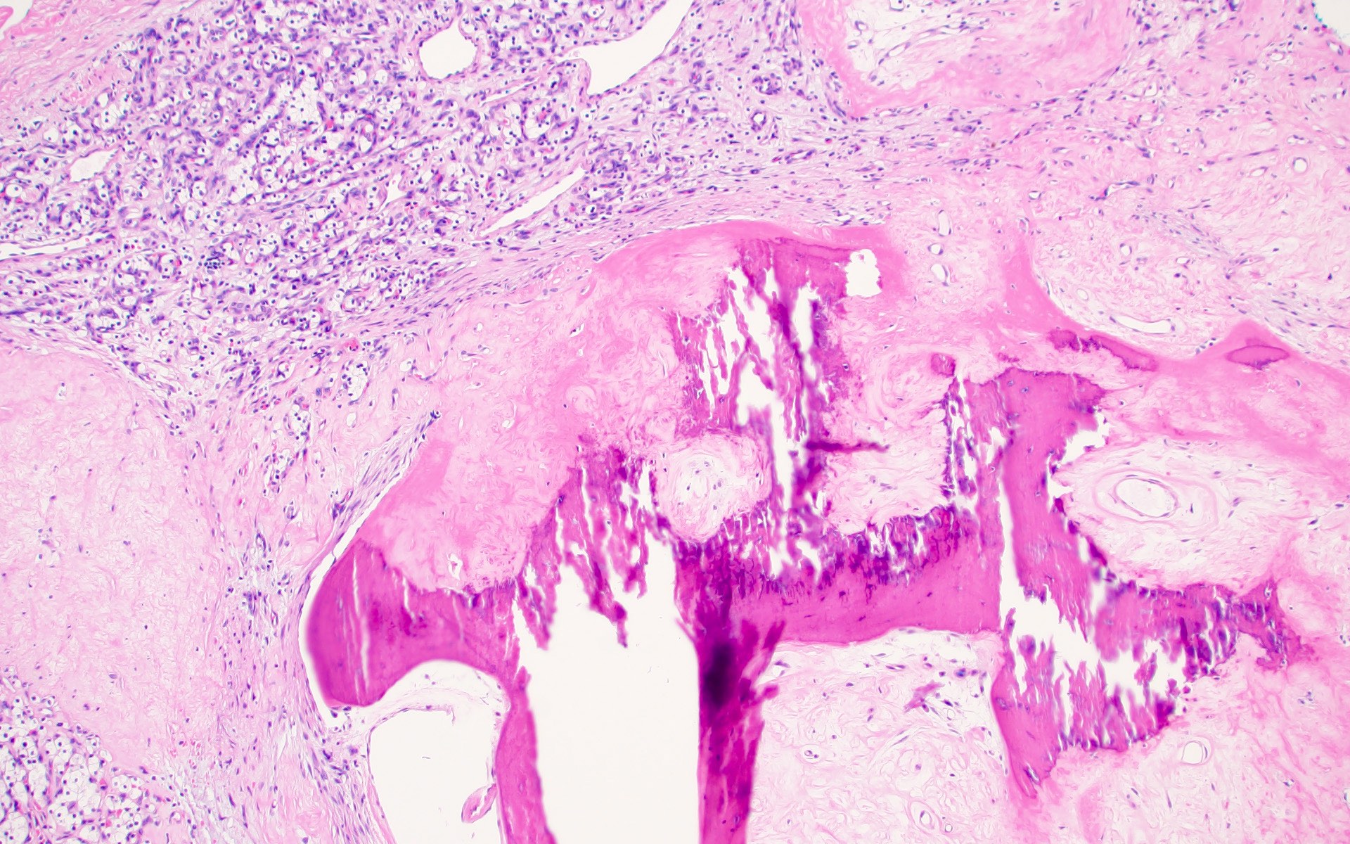
Heterotopic bone formation


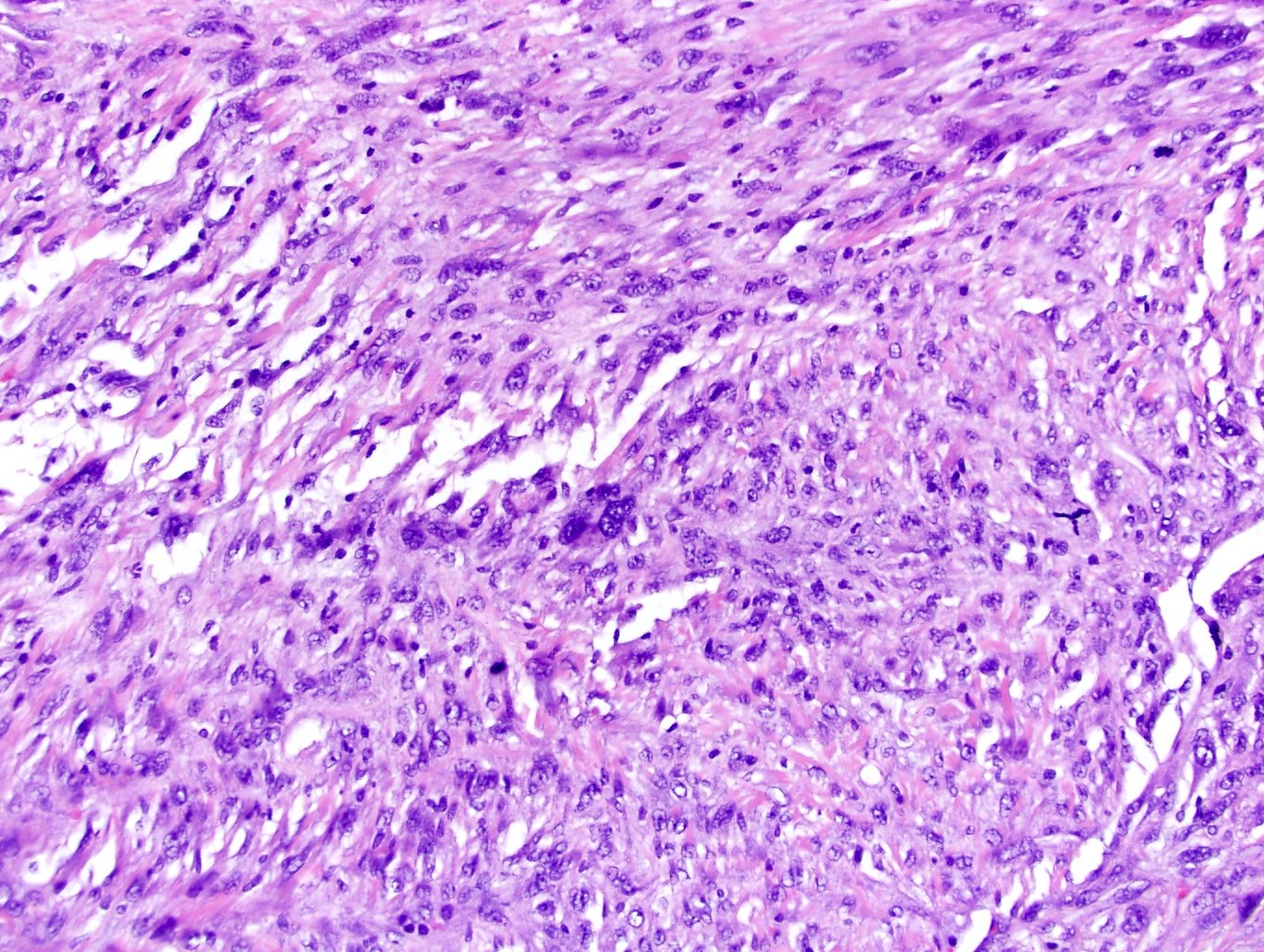
Clear cell RCC could have areas of eosinophilic cytoplasm mimicking other RCC sybtypes
Intramural granulomatous reaction
Involving pelvicalyceal system (pT3a)
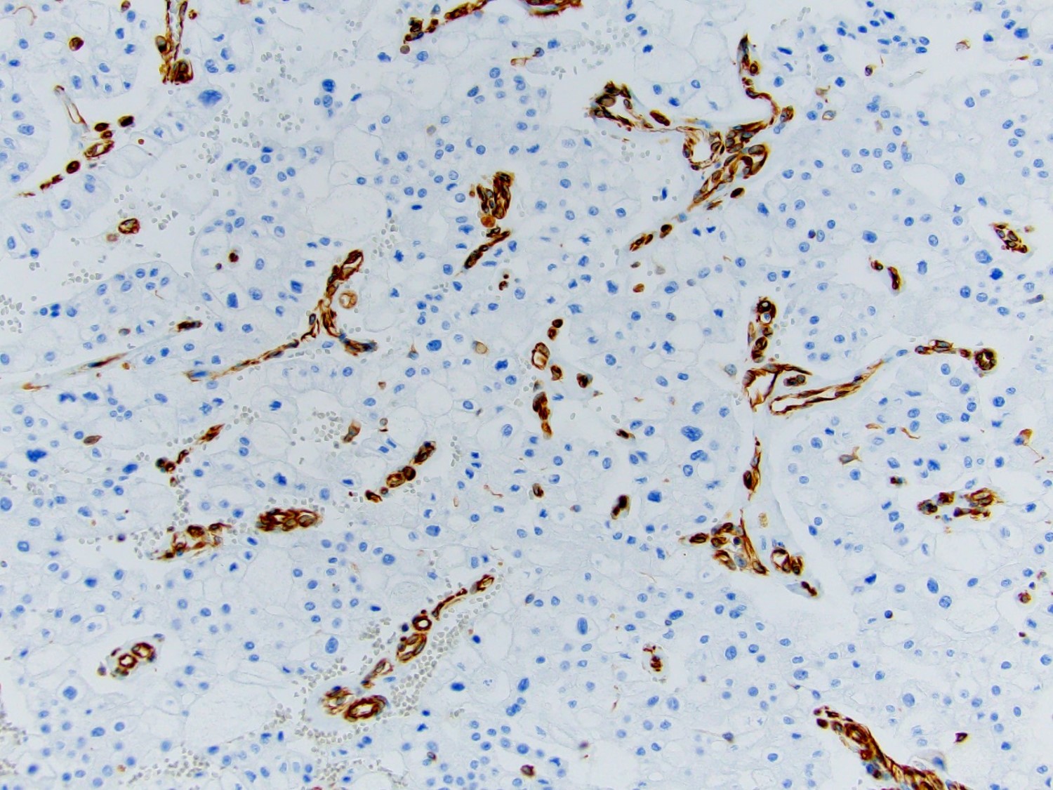
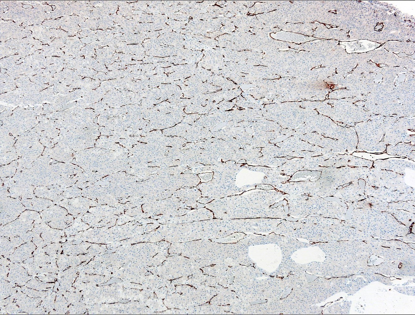
CD10 IHC staining
CAIX IHC staining
Sarcomatoid, pancytokeratin
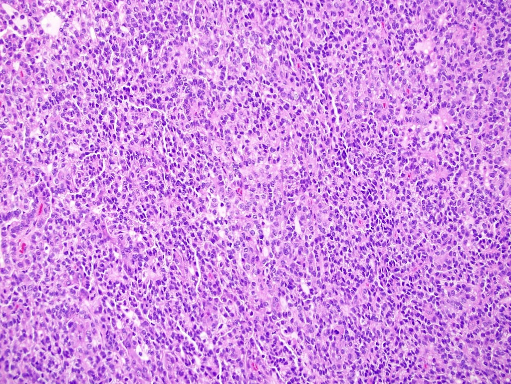
Contributed by Gregory T. MacLennan, M.D.
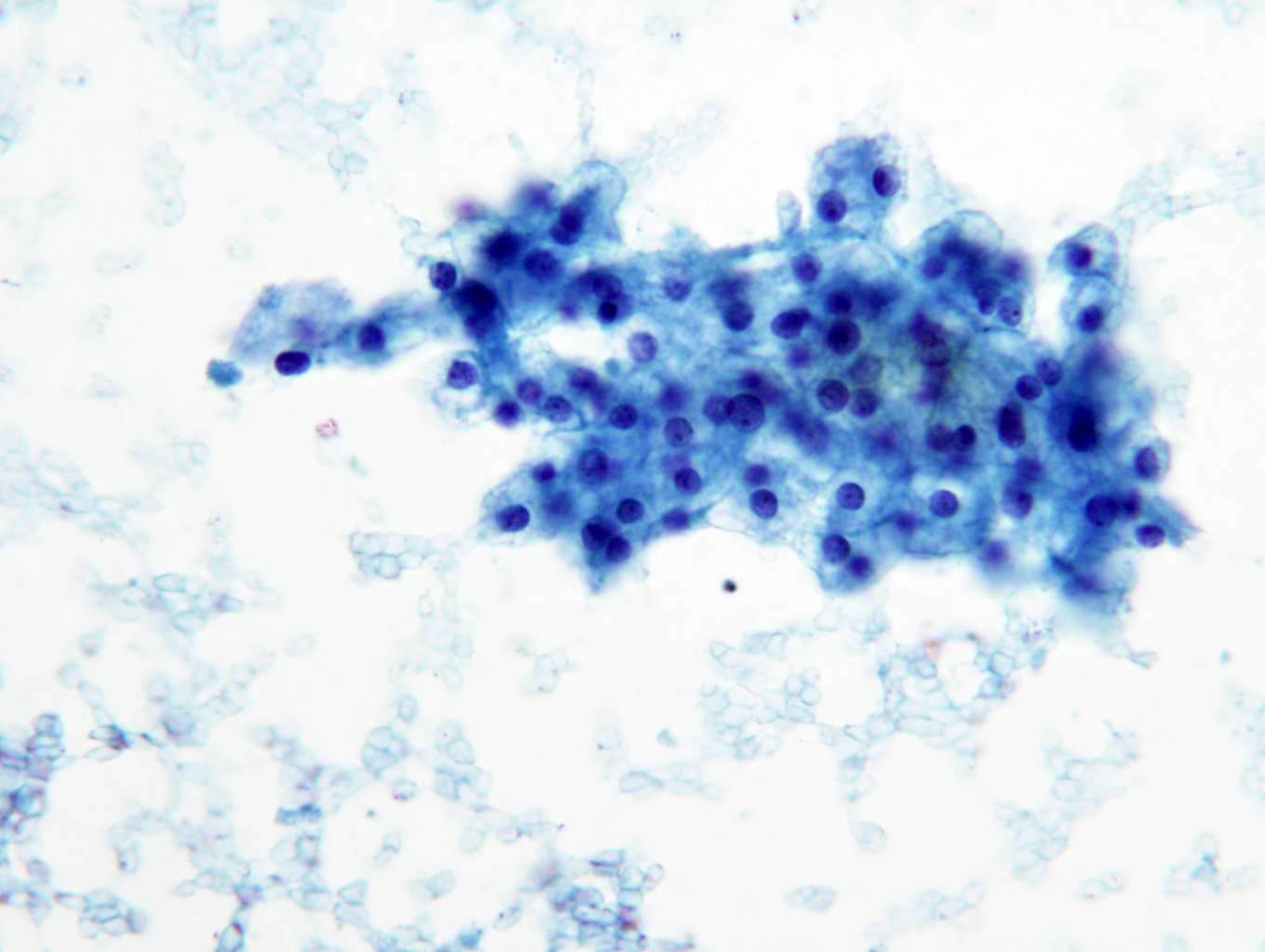
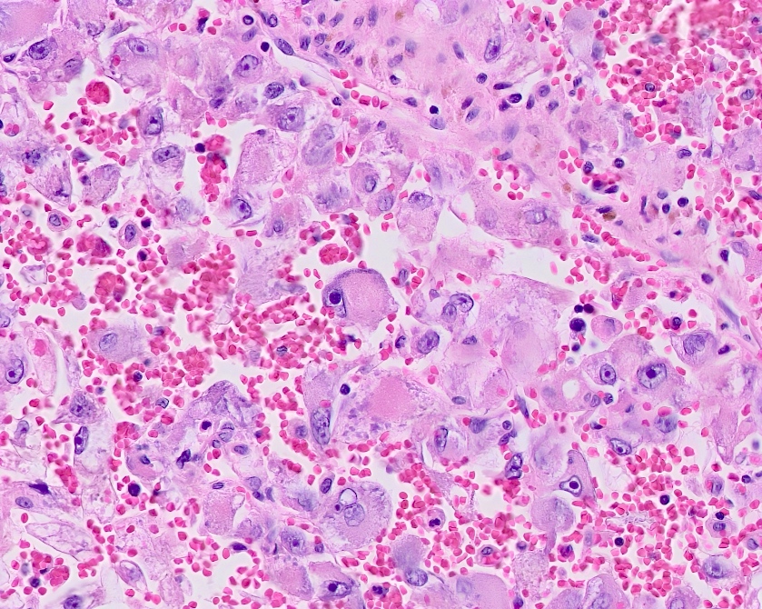
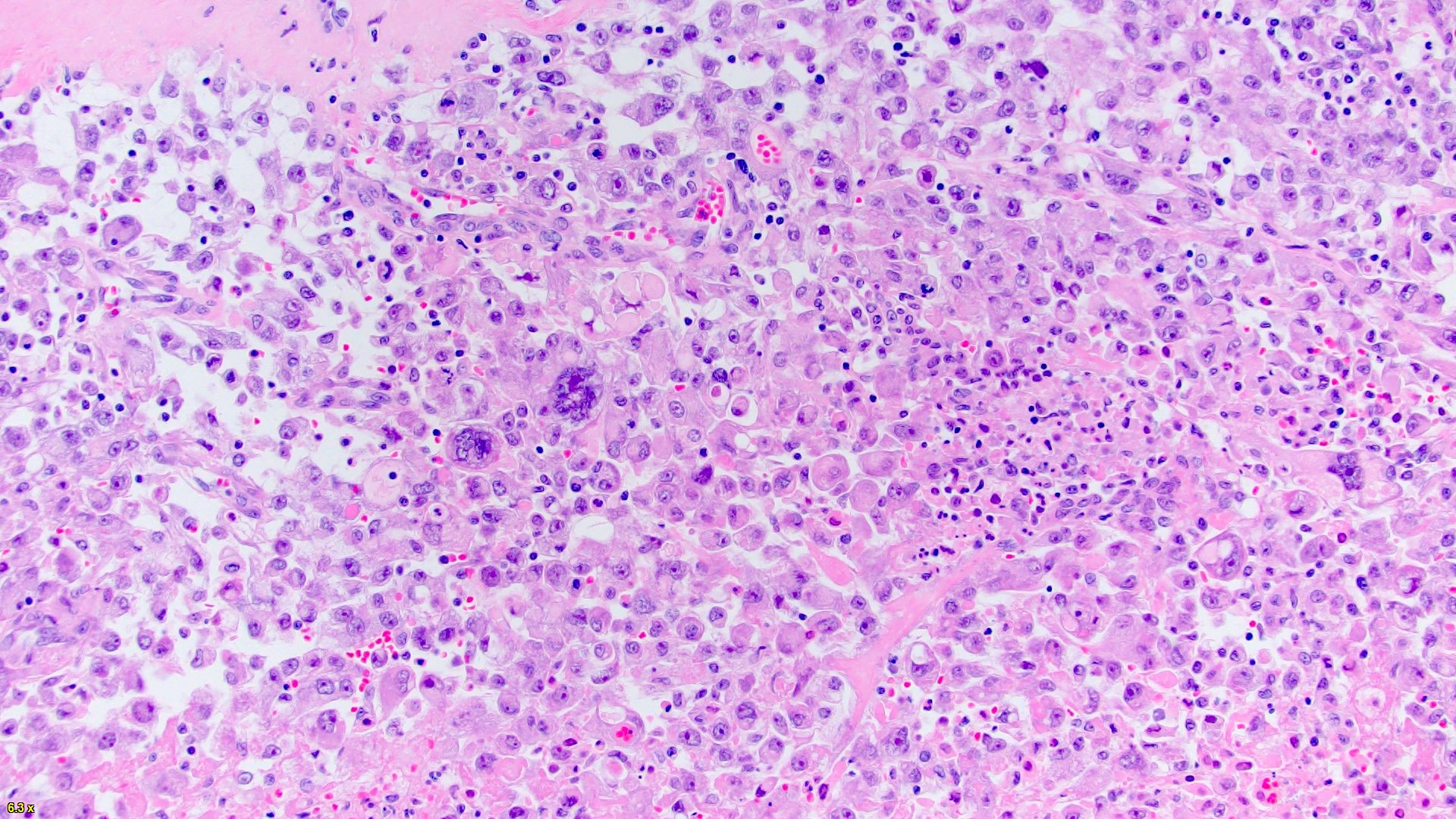
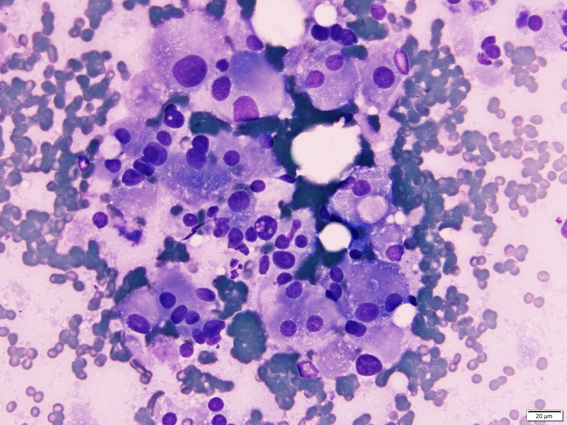
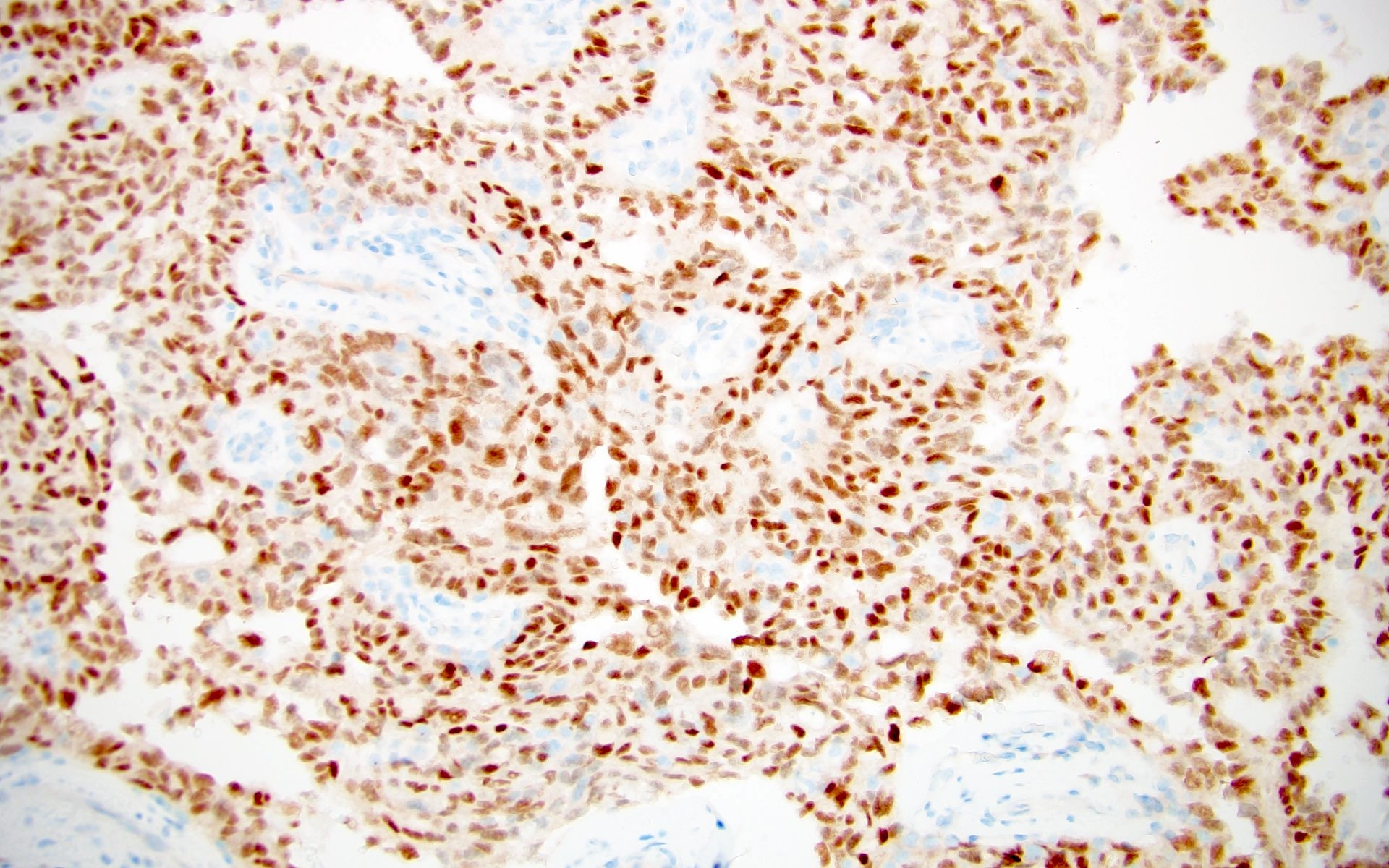
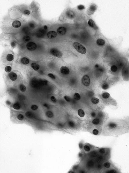
FNA cytology
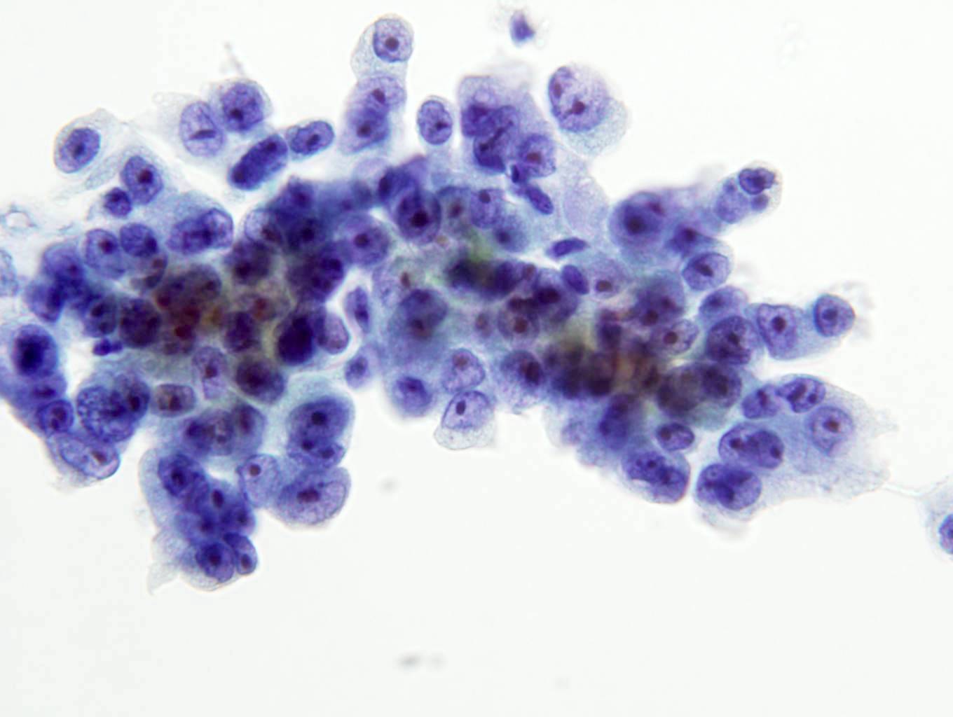
FNA high grade
Contributed by Behtash Nezami, M.D.
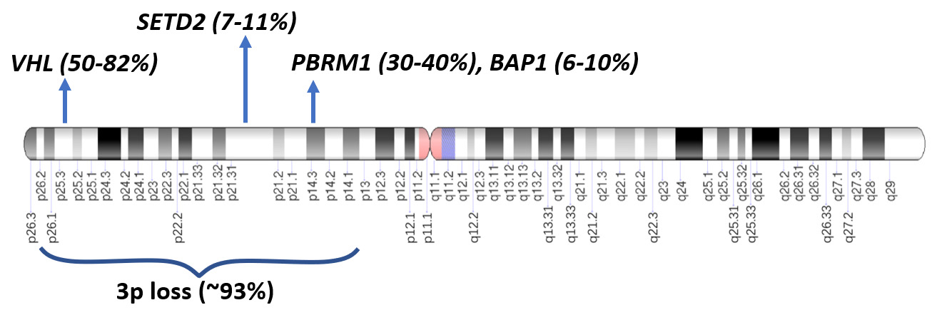
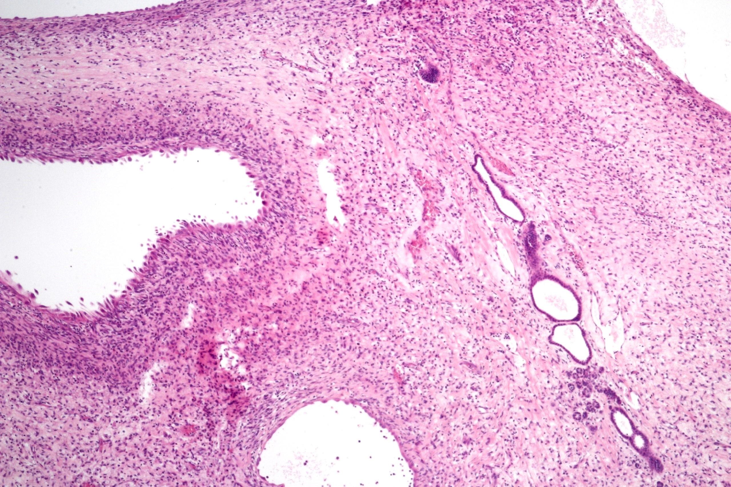
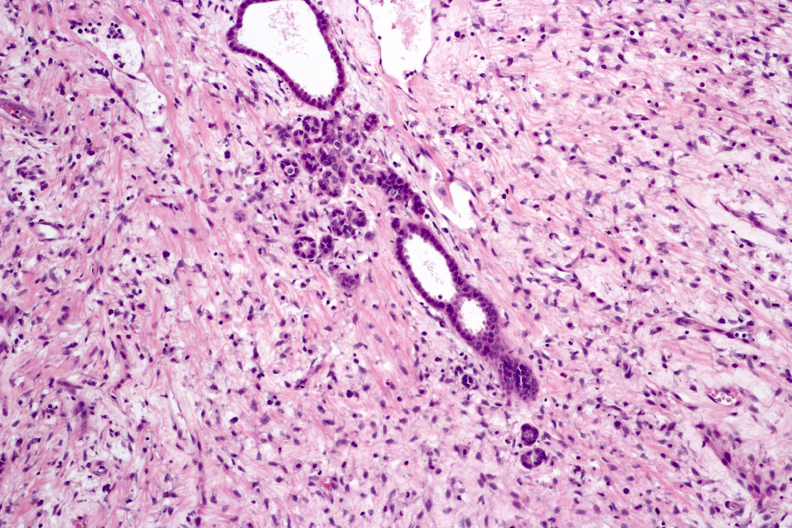
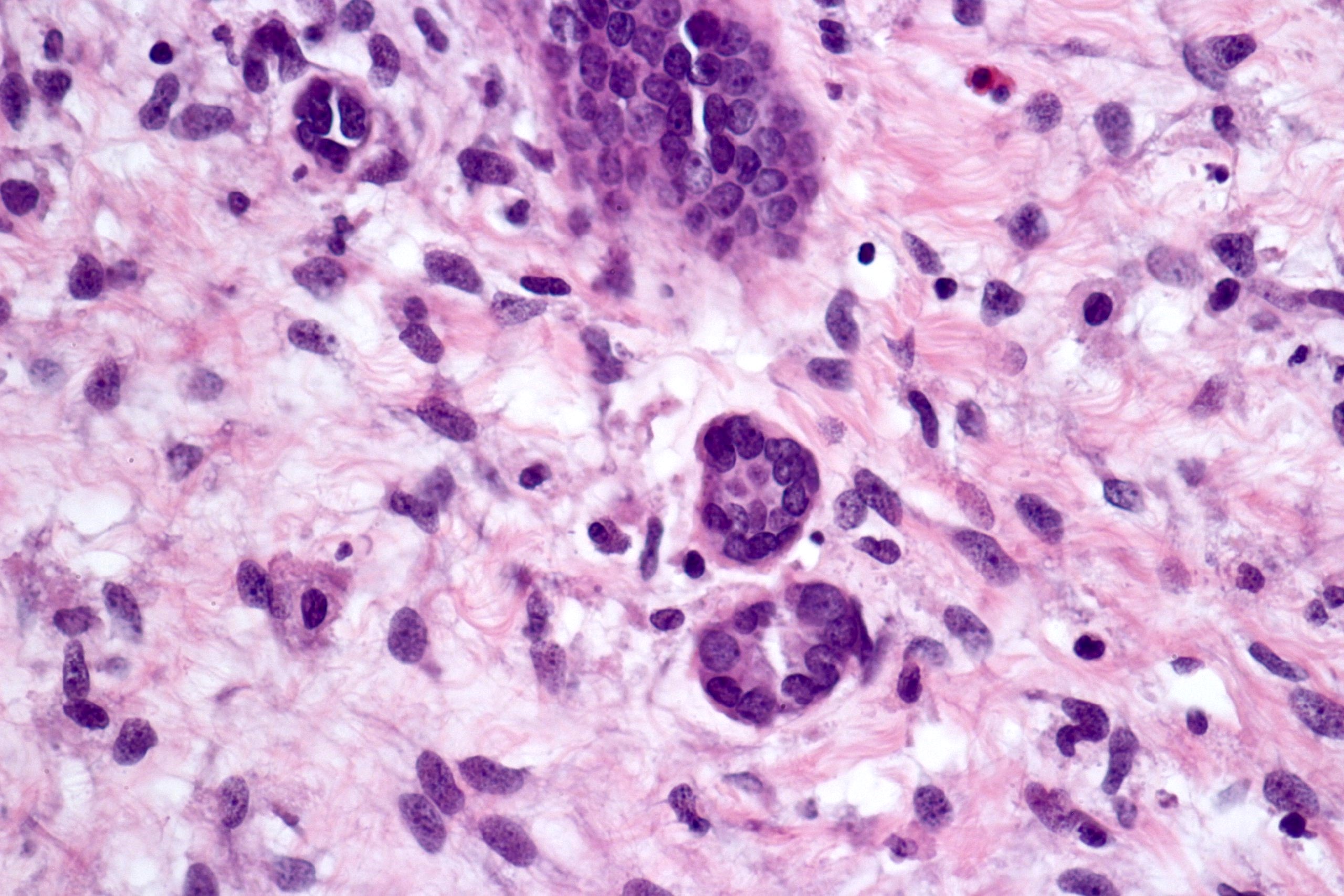
Genetic alterations
Contributed by Gregory MacLennan, M.D.
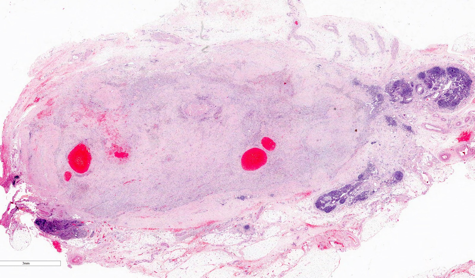
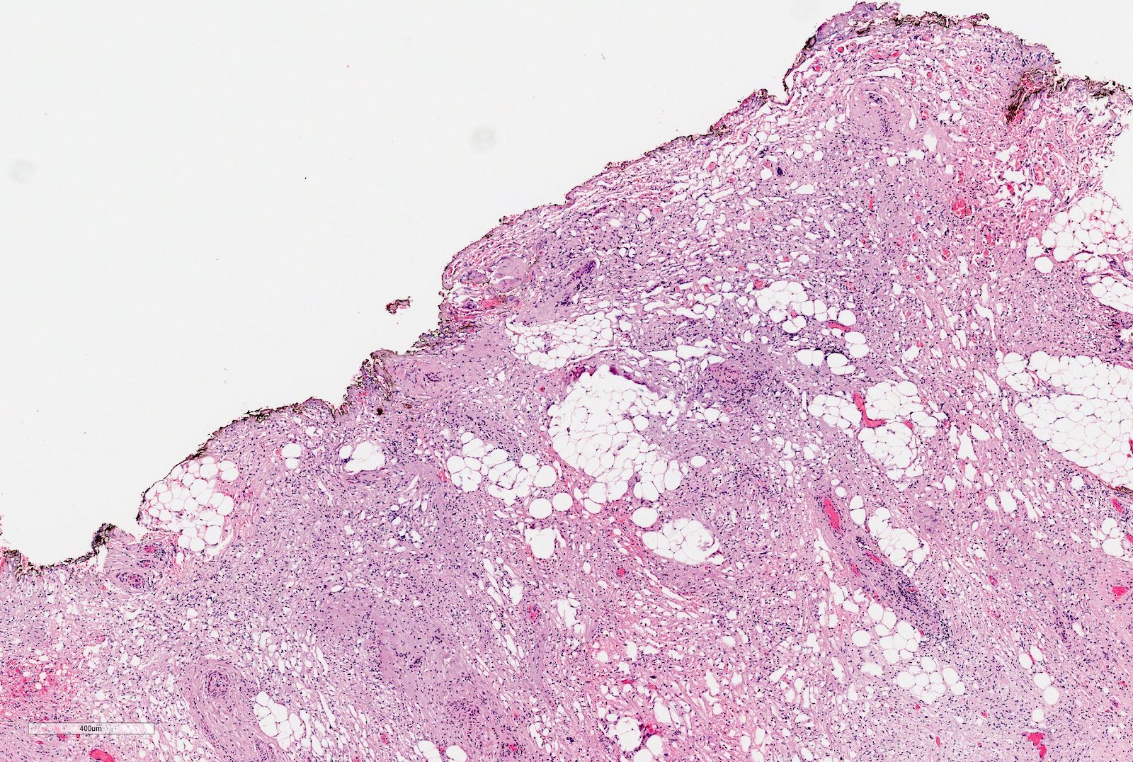
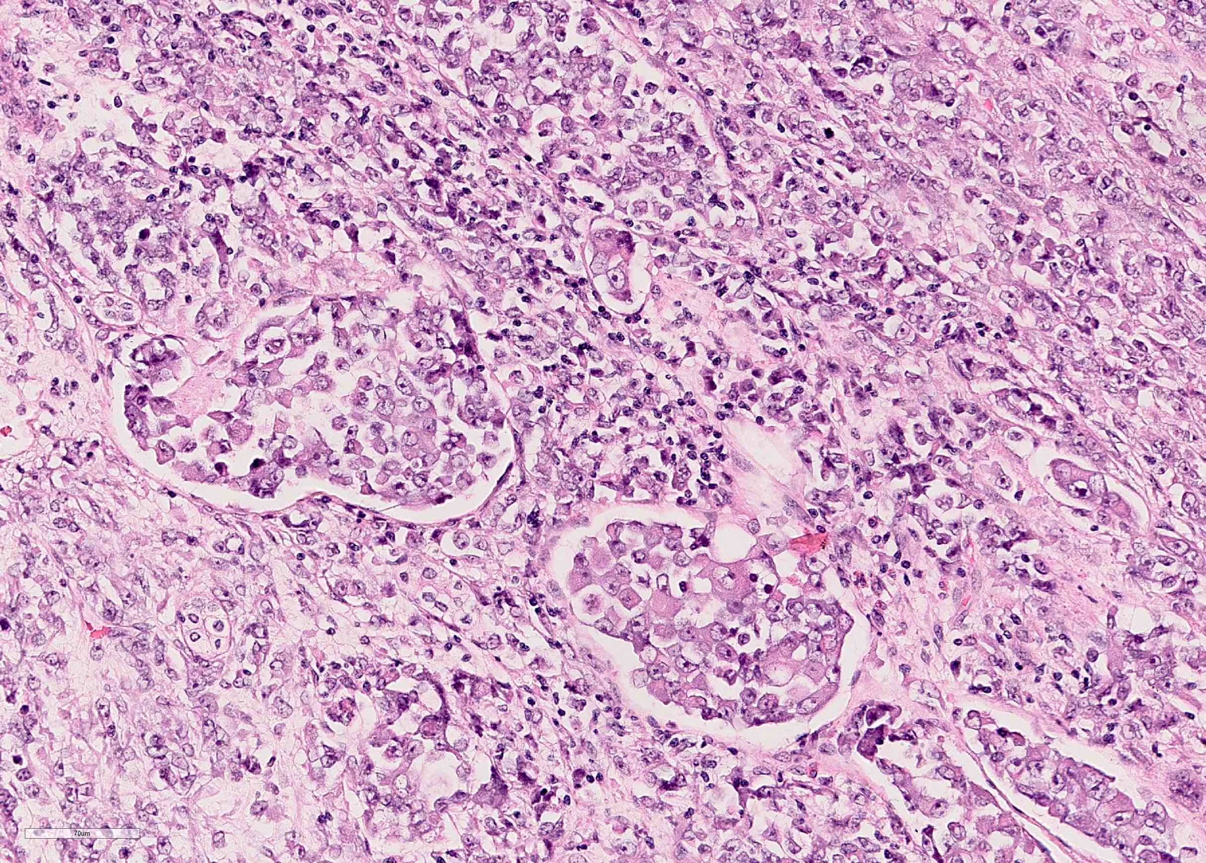
Partial nephrectomy specimens

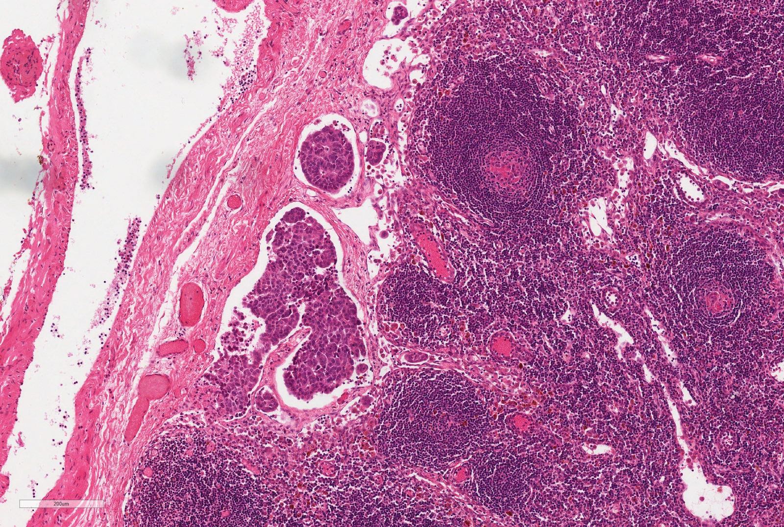
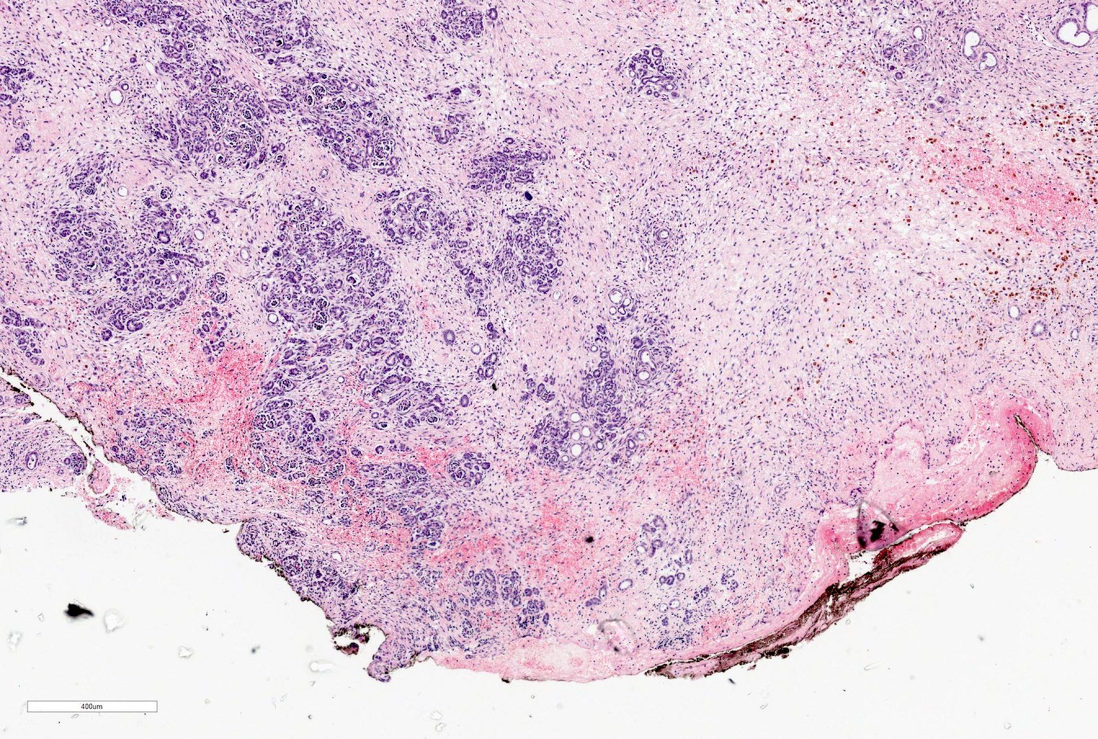
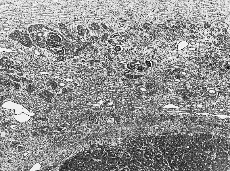
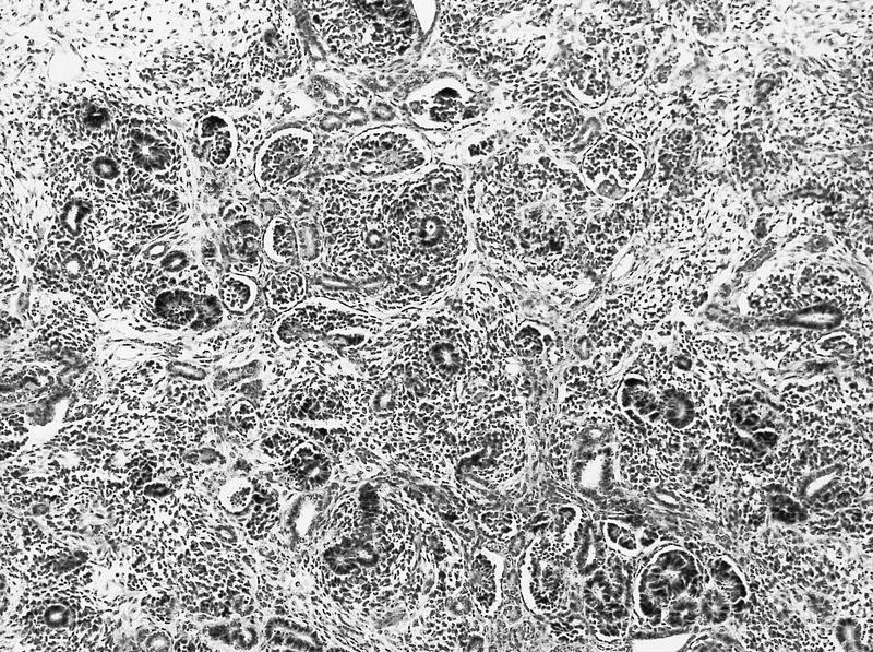
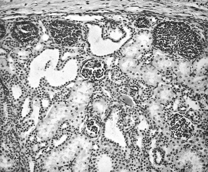
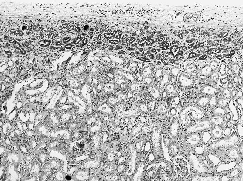
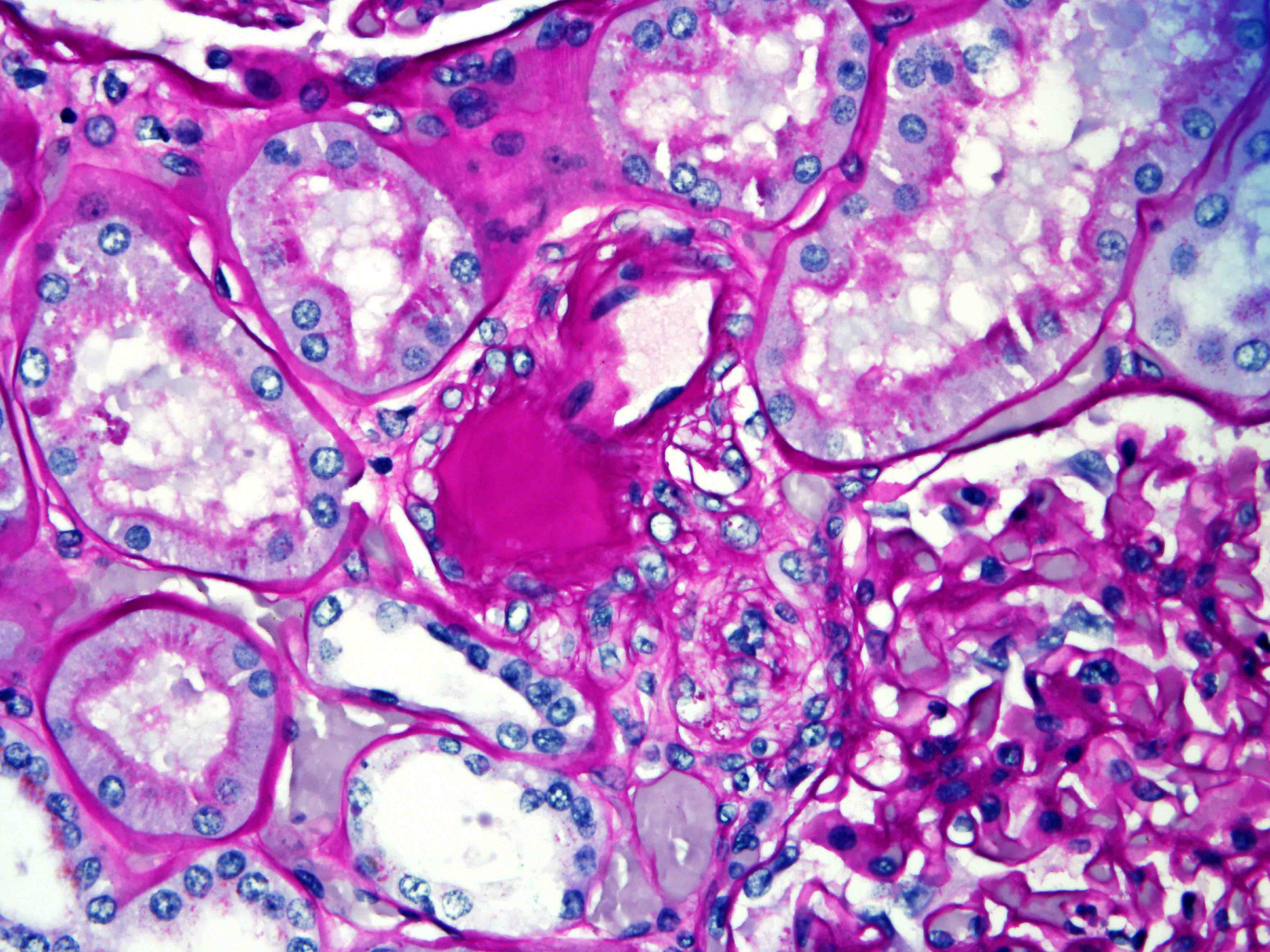
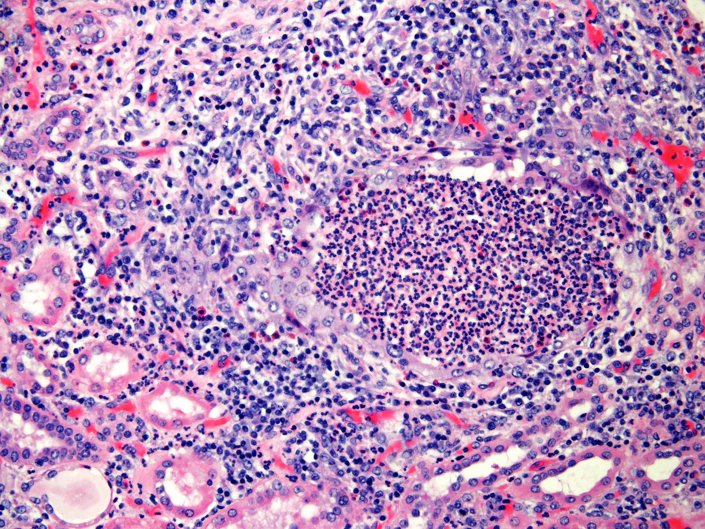
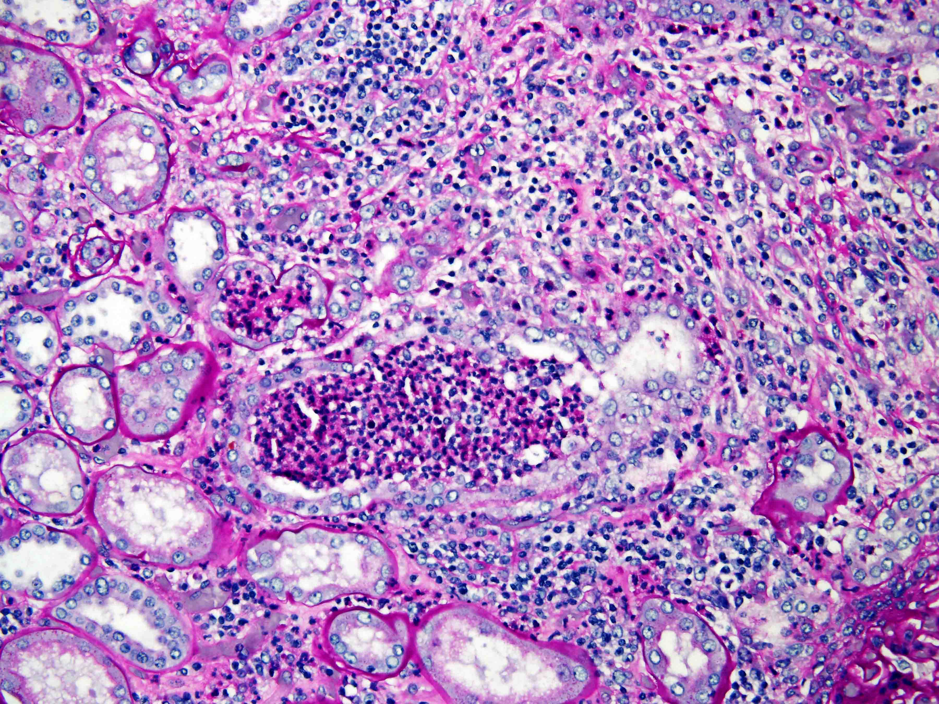
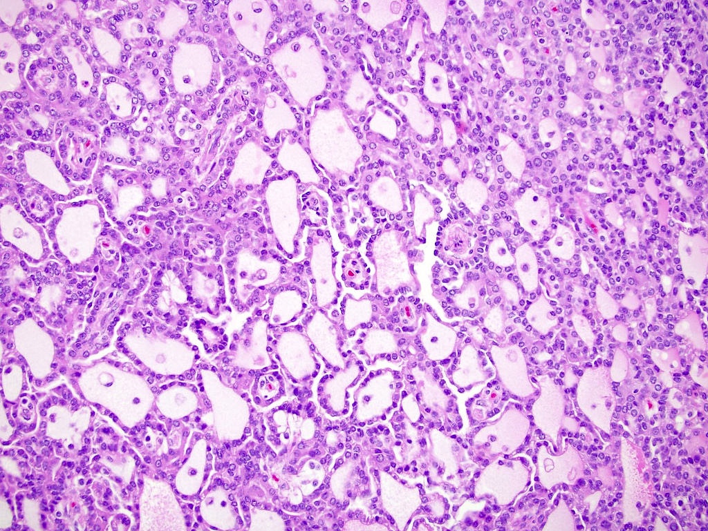
Acquired cystic kidney disease
Contributed by Gregory MacLennan, M.D. and @katcollmd on Twitter
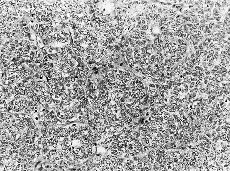
Overall architecture
Papillary architecture
Tubular architecture
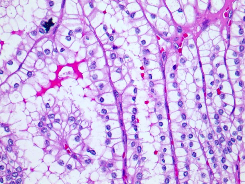
Low grade nuclear features
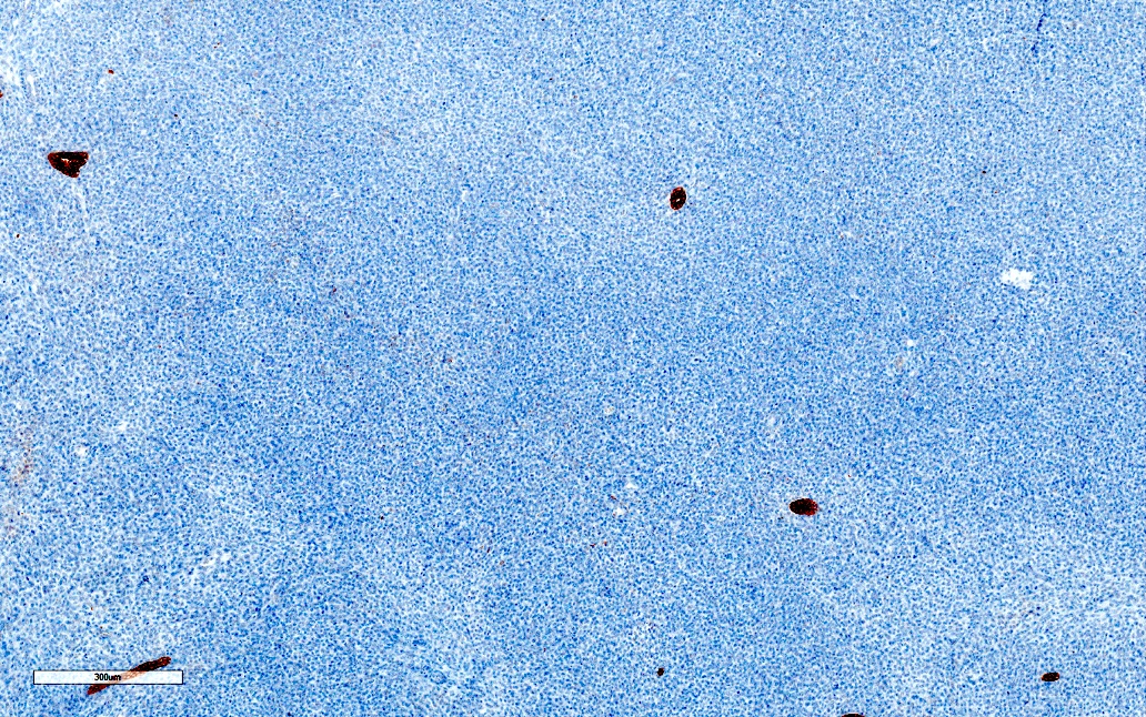
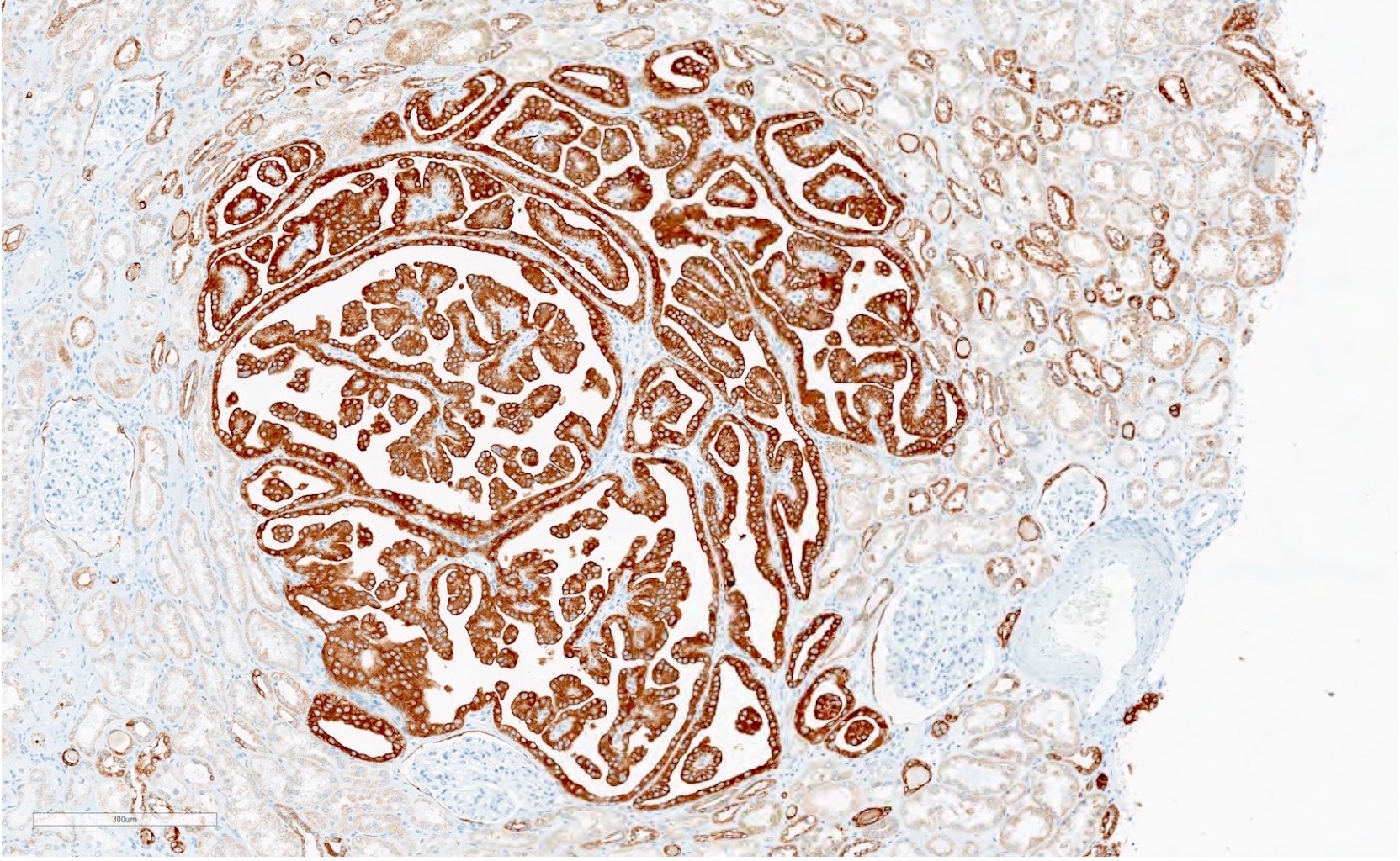
Clear cell papillary renal cell tumor
Clear cell papillary renal cell tumor
CAIX
CK7
CD10
AMACR
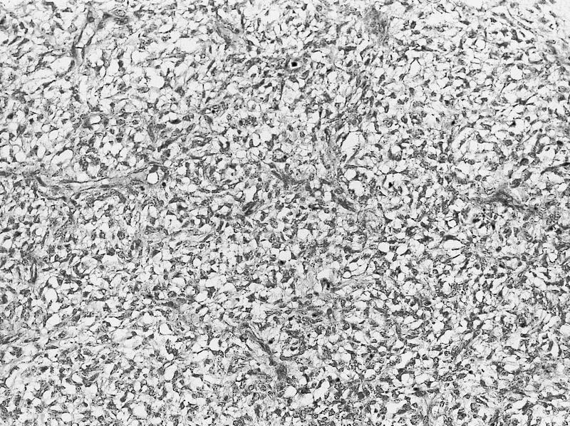
AFIP images

Age distribution of 156 cases
Images hosted on other servers:

Clear cell sarcoma

Large tan to tannish yellow tumor
Contributed by Maxwell Rollins, M.D.


Various images
AFIP images
3rd Series, Vol. 11
Images hosted on other servers:

Small oval cells

Clear and spindle cells, fibrovascular septas

Renal mass

Renal mass in a 19 month old boy

EGFR+
Contributed by Maxwell Rollins, M.D.
Touch prep
AFIP images
Fine needle aspiration
Images hosted on other servers:

Morphological patterns
Images hosted on other servers:

MRI, T2 and T1 weighted
Contributed by Daniel Anderson, M.D., M.B.A. and Debra L. Zynger, M.D.

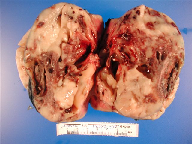
Involving kidney and pelvis
Partially necrotic
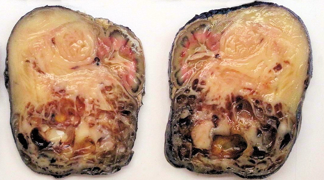
Large tumor
Images hosted on other servers:

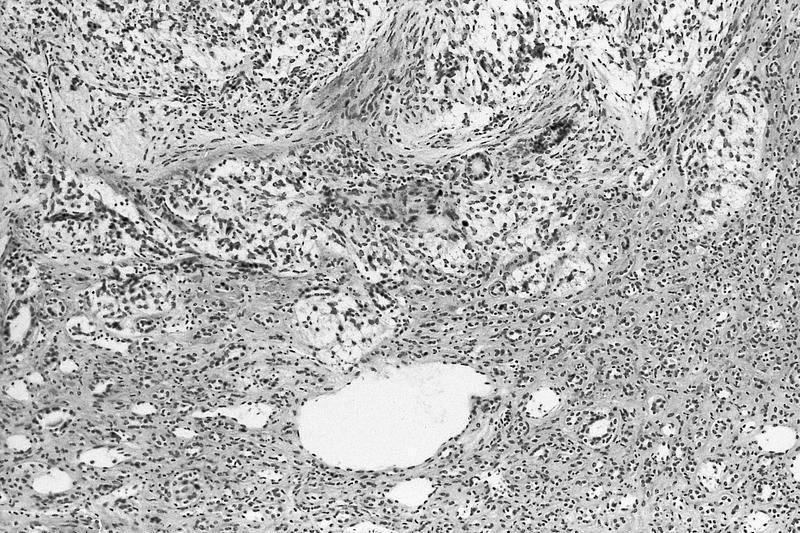
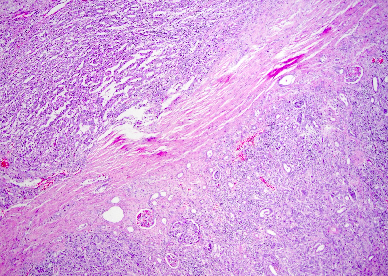
Infiltrative gray-white tumor

Tumor invades renal capsule and infiltrates adipose tissue
Contributed by Daniel Anderson, M.D., M.B.A. and Garrison F. Pease, M.D.
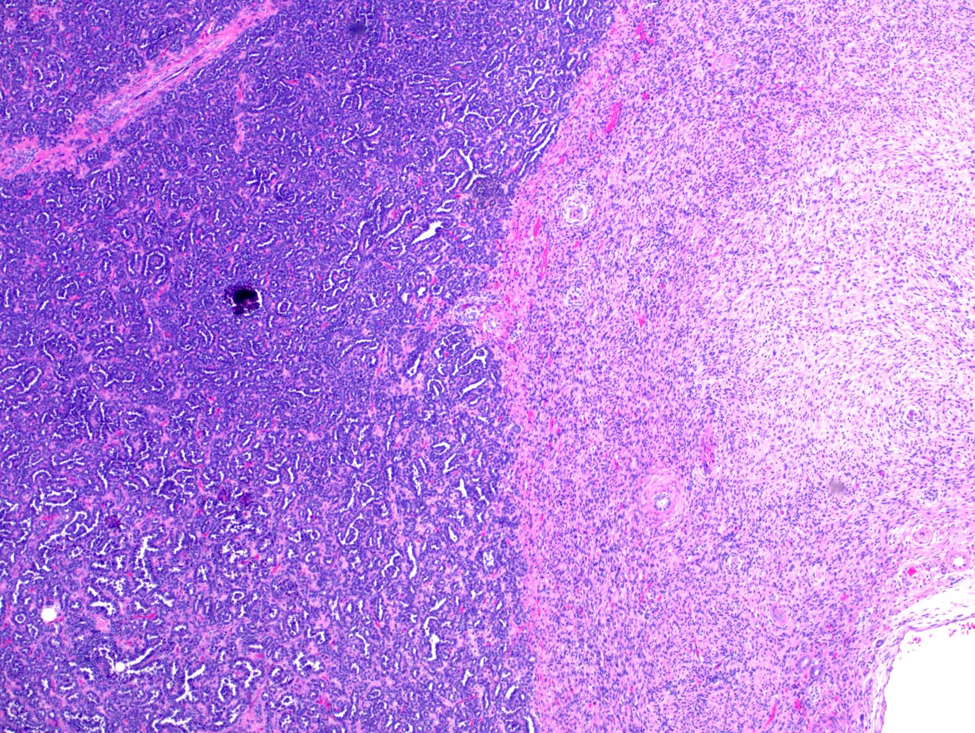
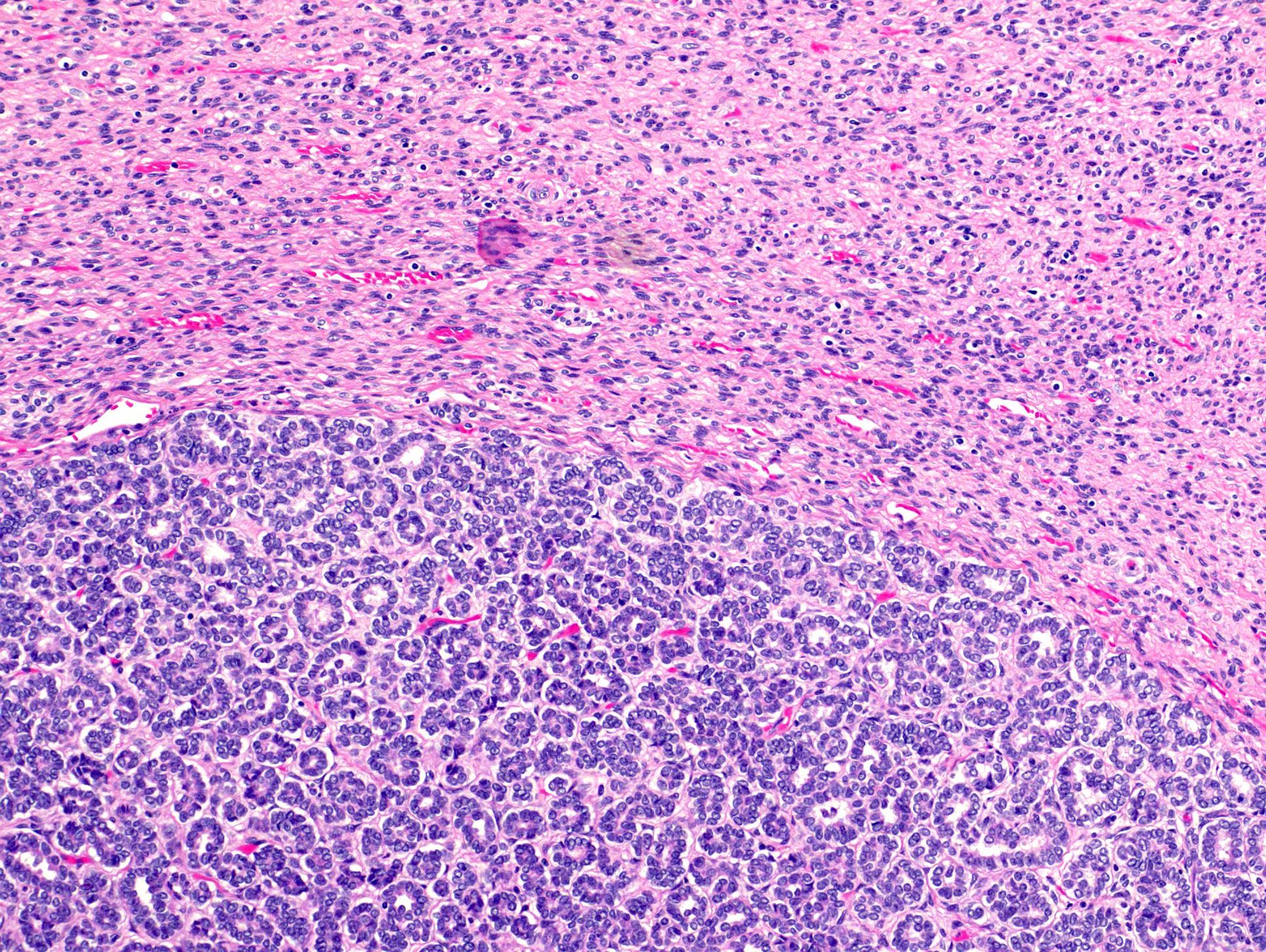
Adjacent renal parenchyma

Architecture
Infiltrative border
Prominent nucleoli
Tumor and nonneoplastic
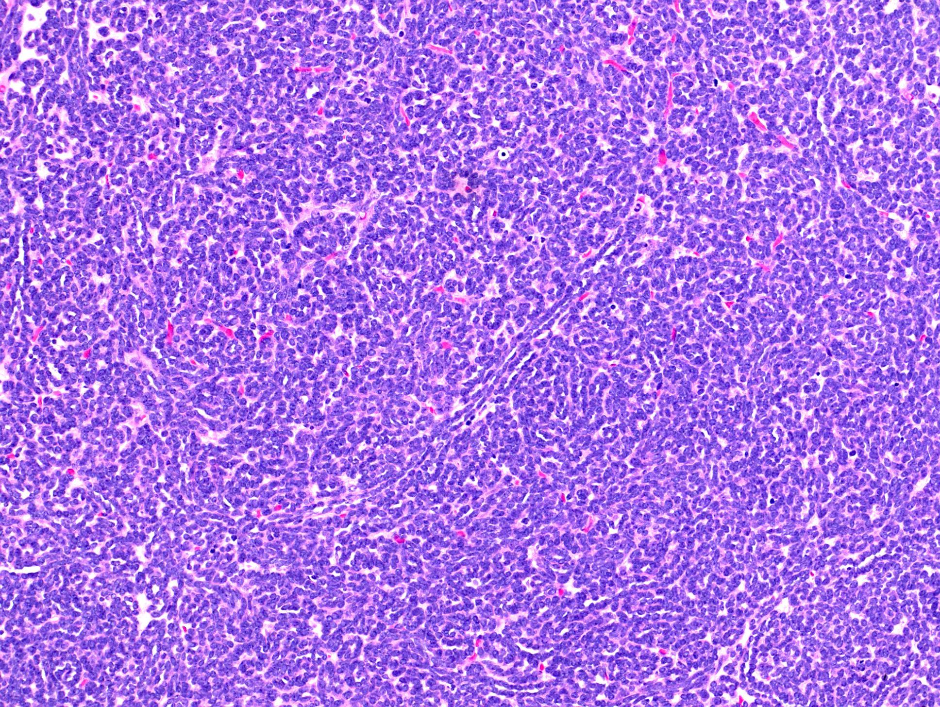
Tumor tubules and cords
Cellular details
Cellular and nuclear details
Border tumor and nonneoplastic
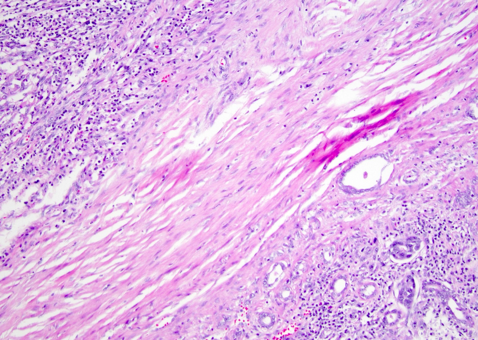
Possible dysplastic tubule epithelium
Collecting duct carcinoma
Images hosted on other servers:

Large renal mass

Upper pole mass

CT large mass
Contributed by Ellen D’Hooghe, M.D., Gordan M. Vujanic, M.D., Ph.D. and Case #57

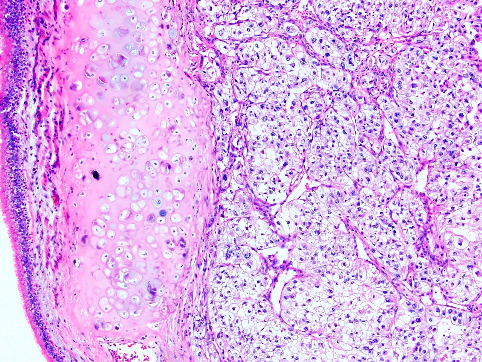
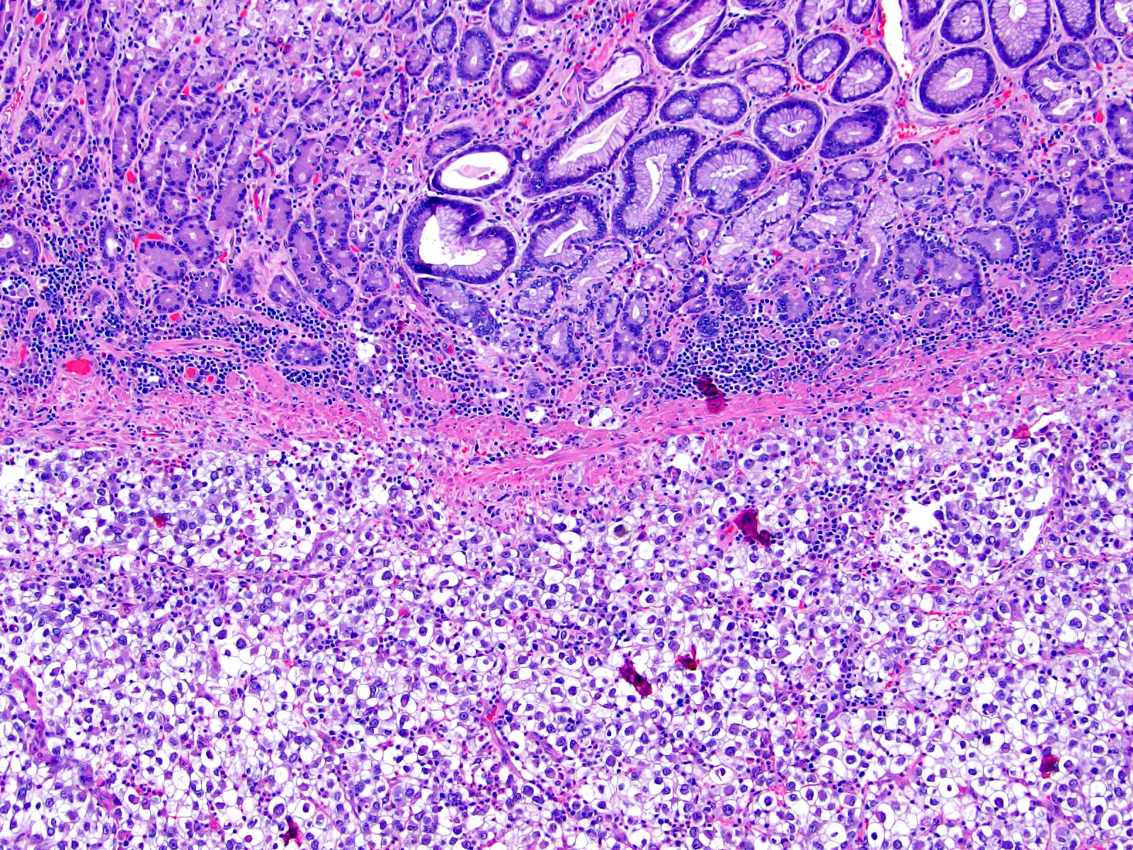
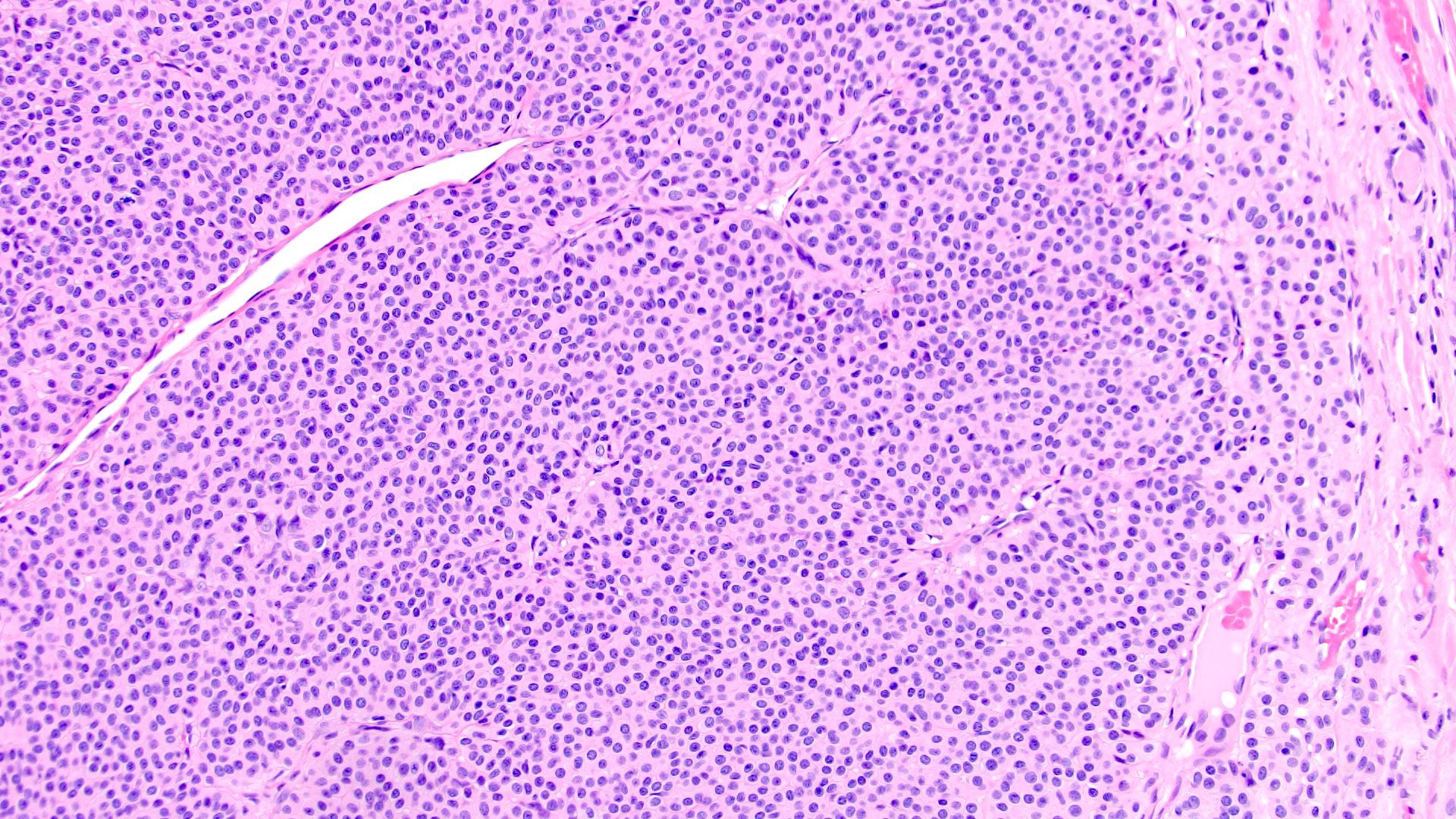
Classic type
Mixed type
Cellular type
Contributed by Ellen D’Hooghe, M.D. and Gordan M. Vujanic, M.D., Ph.D.
Classic type
Classic type vessels
Entrapped normal renal parenchyma

Extramedullary hematopoiesis
Islands of cartilage
Cellular type

Cellular type
Single entrapped tubule
Tumor-kidney interface
Mixed type

Sinus invasion
Perirenal fat invasion
Images hosted on other servers:

Ultrasonography of multilocular cystic mass

MRI of multilocular cystic mass

Abdominal CT with bilateral CPDN
Contributed by Americo Brilhante, M.D. and Daniel Athanazio, M.D., Ph.D.
Multilocular cyst of the kidney
Contributed by Americo Brilhante, M.D. and Daniel Athanazio, M.D., Ph.D.
Multilocular cystic tumor
Nephroblastomatous elements
Transition between tumor and renal cortex
Fibrous septa and epithelial lining
Small tubules with hobnail morphology
Abortive / immature tubules
Abortive / immature tubules
Primitive blastema and tubules
AE1 / AE3
WT1
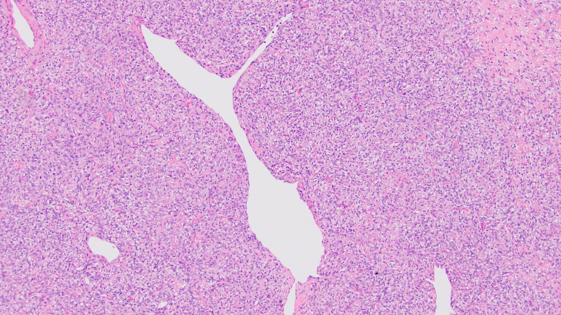
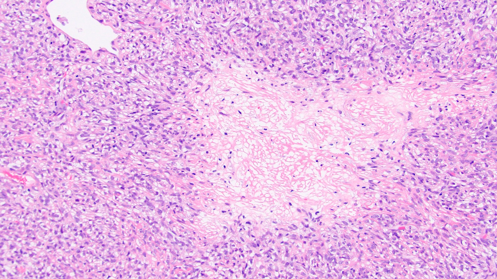
Contributed by Maria Tretiakova, M.D., Ph.D.
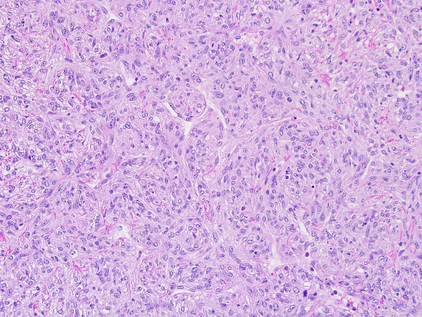
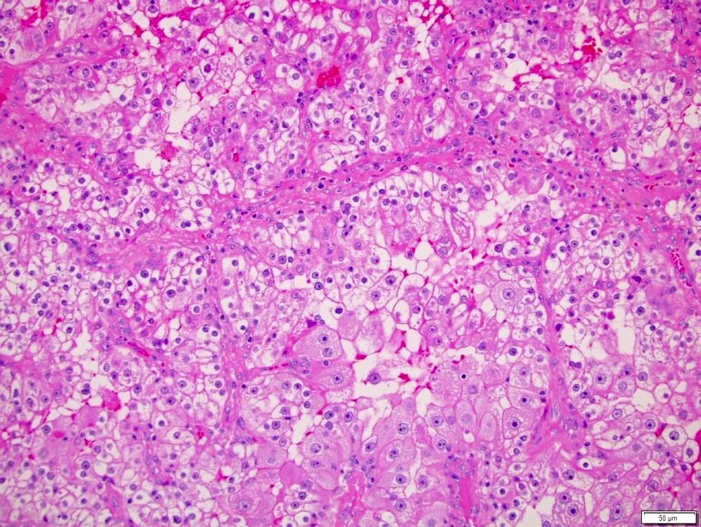
TCEB1 mutated RCC
CAIX
CK7
CD10
Clear cell RCC with TCEB1-like features
Images hosted on other servers:

TCEB1 mutations and HIF accumulation

TCEB1 mutated RCC with monosomy 8
Images hosted on other servers:

Circumscribed,
solid tumor with
gray-tan to brown
cut surface
Contributed by Maria Tretiakova, M.D., Ph.D.
Vessels at periphery
Solid nested growth pattern
Entrapped tubules
Clear vacuolated to granular cytoplasm
High grade nuclei
KIT
Cathepsin K
CD10
CK7
Vimentin
Images hosted on other servers:

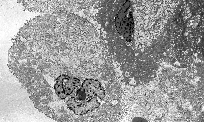
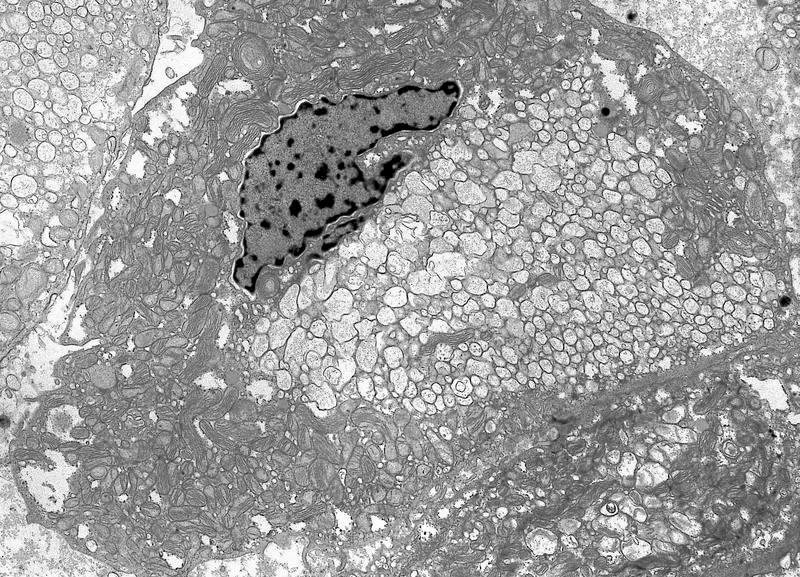
Dilated cisterns of RER and numerous mitochondria
Images hosted on other servers:

mTOR mediated pathways
Contributed by Doreen Palsgrove, M.D.
CT abdomen and pelvis
Images hosted on other servers:


CT and MRI with gross specimen
Images hosted on other servers:

Nephrectomy
Contributed by Doreen Palsgrove, M.D. and @katcollmd on Twitter
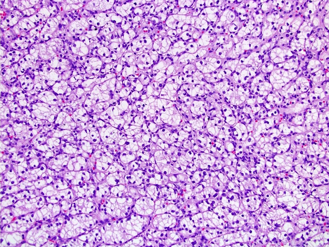
Solid growth
Abundant cytoplasm
Basophilic cytoplasmic inclusions
CK20


Eosinophilic, solid and cystic renal cell carcinoma
Images hosted on other servers:

Copy number alterations
Images hosted on other servers:

Potential pathogenesis
Images hosted on other servers:

CT, MRI

Case with TSC

Large renal mass

Heterogeneous mass
Contributed by Judy Sarungbam, M.D.
Circumscribed, pink-tan mass
Images hosted on other servers:

Circumscribed, lobulated cut surface
Contributed by Anil Parwani, M.D., Ph.D., M.B.A.

Diffuse growth
Cytologic and nuclear features

Cytologic features

Carcinoma-like growth pattern

Cytologic features
MelanA
HMB45
Images hosted on other servers:

Tumor cells with premelanosomes
Images hosted on other servers:

Molecular analysis of TFE3 gene

TSC2 mutation
Images hosted on other servers:

Proposed
mechanisms of
tumorigenesis
in HLRCC
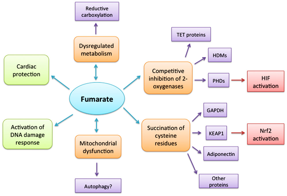
Consequences
of elevated
cellular
fumarate

TCA cycle enzyme
Contributed by Debra Zynger, M.D., Nicole K. Andeen, M.D. and Maria Tretiakova, M.D., Ph.D.

FH deficient RCC

FH
CAIX

PAX8
HLRCC with
tubulopapillary
growth and
eosinophilic cells

HLRCC with pale
eosinophilic cytoplasm
and inclusion-like
nucleoli

Uterine leiomyoma
from a patient
with HLRCC, with
multinucleation
Images hosted on other servers:


Findings in patient with HLRCC and RCC


HLRCC renal tumors

Clear cell RCC

Uterine leiomyomas in HLRCC

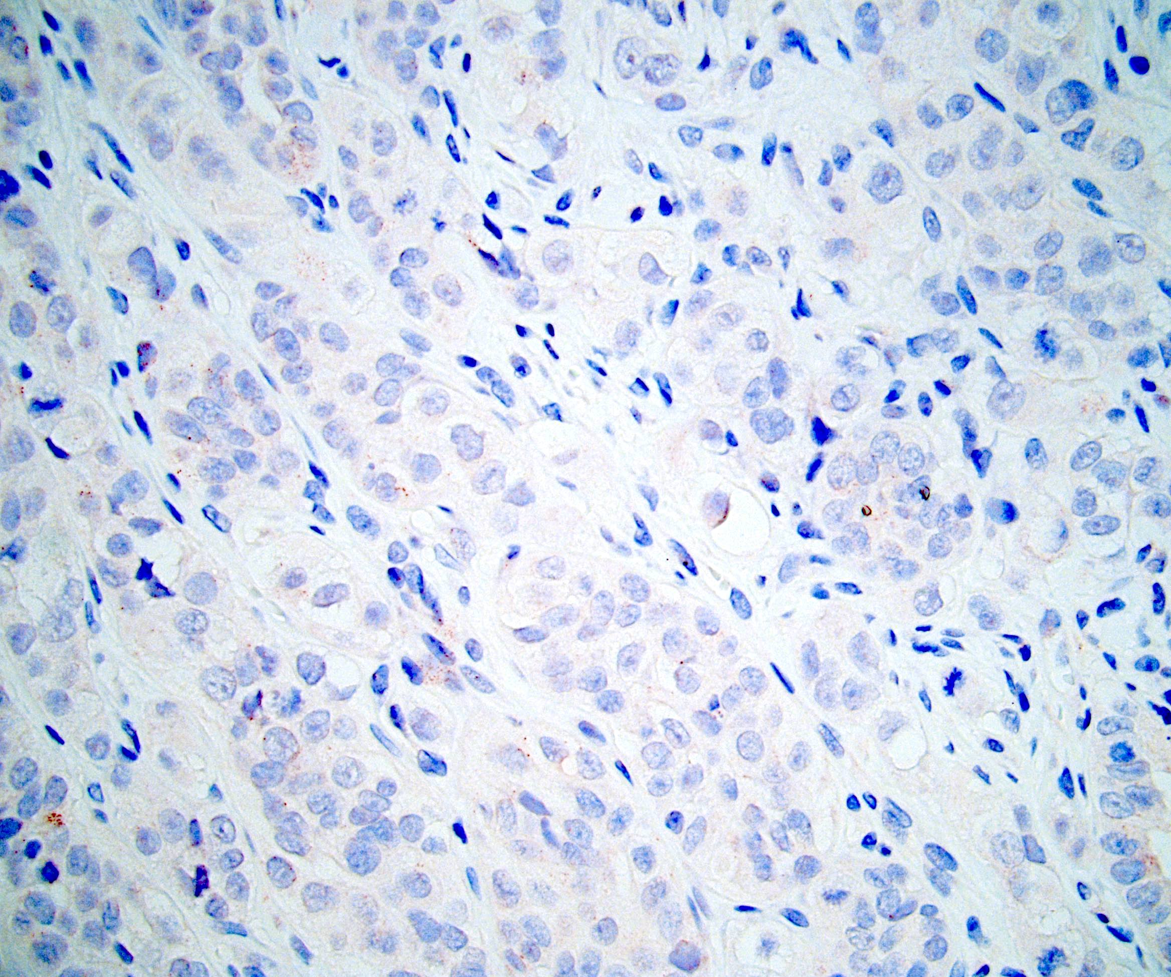
2SC IHC shows
diffuse and strong
reactivity in HLRCC
renal cancer

2SC IHC shows
cytoplasmic staining
pattern in a few
unclassified RCC
Contributed by Maria Tretiakova, M.D., Ph.D.
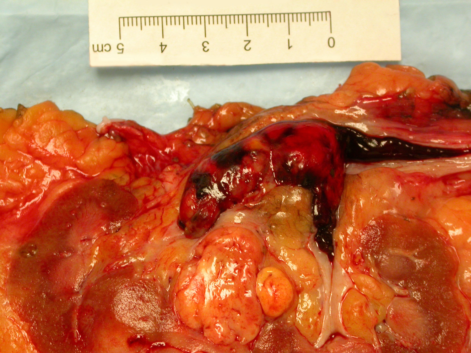
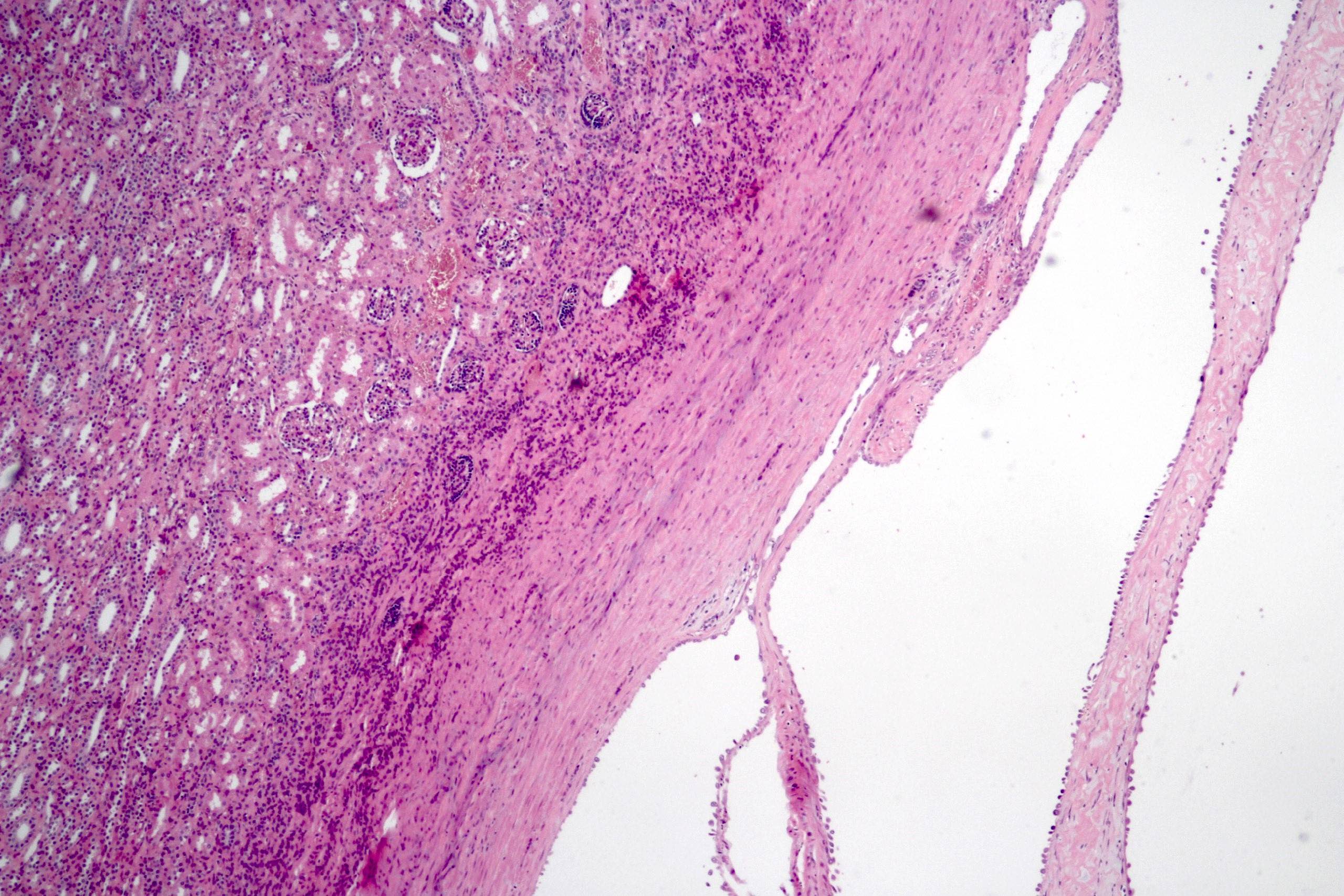
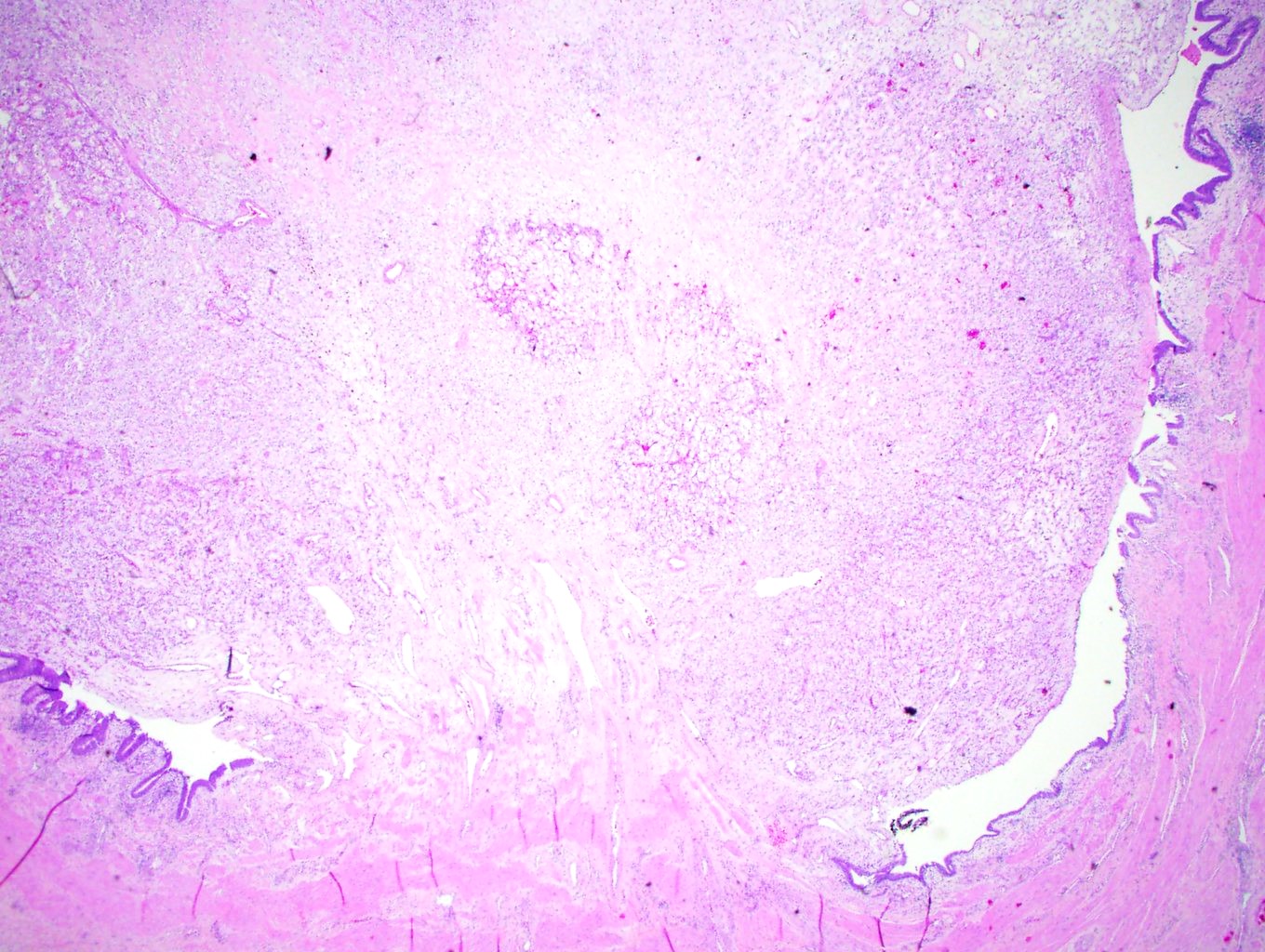
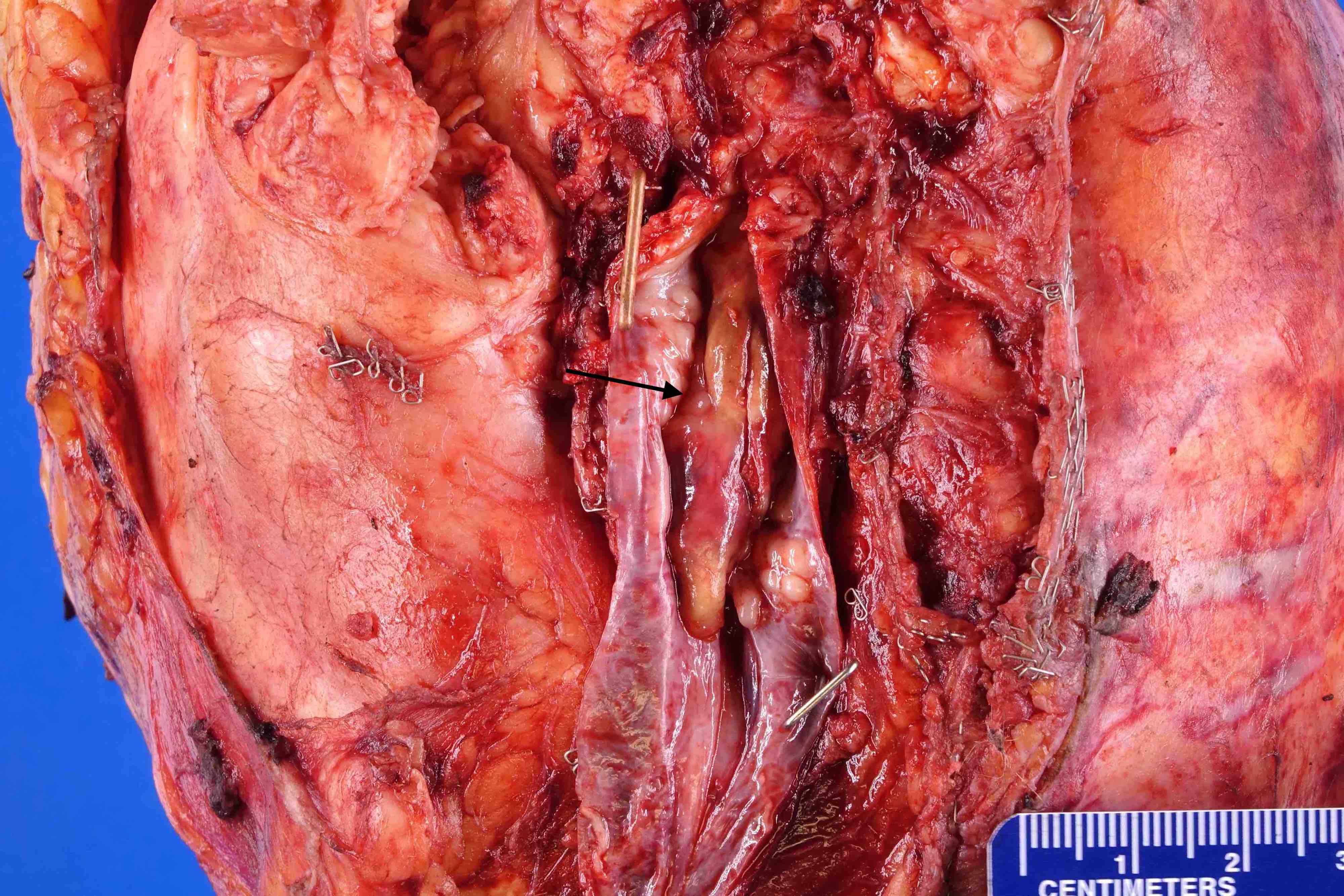
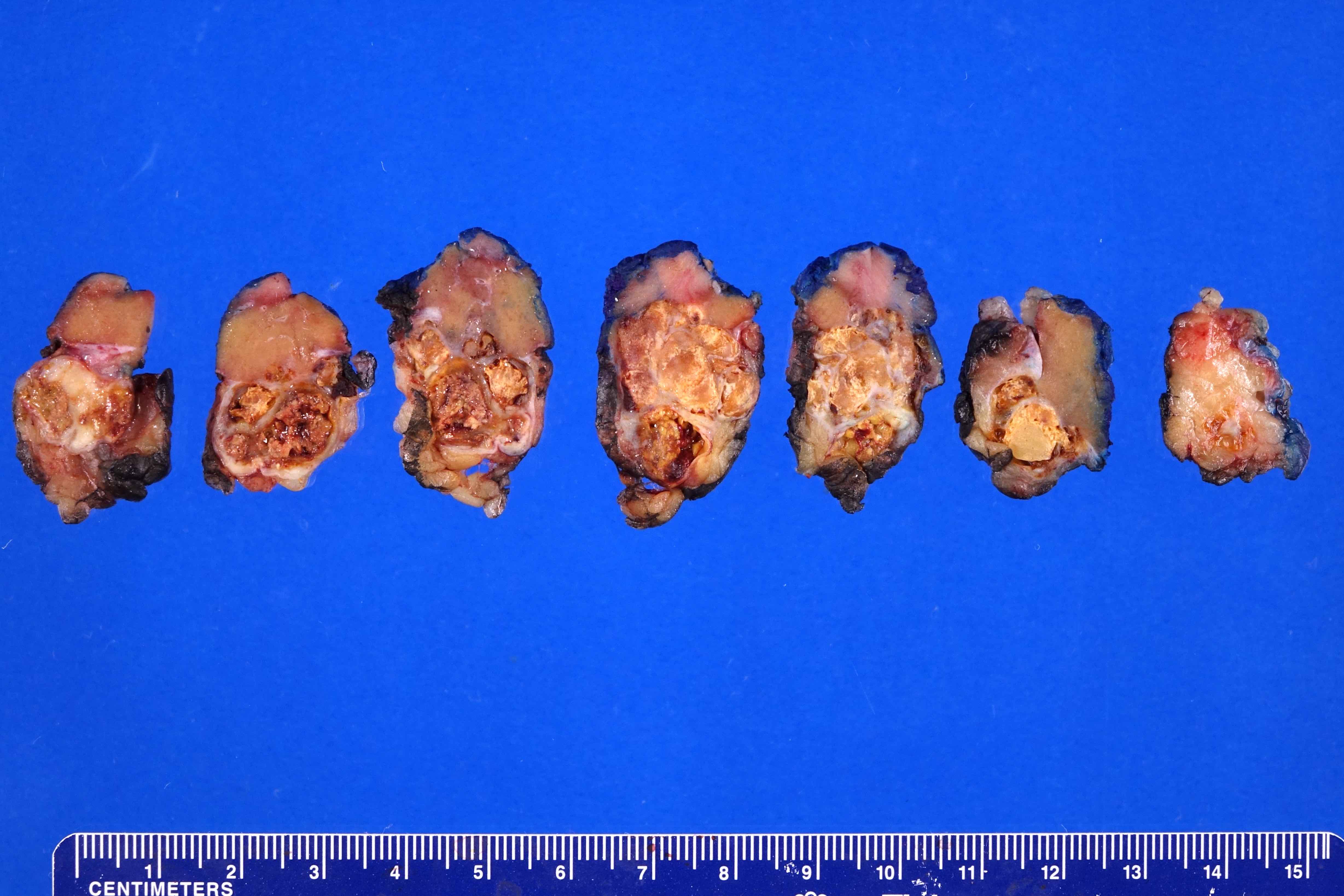
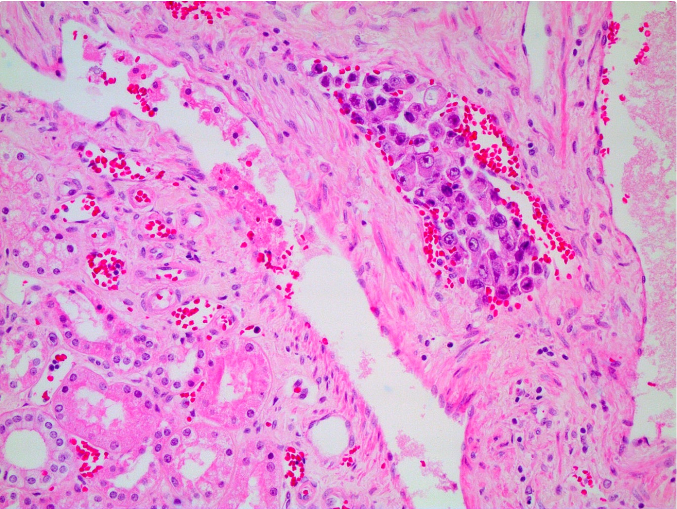
Renal sinus invasion (pT3)
Contributed by Maria Tretiakova, M.D., Ph.D.
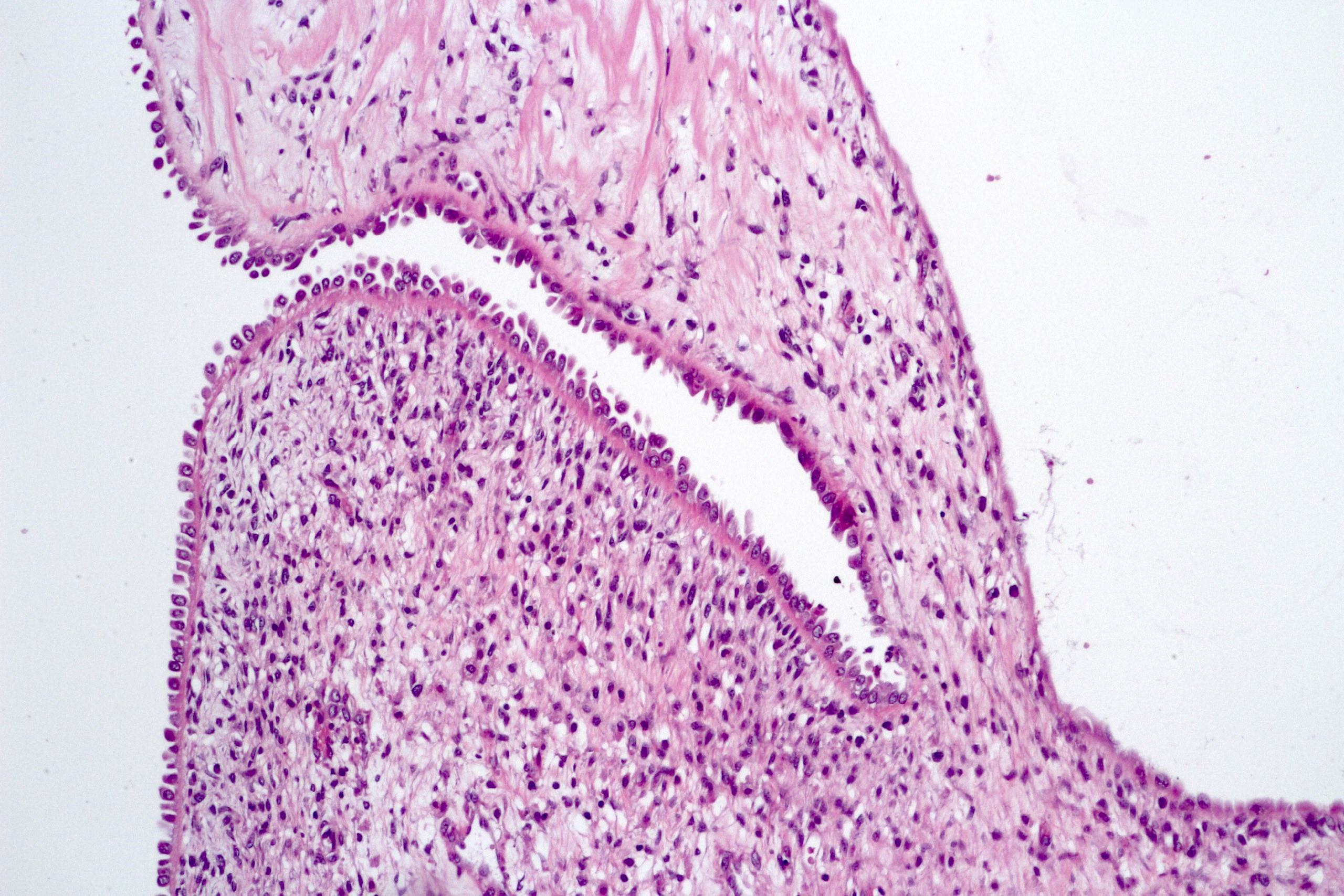
Positive margin, invasion of ureter
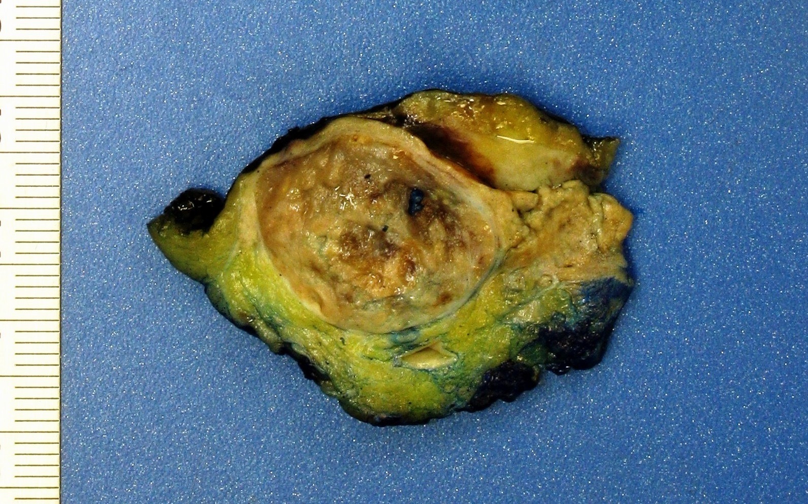
Coagulative tumor necrosis
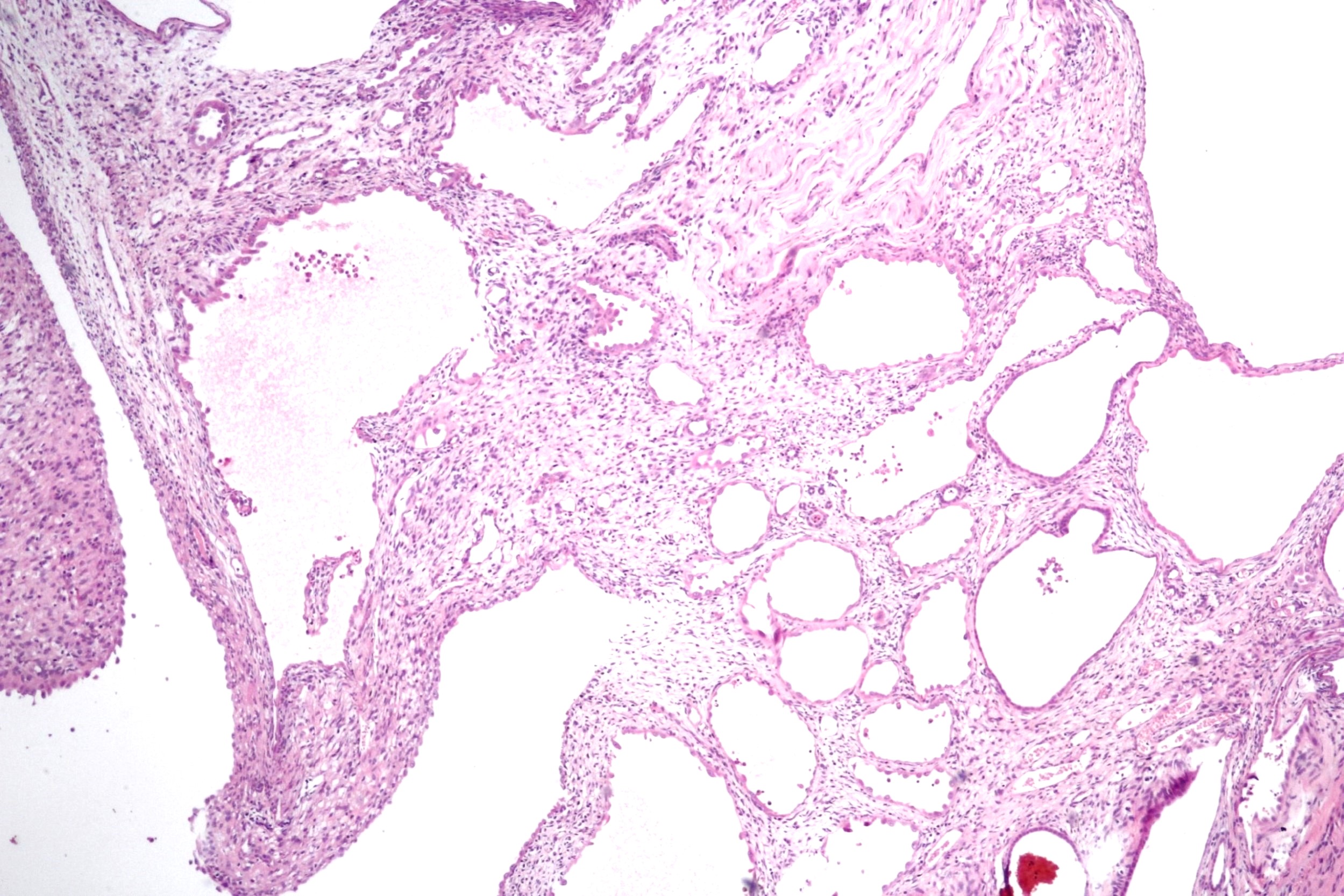
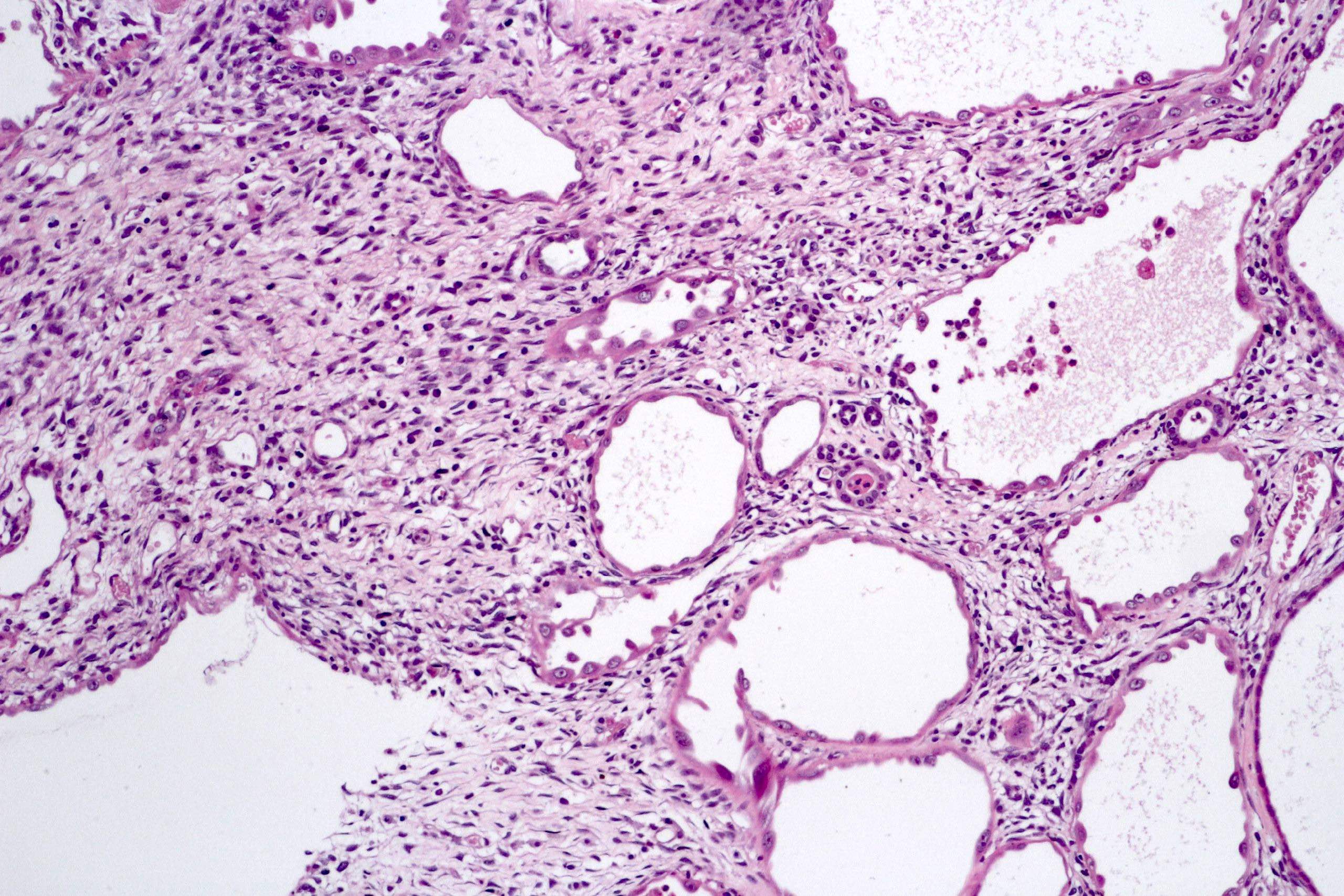
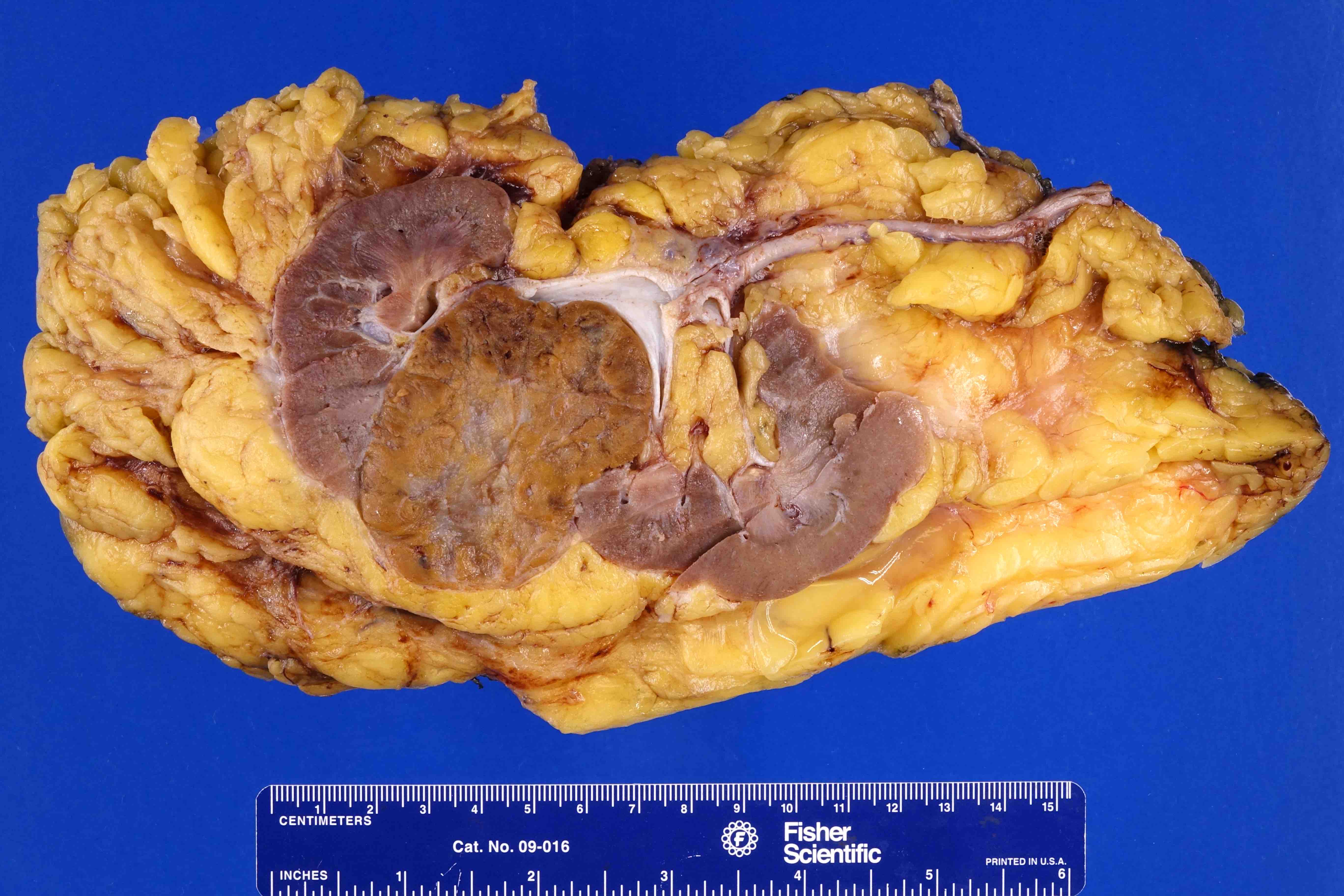
RCC in fat (pT3)
RCC in fat, microscopic invasion
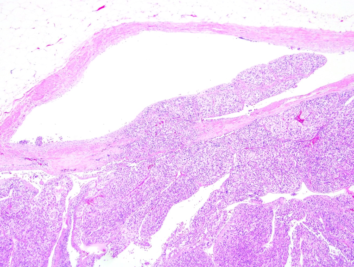
RCC involving a vein (pT3)
RCC involving renal vein margin (pT3)
RCC metastasis to bronchus (pM)
RCC metastasis to stomach (pM)
Images hosted on other servers:

Heterogeneous
enhancing mass;
unevenly increased
FDG uptake

Well defined heterogenous lesion

Bilateral inhomogeneous lesions
Images hosted on other servers:

Well defined pale mass

Myxoid areas
Contributed by Maria Tretiakova, M.D., Ph.D.
Capsule and nests
Small uniform tumor cells
Abundant vasculature
Rare mitotic figure

Positive SMA
Positive collagen IV
Negative S100
Contributed by Debra L. Zynger, M.D.
Basal lamina
Images hosted on other servers:

Thin microfilament, dense bodies
Contributed by Maria Tretiakova, M.D., Ph.D.
Small, dark nuclei
Nucleoli are barely visible
Nucleoli are easily seen
Large pleomorphic nuclei
Sarcomatoid differentiation
Anaplastic giant and rhabdoid cells
Anaplastic giant cells

Nonanaplastic giant cells

Dual low and high grade RCC
Contributed by Debra Zynger, M.D.

RCC, clear cell type
RCC, papillary type 2
RCC, clear cell papillary type
RCC, chromophobe type

Oncocytoma
Images hosted on other servers:

Left kidney mass
Images hosted on other servers:

Well demarcated tumor
Contributed by Maria Tretiakova, M.D., Ph.D.
Lipoblast-like cells
Nuclear pleomorphism
Capillary network
Spindle tumor cells
Inhibin positive
CAIX positive
Images hosted on other servers:

Kidney ultrasound

Noncontrast CT

Contrast CT
Contributed by Maria Tretiakova, M.D., Ph.D. and AFIP images

Cavernous hemangioma
Hypocellular AH
Anastomosing hemangioma
Sinusoidal anastomosing pattern
Fibrin thrombi (AH)
Extramedullary hematopoiesis
CD31 (AH)

Renal hemangioma
Images hosted on other servers:

Heterogenous pattern

Stellate central hypodensity

2 abutting tumors

Preoperative CT
Contributed by Bonnie Choy, M.D.
Multiple tan-brown tumors
Contributed by Chin-Lee Wu, Ph.D. and Maria Tretiakova, M.D., Ph.D.

Dual population of eosinophilic cells
BHD related hybrid tumor
Contributed by Roula Albadine, M.D. and Julie Guilmette, M.D.
Oncocytic tumor cells

Oncocytic cells with granular eosinophilic cytoplasm
Hale colloidal iron
Contributed by Bonnie Choy, M.D.
Touch preparation
Images hosted on other servers:

CT


MRI
Images hosted on other servers:

Well circumscribed, solid cut surface

Encapsulated

Hemorrhage and hemosiderin deposition
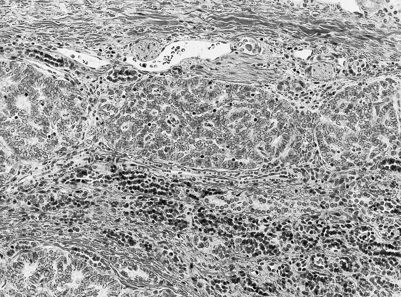
Contributed by Sean Williamson, M.D. and Matthew Wasco, M.D. (Case #467)
Glomoid appearance
Papillary architecture

Prominent vasculature

Polygonal to spindle cells
Contributed by Alcino Pires Gama, M.D.
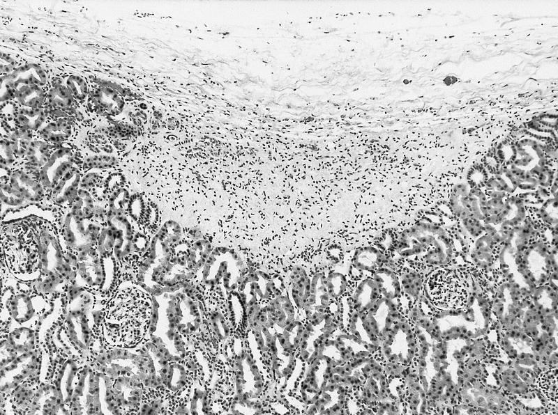
Normal juxtaglomerular apparatus
Contributed by Sean Williamson, M.D. and Matthew Wasco, M.D. (Case #467)

Rhomboid shaped renin protogranules
Images hosted on other servers:


Ultrasound and CT

CT

CT

CT
Images hosted on other servers:

Solid, tan-white mass

Whorled, bulging cut surface
Contributed by Alcino Pires Gama, M.D. and Bonnie Choy, M.D.
Intersecting fascicles
Bland spindle cells
h-caldesmon
SMA
Contributed by Garrison Pease, M.D.


Well circumscribed tumor
Contributed by Garrison Pease, M.D. and Maria Tretiakova, M.D., Ph.D.


Solid with boats in a bay pattern

Boats in a bay and low grade cytomorphology

Solid pattern and low grade cytomorphology


Solid with boats in a bay pattern


Solid with boats in a bay pattern


Solid with boats in a bay pattern

Solid compact component

CD117 / KIT negative


CK7 positive expression

CK7 positive expression

CD117 / KIT negative expression

GATA3 positive expression

Vimentin negative

Cathepsin K negative
Images hosted on other servers:

Homogenous solid mass, no calcification or necrosis
Images hosted on other servers:

Largely solid, tan homogenous renal mass

Fibrous, white grey cut surface

White solid and cystic cut surface
Contributed by Rose Chami, M.D.
Biphasic tumor with epithelial and stromal components
Highly cellular tumor
Tubular and glomeruloid structures
Psammoma bodies
Cellular solid appearance
Spindle cell component
AE1/AE3
WT1, both components
WT1, epithelial component
WT1, stromal component
CD34, both components
BRAF V600E, epithelial component
Images hosted on other servers:


CT scan
Contributed by Debra L. Zynger, M.D.
Partial nephrectomy

Radical nephrectomy
Contributed by Tatjana Antic, M.D. and @katcollmd on Twitter
Well circumscribed
Scar
Calcifications
Packed tubules
Loose stroma

Mitotic figure
Pseudopapillary
Pseudopapillary and glomeruloid
Metanephric adenoma
Metanephric adenoma
BRAF
WT1
Contributed by Ellen D’Hooghe, M.D. and Gordan M. Vujanic, M.D., Ph.D.
Solid and cystic tumor with hemorrhage
Contributed by Ellen D’Hooghe, M.D. and Gordan M. Vujanic, M.D., Ph.D.
Nodularity
Alternating cellularity
"Onion skinning"
Cysts
Intratubular growth
Angiodysplasia
Tumor - kidney interface
CD34
Images hosted on other servers:

Metastatic oral squamous cell carcinoma

Metastatic phyllodes tumor
Contributed by Debra Zynger, M.D.
Lung squamous cell carcinoma
Images hosted on other servers:

Myeloma

Small cell lung cancer

Solitary fibrous tumor
Contributed by Maria Tretiakova, M.D., Ph.D., Nicole K. Andeen, M.D.

Breast carcinoma
Gastric carcinoma

Well differentiated neuroendocrine tumor
Images hosted on other servers:

MRI
Contributed by Bonnie Choy, M.D.
Solitary, well circumscribed
Mixed solid and cystic
Contributed by Bonnie Choy, M.D. and @katcollmd on Twitter
Predominantly cystic area
Predominantly solid area
Hypocellular stroma
Hypercellular stroma
Cuboidal epithelium

Hobnailed epithelium


Mixed epithelial and stromal tumor
ER
PR
CD10
PAX8
Images hosted on other servers:

Isodense mass in the renal medulla

Hyperintensity on T2 weighted image
Images hosted on other servers:

Cut surface with focal hemorrhage
Contributed by J. Cody Craig, M.D., Aida Valencia, M.D., Jennifer B. Gordetsky, M.D. and @katcollmd on Twitter
Bland tubules and spindle cells
Whorled tubules
Bland cuboidal and spindle cells
Compact tubules
Elongated tubules
Bland spindle cells
Mucinous tubular and spindle cell carcinoma
PAX8
CK7
p504s / AMACR
Images hosted on other servers:

Most common genetic alterations

SNP array analysis
Images hosted on other servers:

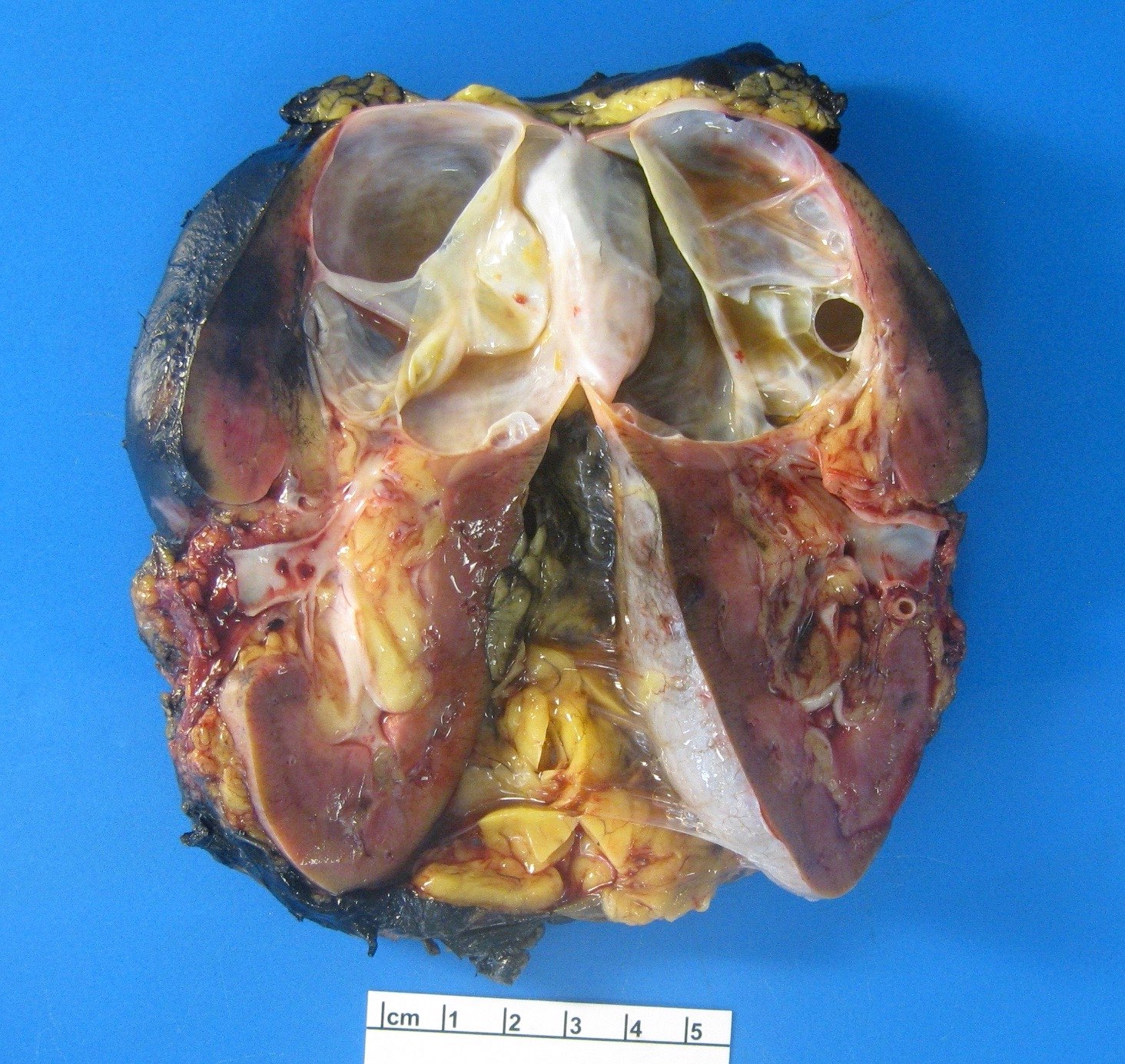
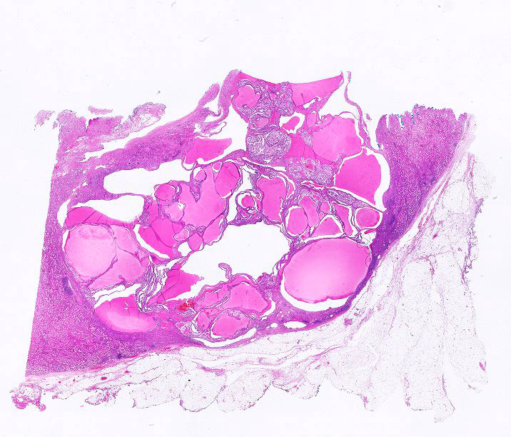
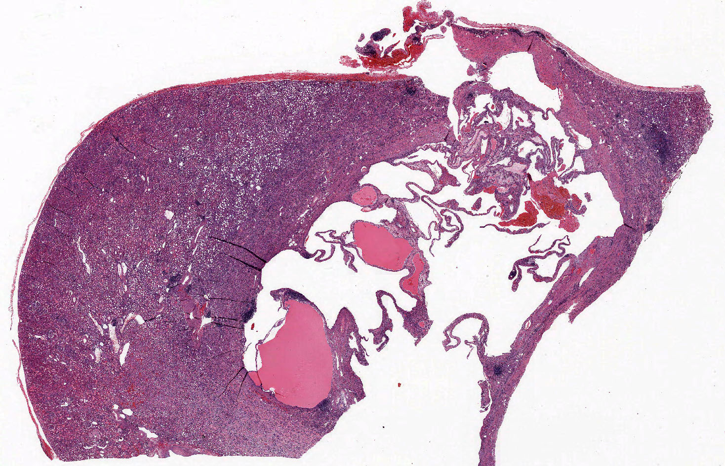
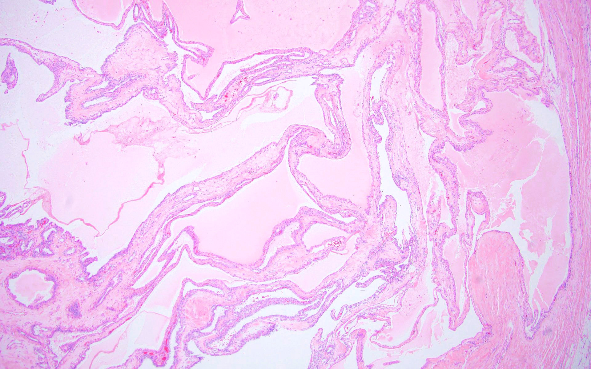
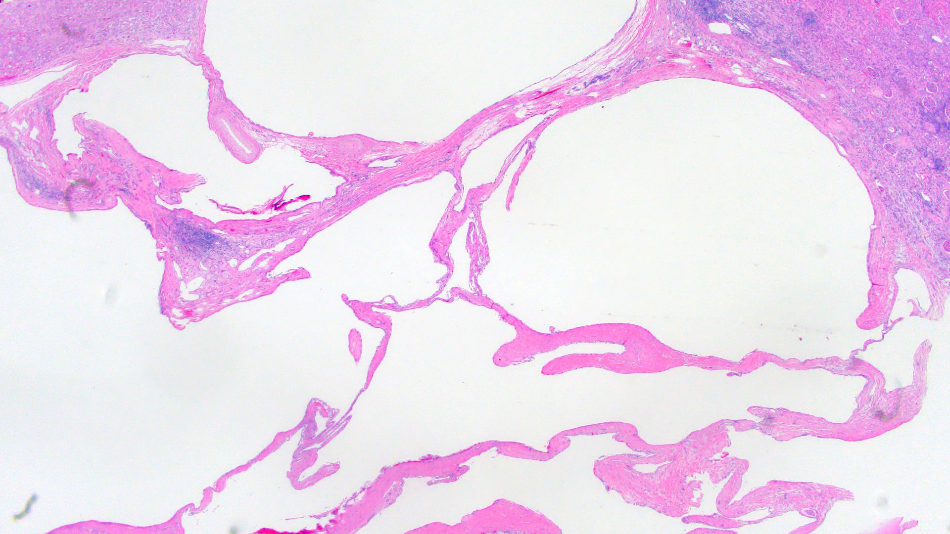
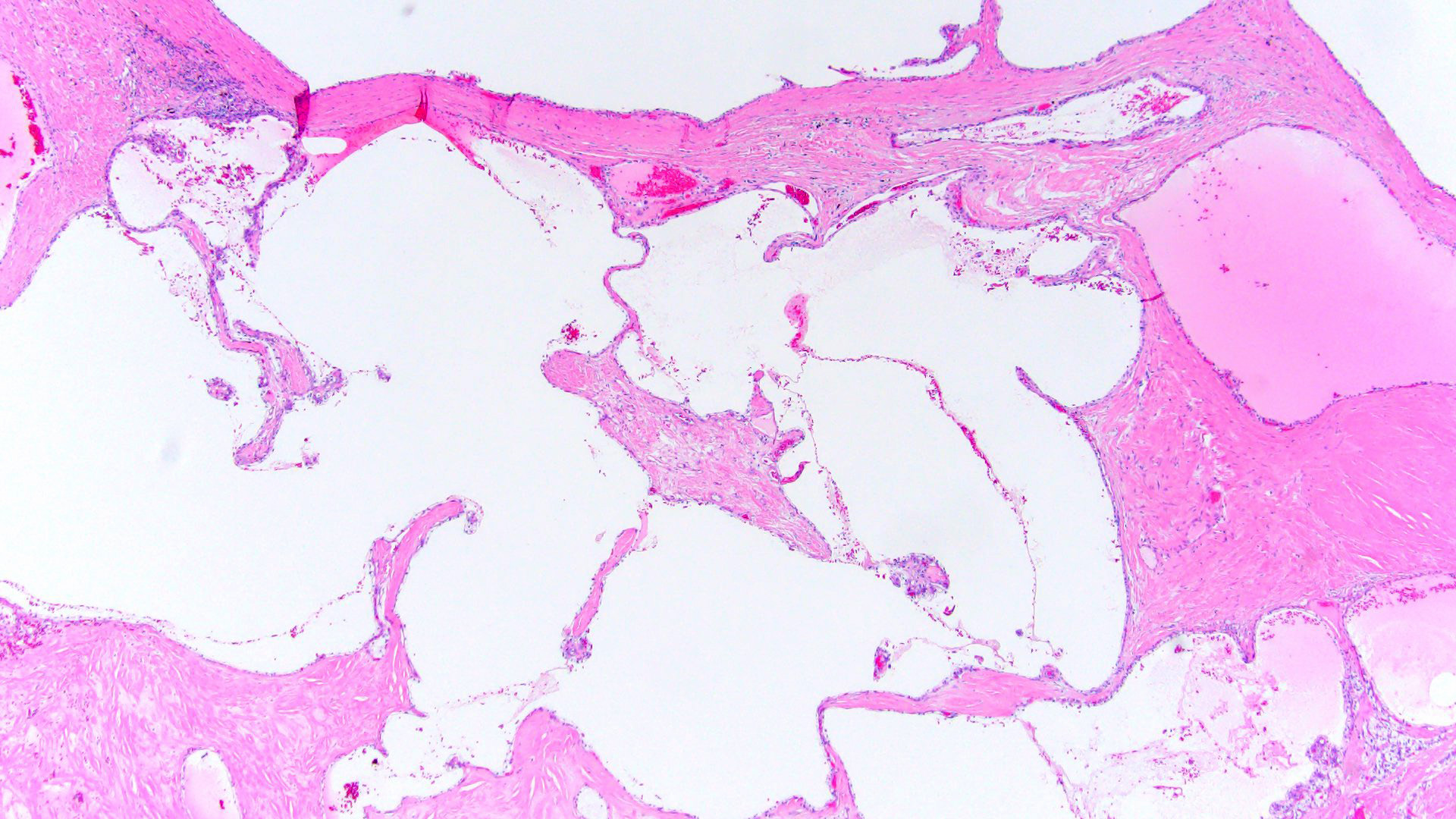
Pure multilocular cystic mass
Contributed by Maria Tretiakova, M.D., Ph.D.
Cortical multilocular cystic tumor
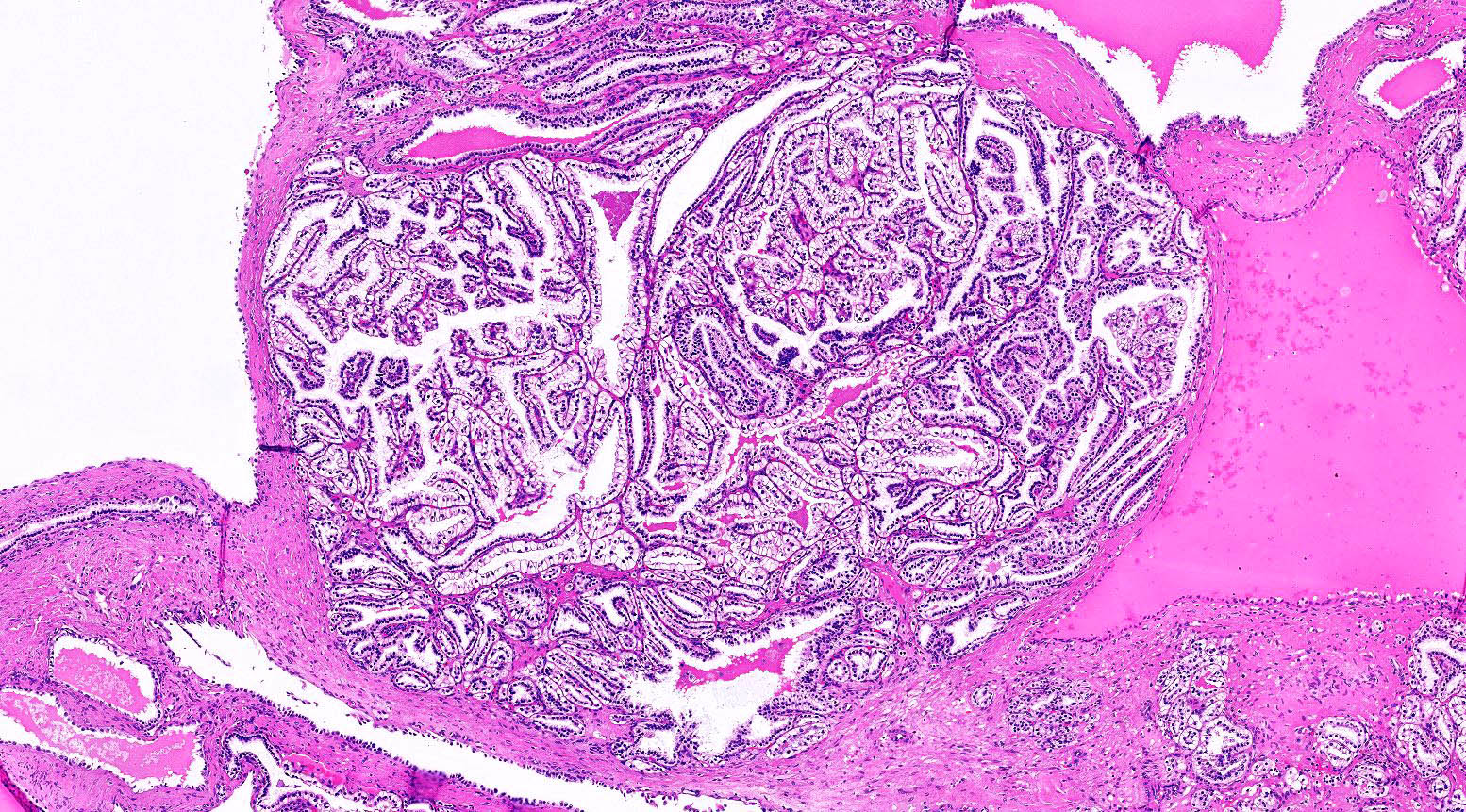
Irregular tumor pushing into renal hilum
Contributed by Maria Tretiakova, M.D., Ph.D. and @ahmsab_MD on Twitter
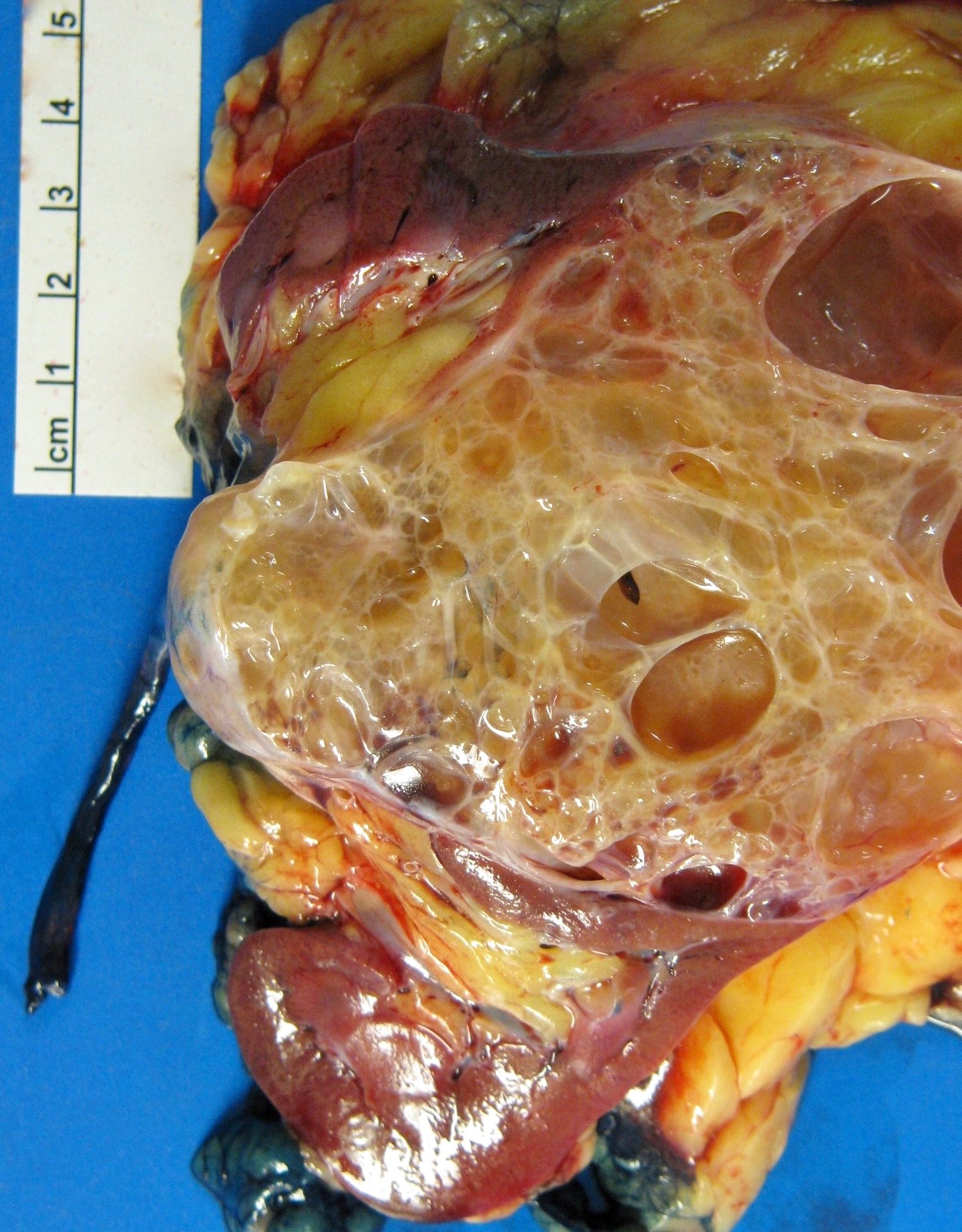
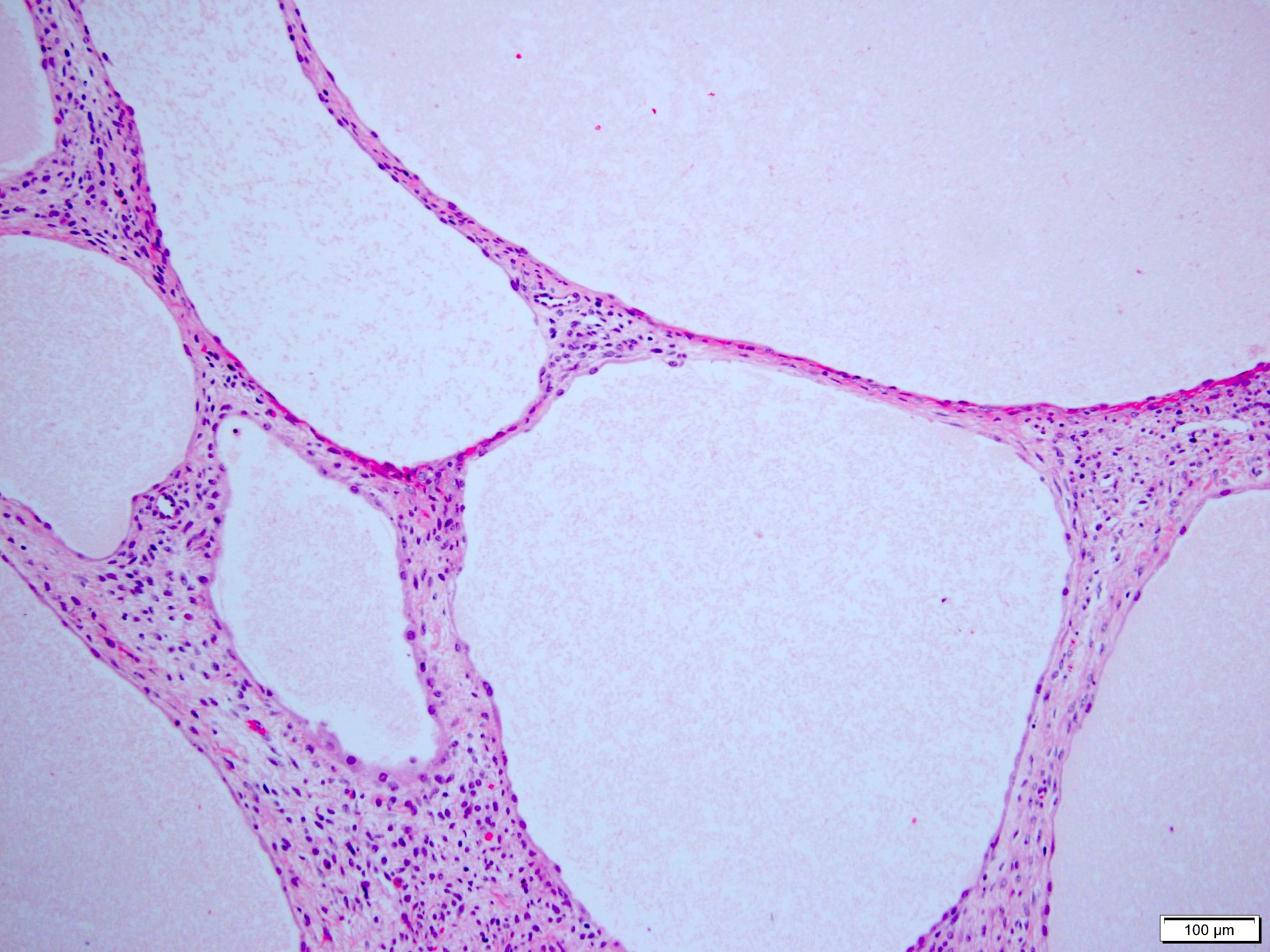
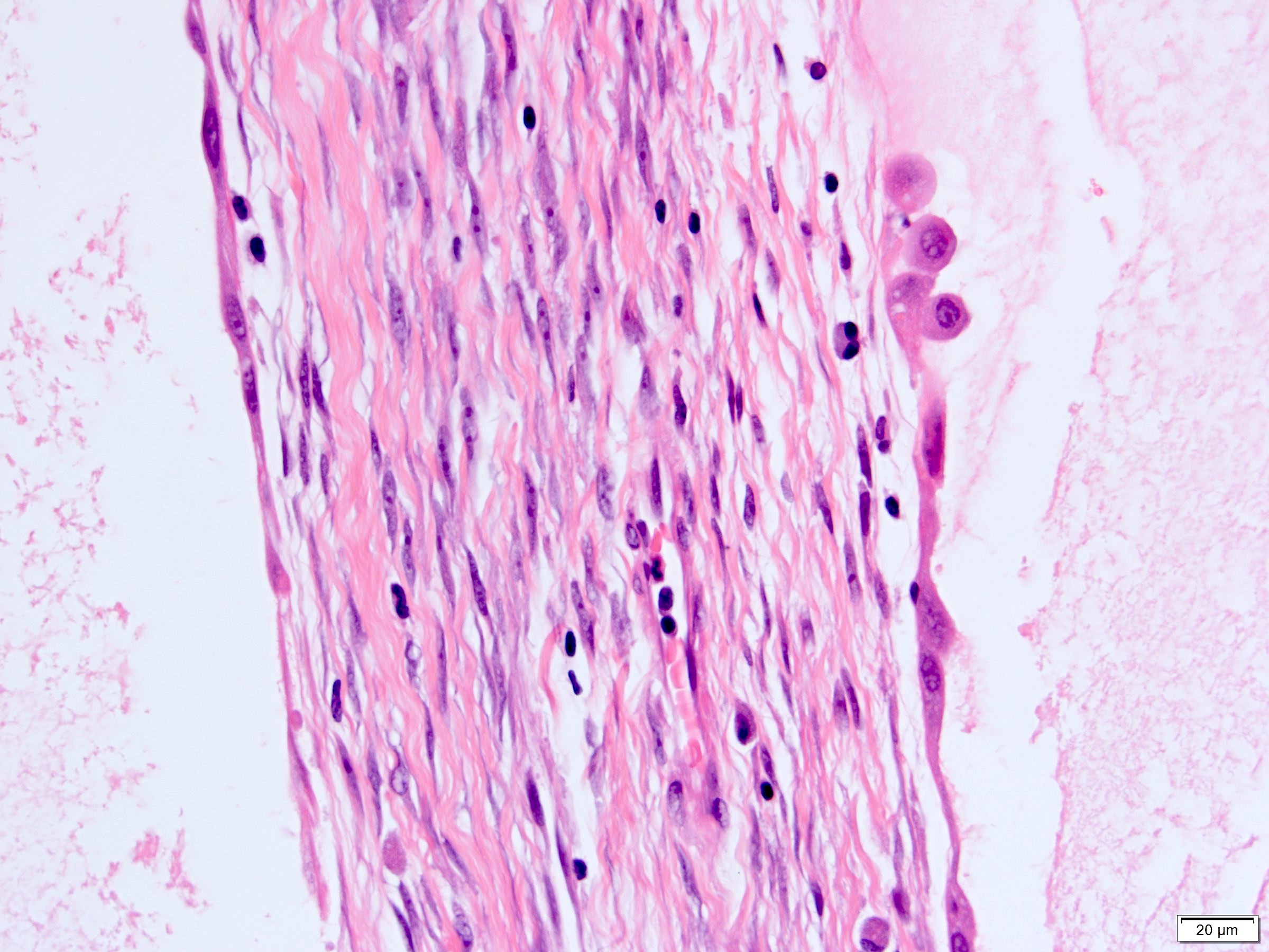
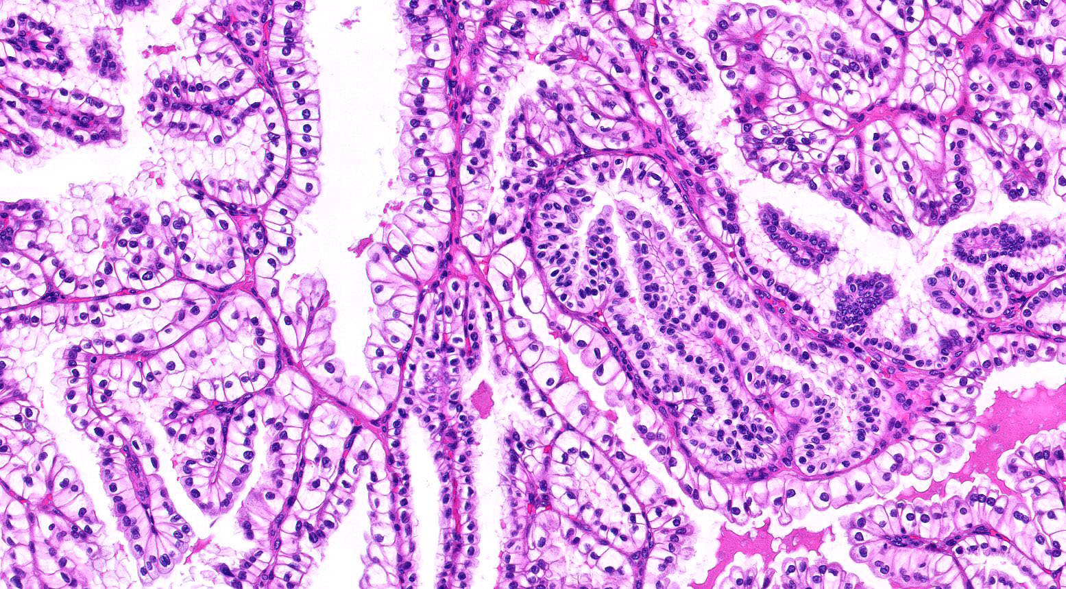
Variably sized cysts with low grade clear cell lining
Variably sized cysts with low grade clear cell lining
renal neoplasm
of low malignant
potential
Images hosted on other servers:

Large renal mass

Renal mass
Images hosted on other servers:

Intraoperative appearance
Contributed by Ellen D’Hooghe, M.D. and Gordan M. Vujanic, M.D., Ph.D.
Encapsulated tumor
Lobulated tumor

Necrotic tumor
Postchemotherapy tumor
Extrarenal growth of tumor
Multifocal tumor
Contributed by Ellen D’Hooghe, M.D. and Gordan M. Vujanic, M.D., Ph.D.

Blastemal component

Epithelial component
Squamous epithelium

Undifferentiated stroma
Differentiated stroma

Adipose tissue
Mature cartilage
Subtotally necrotic tumor
"Dying" blastema
Chemotherapy induced changes
Anaplasia
Focal anaplasia
Pseudoanaplasia

Renal sinus involvement
Tumor thrombus

Perirenal fat invasion
Lymph node metastasis - viable
Lymph node metastasis - nonviable
Resection margin - nonviable tumor
Incompletely resected tumor
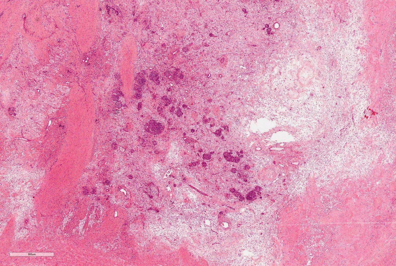
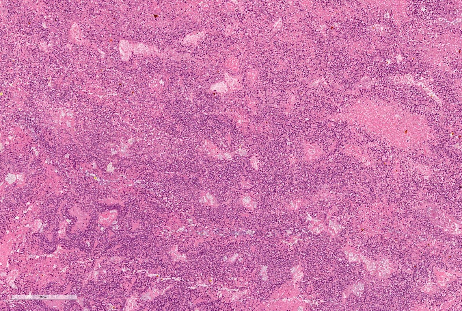
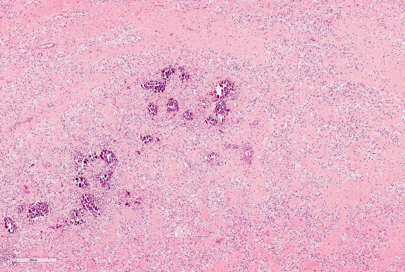
Case of the Week #338

Various images
AFIP images
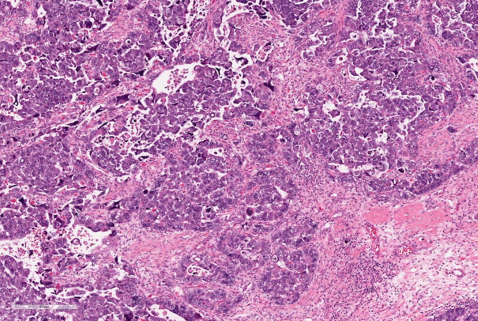
Intralobar

Intralobar nephrogenic rest
Neoplastic intralobar rest
Nephroblastomatosis
Perilobar

Microscopic rests composed of blastemal cells, are termed "incipient" in neonates and "dormant" otherwise
Sclerosing perilobar nephrogenic rest
Focal scarring

Hyperplastic perilobar rest in sclerosing stage
Multifocal hyperplastic perilobar rests

Nearly confluent rind of perilobar rest tissue; thick rind of perilobar tissue

Neoplastic perilobar rest
Case of the Week #338


Various images
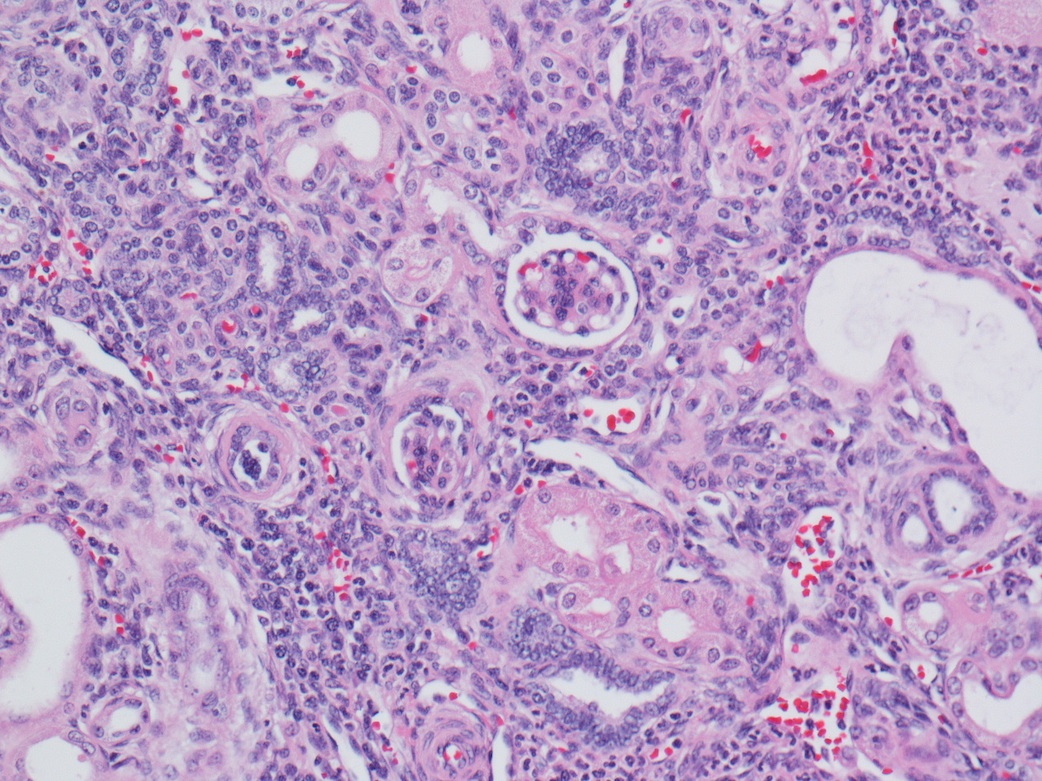
Contributed by Ian W. Gibson, M.B.Ch.B., M.D.
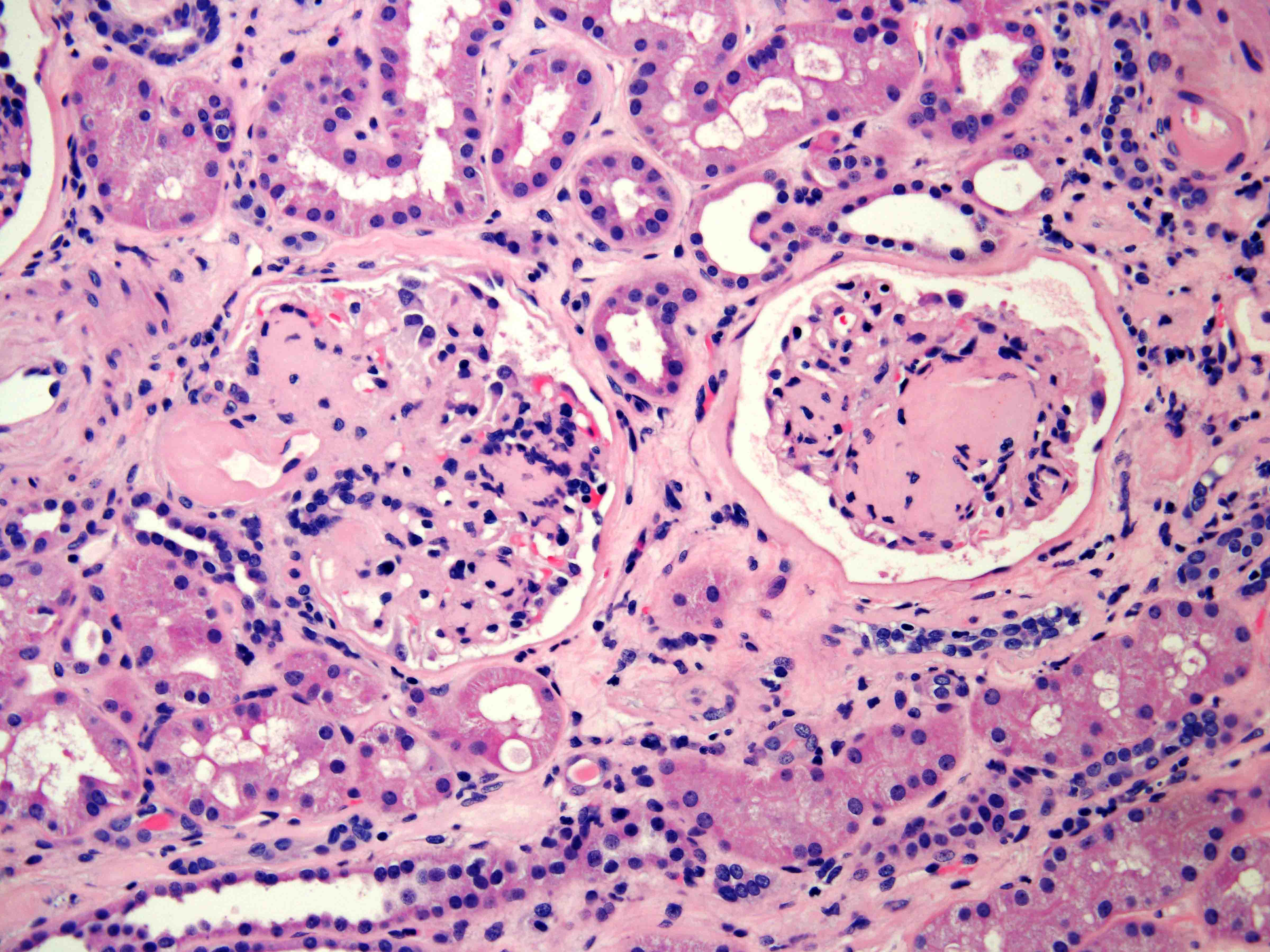
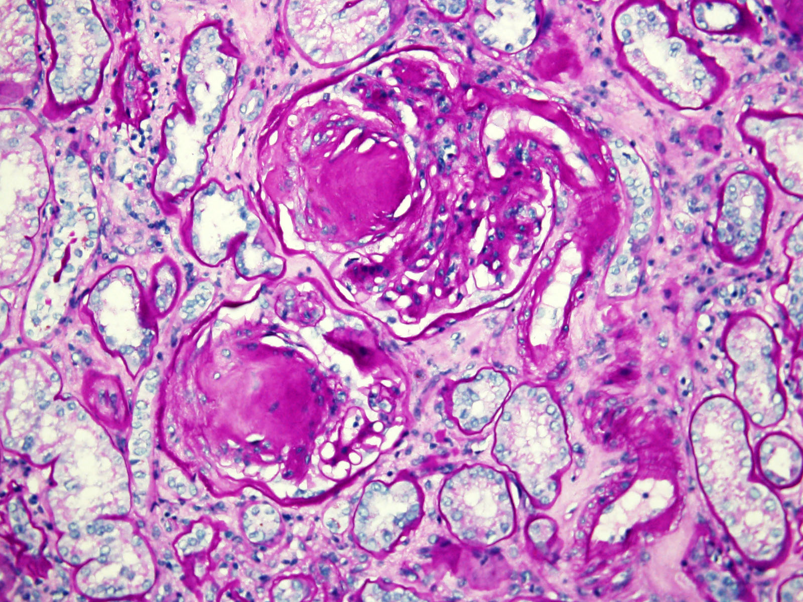
Kimmelstiel-Wilson nodules, hyaline arteriolosclerosis

Nodular hyaline arteriolosclerosis
Organized atheromatous embolism
Tubular neutrophilic casts
Monotonous dense small lymphocytes
AFIP images
Renal oncocytoma
MRI shows same tumor with prominent central scar
Contributed by Debra Zynger, M.D.
Tan, central scar

Pale brown, central scar
Pale brown, central scar
Mahogany

Congested, dark brown

Hemorrhagic, red
Contributed by Rola Saleeb, M.D., Ph.D.

Small nested morphology
Degenerated cells
Loose stroma

c-KIT

CK7
Hale colloidal iron
Contributed by Maria Tretiakova, M.D., Ph.D.
Compact, nested
Central scar
Nested, cystic
Lack of prominent membranes
Oncoblasts (20x)
Degenerate atypia
Edematous stroma
Microcystic, hobnailed
Atypia multinucleation
Fat extension
Rosettes

Vascular extension
S100A1
CK7
AFIP images
Abundant granular cytoplasm
Images hosted on other servers:

Small clusters and single cells with uniform cytoplasm

Diff-Quik shows
homogeneous granular
cytoplasm and round, regular
nuclei without grooves
AFIP images
Cytoplasm is packed with large mitochondria
Images hosted on other servers:

FISH: loss of chromosomes 1 and 17
Contributed by Ellen D’Hooghe, M.D. and Gordan M. Vujanic, M.D., Ph.D.
Polypoid / pedunculated calcified mass
Contributed by Ellen D’Hooghe, M.D. and Gordan M. Vujanic, M.D., Ph.D.
Osteoid and cellular components
Osteoid component
Cellular component
Vimentin+
WT1+
AE1 / AE3-
Images hosted on other servers:

Hypoenhacing lesion on CT

CT scan upper pole tumor
Contributed by Nicole K. Andeen, M.D. and Maria Tretiakova, M.D., Ph.D.
Granular cut surface
Images hosted on other servers:

Circumscribed tumor with extensive hemorrhage

Encapsulated tumor with hemorrhage and necrosis

Tumor with capsular invasion

Tan-pink mass with satellite nodules

Renal vein extension
Contributed by Rola Saleeb, M.D., Ph.D., Vincent Francis Castillo, M.D., Nicole K. Andeen, M.D.,
Maria Tretiakova, M.D., Ph.D., Semir Vranić, M.D., Ph.D. and @SueEPig on Twitter
Circumscribed mass
Papillary architecture
Low grade PRCC
PRCC with foamy histiocytes and hemosiderin
Foamy macrophages
PRCC with clear cytoplasm
High grade PRCC
High grade eosinophilic cells
Pseudosolid pattern
Psammoma bodies
Tubulopapillary architecture
PRCC with low grade morphology
PRCC with high grade morphology
Papillary renal cell carcinoma

Encapsulated PRCC

Solid growth

Glomeruloid growth pattern

Biphasic pattern

Cyclin D1

AMACR positive
CK7 focally positive
Retained FH expression
Contributed by Debra L. Zynger, M.D.

Papillary structures and foamy histiocytes
Images hosted on other servers:

Foamy macrophages

Papillary tissue fragment
Images hosted on other servers:

MRI of renal adenomatosis
AFIP images
1.5 cm subcapsular tumor
Contributed by Rola Saleeb, M.D., Ph.D. and Vincent Francis Castillo, M.D.

No fibrous capsule

Tubulopapillary architecture

Subcapsular location
Small unencapsulated tumor

Cytoplasmic clearing
Foamy histocytes

Hemosiderin pigments
Basophilic scant cytoplasm
CK7
Images hosted on other servers:

Chromosome 12 trisomy
Contributed by Nicole K. Andeen, M.D.
Papillary renal cell carcinoma with oncocytic cells
Contributed by Nicole K. Andeen, M.D. and Maria Tretiakova, M.D., Ph.D.


Papillary type 1 oncocytic renal cell carcinoma
Contributed by Semir Vranić, M.D., Ph.D., University of Sarajevo (Bosnia)

Oncocytic "variant" of papillary renal cell carcinoma
Images hosted on other servers:

Racemase, CD10, vimentin, MET
Contributed by Hsin-Yi Chang, M.D. and Jen-Fan Hang, M.D.
Hyperintense in T1 weighted MRI
Hypointense in T2 weighted MRI
Isoechoic mass under ultrasound
Contributed by @katcollmd on Twitter
Papillary renal neoplasm with reverse polarity
Images hosted on other servers:
Courtesy of Katrina Collins, M.D.

In polycystic kidney disease

Delicate papillae
Contributed by Hsin-Yi Chang, M.D., Jen-Fan Hang, M.D., @ahmsab_MD on Twitter and @katcollmd on Twitter
Partially cystic growth
Papillary pattern
Tubulopapillary pattern
Stromal hyalinization
Low grade inverted nuclei
Type D papillary adenoma
GATA3
Papillary renal neoplasm with reverse polarity
Papillary renal neoplasm with reverse polarity
Images hosted on other servers:

Ultrasonography and CT of abdomen

Well circumscribed, huge CN in the right kidney
Contributed by Daniel Anderson, M.D., M.B.A.
Bulging mass
Multicystic
Thin septa and fine vasculature
Images hosted on other servers:

Specimen after nephrectomy
Contributed by Daniel Anderson, M.D., M.B.A.

Cystic nephroma
Adjacent renal parenchyma
Cystic nephroma flat
Cuboidal, hobnail, flat
Cystic nephroma with hobnailing
Images hosted on other servers:

DICER1 protein domains and mutations
Pediatric renal tumors: molecular diagnostics and COG updates
Images hosted on other servers:

Noncontrast coronal CT with 5.7 cm mass

Noncontrast axial CT with 2.5 cm mass

Postcontrast axial CT without evidence of enhancement

Coronal and axial (contrast) CT scans
Images hosted on other servers:

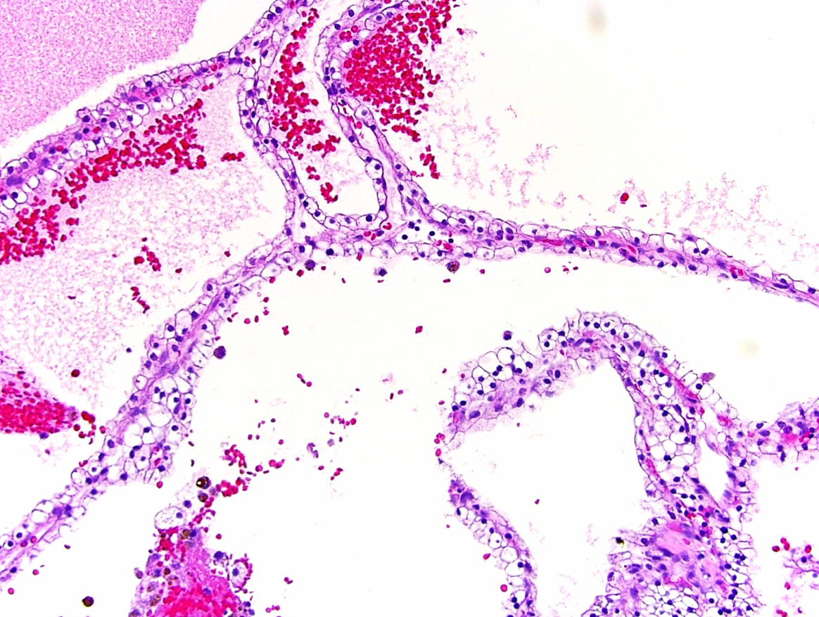
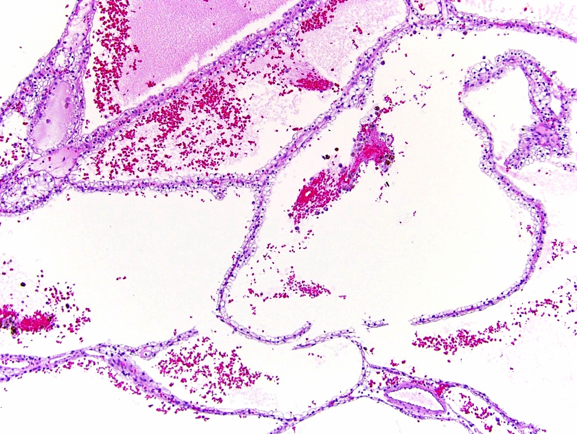
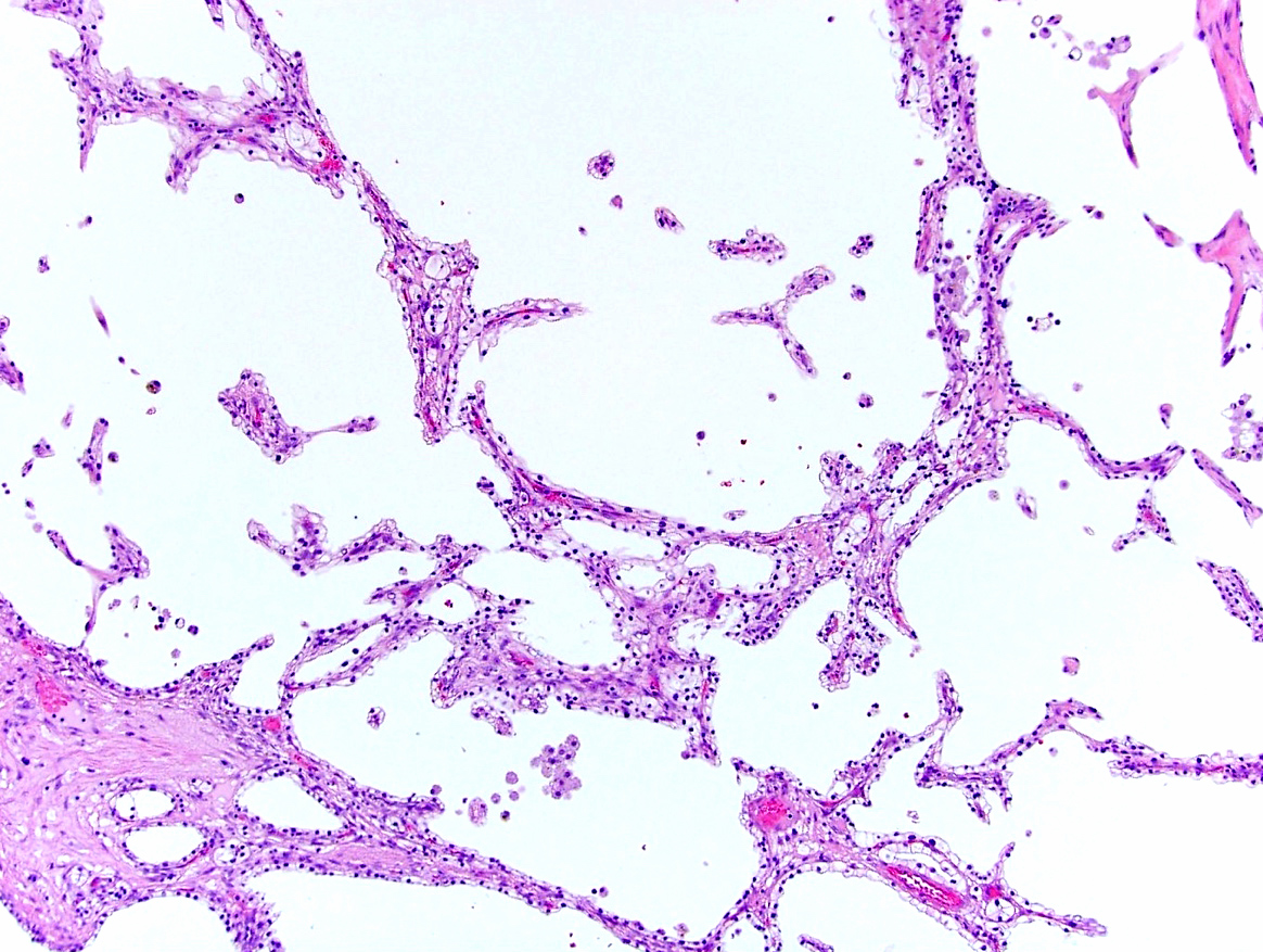
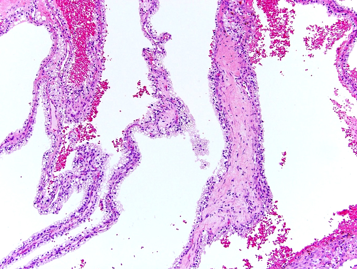
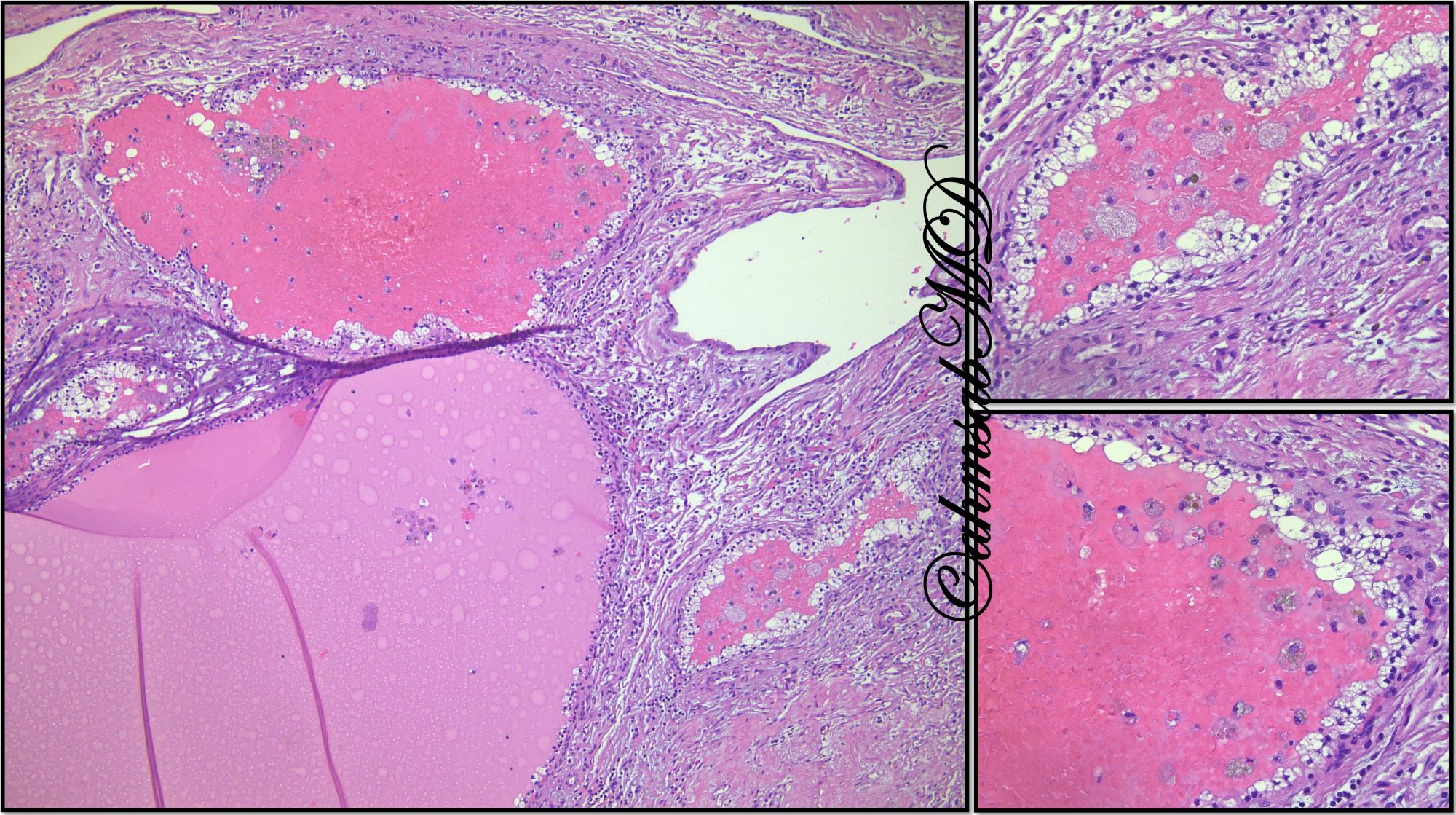
Partially cystic, hemorrhagic mass

Multiloculated cystic mass
Contributed by Maria Tretiakova, M.D., Ph.D.
Atrophic appearance
Variably sized follicles
Images hosted on other servers:

Giemsa

Uniform, medium sized cells
Images hosted on other servers:

EWSR1::PATZ1 schematic
Contributed by Rajal B. Shah, M.D., Ph.D.
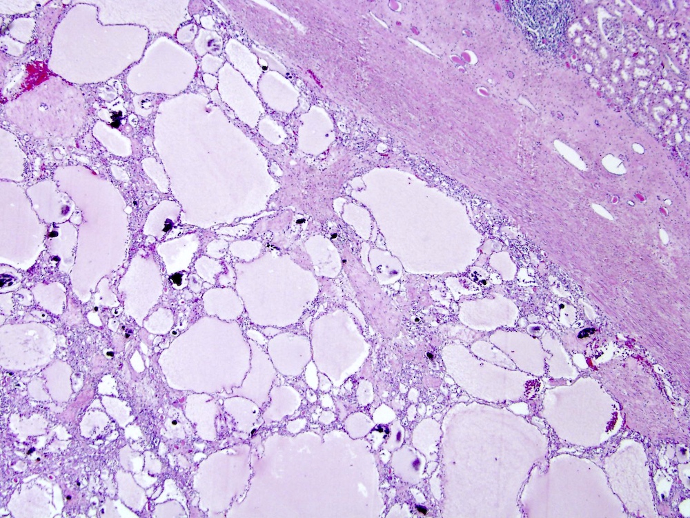
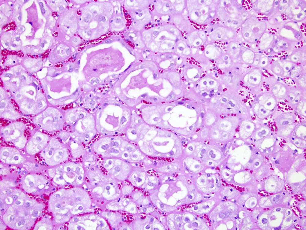
Clear cell renal tumors
IHC of clear cell renal tumors
Contributed by Jean-François Aita, M.D.
Large clear cell RCC
2 small RCCs
RCC with venous invasion
Cystic Bosniak 4 RCC
Multilocular cystic renal carcinoma
Contributed by Debra Zynger, M.D. and AFIP
pT2b

pT3a

pT4
Tumor less than 3 cm
Arising in adult type polycystic renal disease
Diffusely infiltrative
Massive tumor extension
Metastases to small intestine
Nodal metastases
Retrograde extension of tumor
Contributed by Maria Tretiakova, M.D., Ph.D., Miruna Claudia Popescu, M.D. and Katrina Collins, M.D.
Clear cell RCC histology
Papillary RCC histology
Chromophobe RCC histology
FH deficient RCC histology
SDH deficient RCC histology
RCC metastatic to stomach
RCC metastatic to bronchus
RCC metastatic to duodenum
Box-like CAIX in CCRCC
CK7 positivity in PRCC
AMACR positivity in PRCC
Vimentin positivity in PRCC
CD10 positivity in PRCC
CD117 in chromophobe RCC
CK7 in chromophobe RCC
Vimentin in chromophobe RCC
Hale colloidal iron in ChRCC
PAX8 in FH deficient RCC
FH loss
PAX8 in SDH deficient RCC
SDHB loss
AFIP images
Cells in alveolar arrangement
Pathology resident led live unknown slide session: pink renal tumors (part 1)
A dummies guide to diagnosis of renal tumors using pattern based approach by Dr. Rajal B. Shah
A dummies guide to diagnosis of clear renal tumors by Dr. Rajal B. Shah
Images hosted on other servers:

Incidental tumor
Contributed by Andrew McLoughlin, M.S., Debra L. Zynger, M.D. and AFIP

Small, white nodule
Multiple small tumors
Images hosted on other servers:

Firm, well circumscribed tumor

White tumor
Contributed by Eduardo Eyzaguirre, M.D.
Medullary based tumor
Small stellate cells
Images hosted on other servers:
Rhabdoid component is solid, white
Contributed by Nicole K. Andeen, M.D.
Discohesive rhabdoid cells with eccentrically placed nuclei, prominent nucleoli and densely eosinophilic cytoplasm
Abrupt transition between conventional clear cell carcinoma and rhabdoid morphology
Rhabdoid cells express CAIX, similar to coexistent conventional clear cell component
Contributed by @SueEPig on Twitter
Adult renal cell carcinoma - rhabdoid variant
- Not relevant to this topic
Images hosted on other servers:

10 cm kidney mass
- Not relevant to this topic
Images hosted on other servers:

Solid to cystic renal mass
Rhabdoid component is solid, white
Contributed by Rola Saleeb, M.D., Ph.D., Nicole K. Andeen, M.D. and @SueEPig on Twitter

Solid sheet pattern


Rhabdoid cells

CCRCC with rhabdoid areas


Rhabdoid cells in renal vein

Discohesive rhabdoid cells


Clear cell carcinoma and rhabdoid morphology
Adult renal cell carcinoma - rhabdoid variant
Adult renal cell carcinoma - rhabdoid variant

Pancytokeratin positive

EMA negative

CAIX positive

CD10 positive


Rhabdoid cells express CAIX
- Not relevant to this topic
- Not relevant to this topic
- Not relevant to this topic
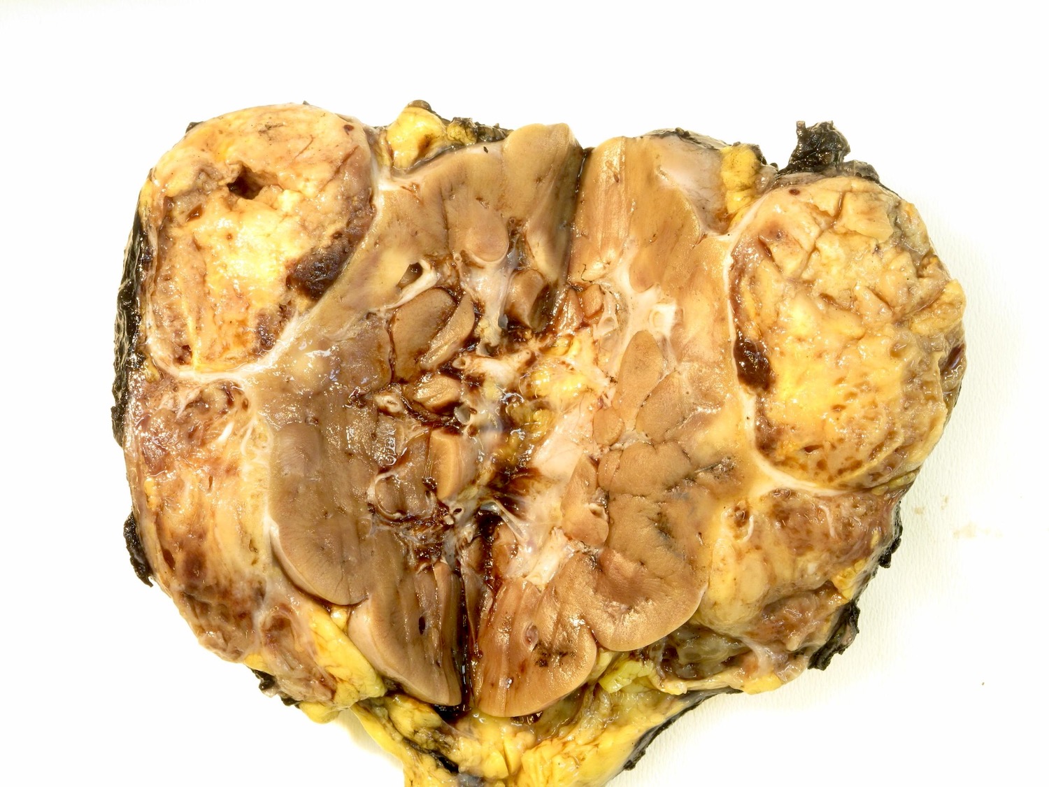
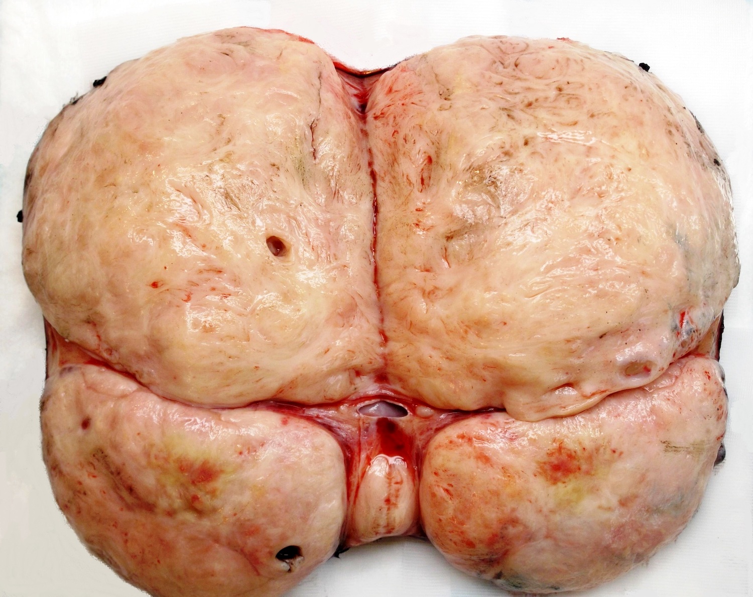
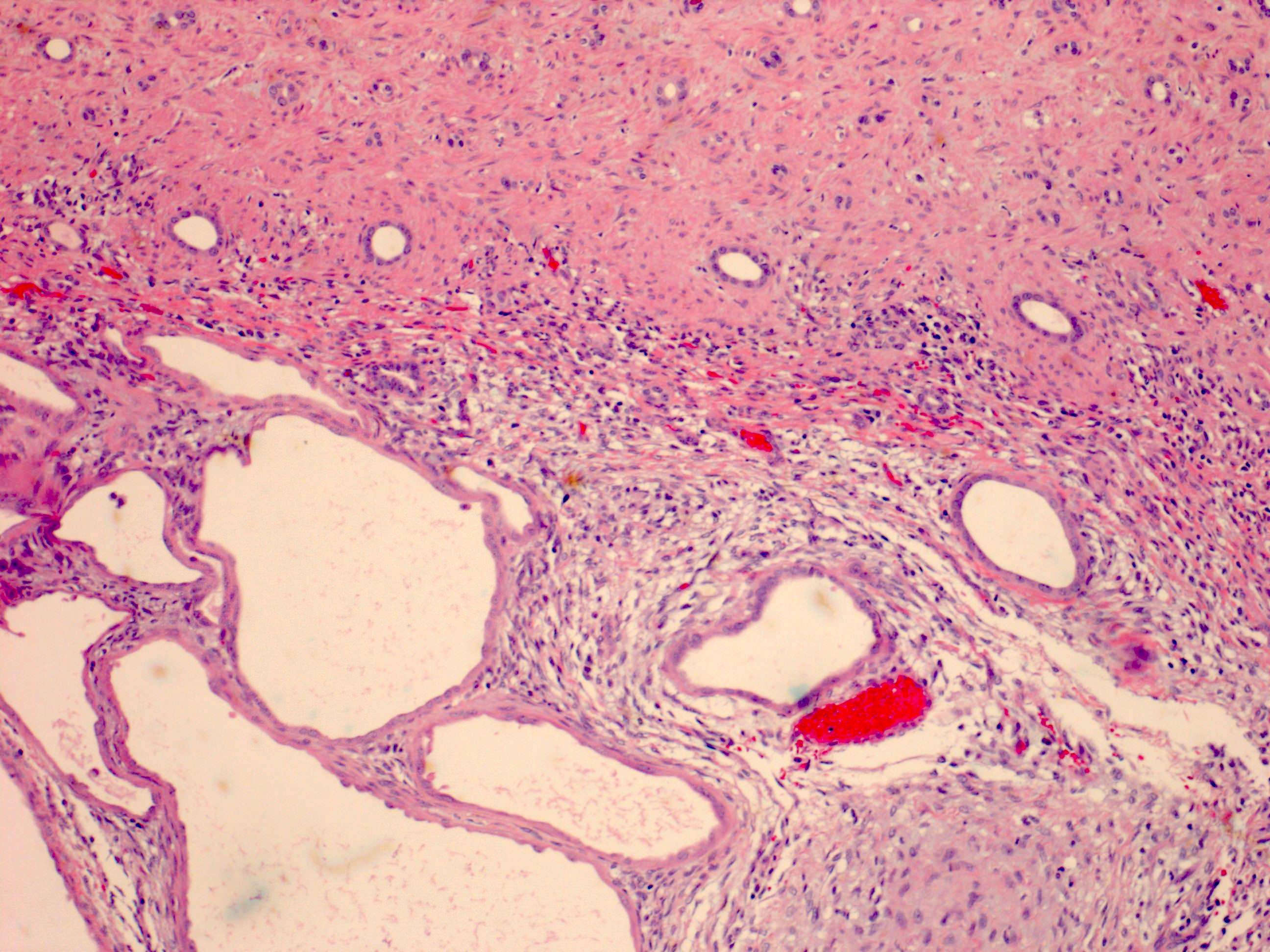
Contributed by Ellen D’Hooghe, M.D. and Gordan M. Vujanic, M.D., Ph.D.
Solid, fleshy tumor
Contributed by Ellen D’Hooghe, M.D. and Gordan M. Vujanic, M.D., Ph.D.

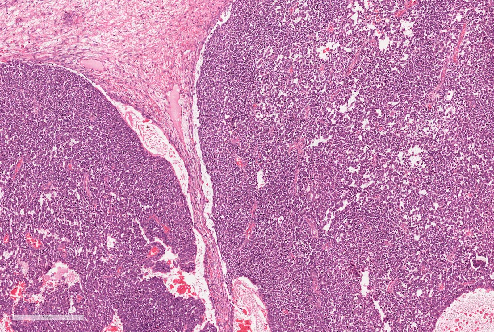
Classic pattern
Classic pattern associated with eosinophils

Hypercellular tumor
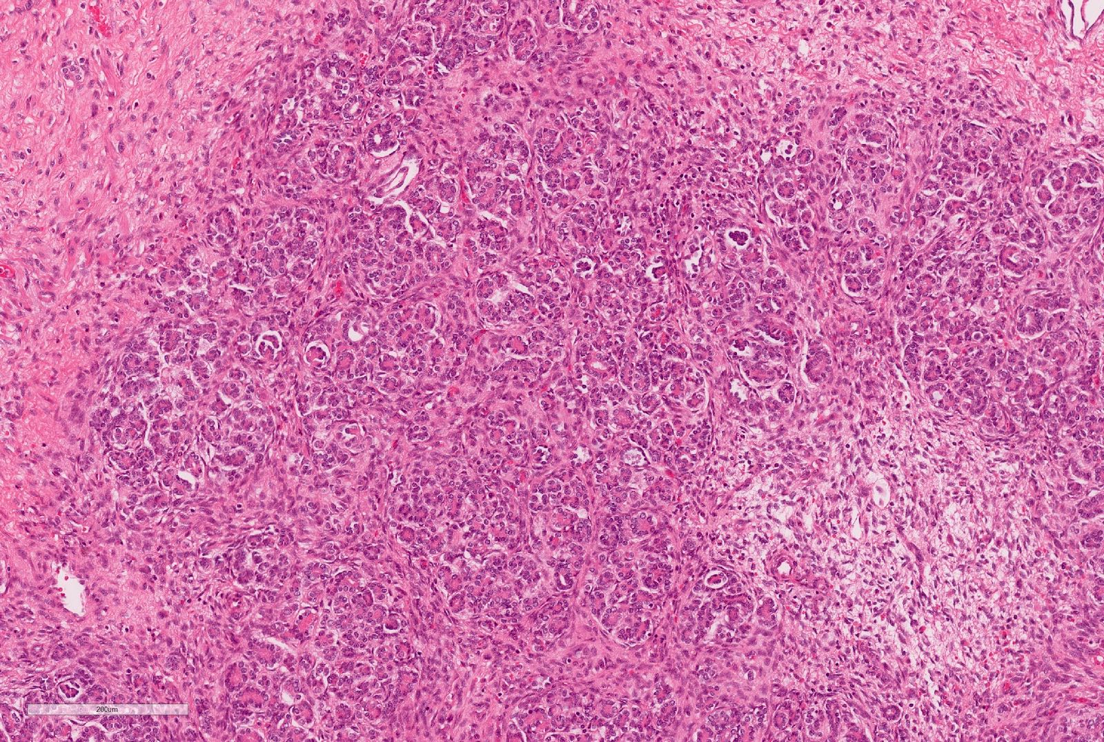
Entrapped normal renal tubules
Epithelioid pattern
Intracytoplasmic
inclusions
Lymphomatoid pattern
Sclerosing pattern
Prominent sclerosing pattern

Spindling growth pattern
Pseudoalveolar pattern
Loss of nuclear INI1
Images hosted on other servers:

Redox alterations
Images hosted on other servers:

Cystic change;
solid neoplasms
Images hosted on other servers:


Various images


SDH deficient RCC
Images hosted on other servers:

Proposed pathogenesis model

Morphologic patterns
Contributed by Daniel Anderson, M.D., M.B.A.
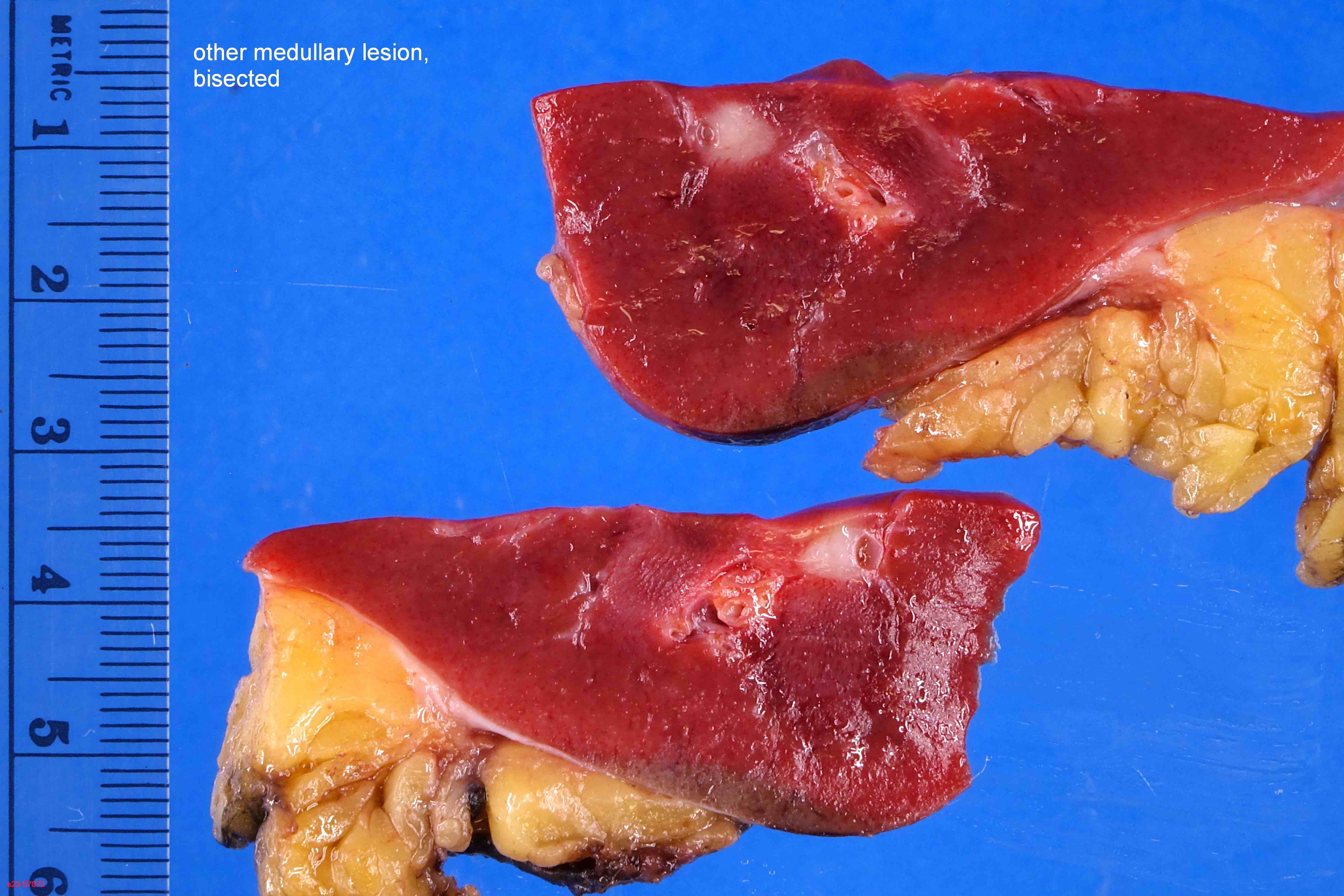
Medulla centered mass
Images hosted on other servers:

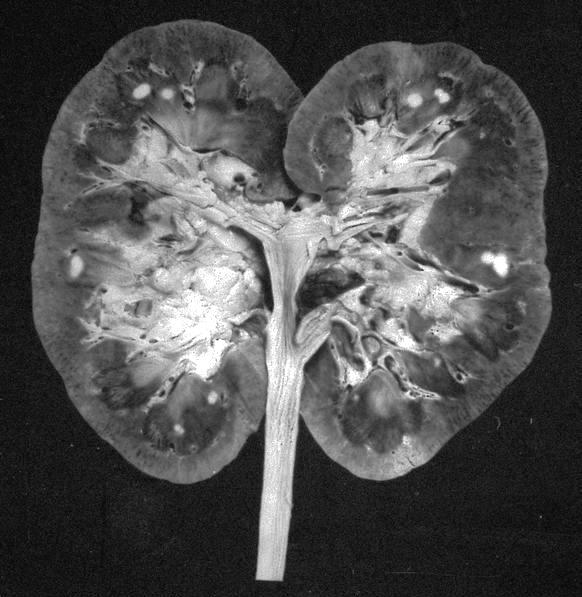
Well circumscribed yellowish white tumor

Focally infiltrative

Lobulated tumor
Contributed by Daniel Anderson, M.D., M.B.A. and @katcollmd on Twitter

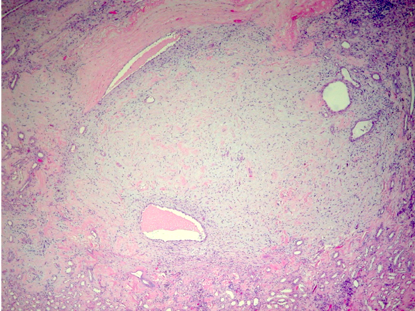
Cribriform, reticular and nested growth
Cribriform nest
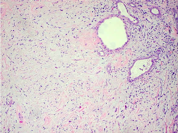
Reticular growth
Nuclear details
SMARCB1 deficient renal medullary carcinoma
Images hosted on other servers:

Loosely cohesive groups
Pathology mini tutorial of renal medullary carcinoma
Cytomorphologic study of renal medullary carcinoma involving serous cavity fluids
Images hosted on other servers:

Signalling pathways involved in EMT reported in sRCC
Images hosted on other servers:

MRI

CT with large renal mass

PET / CT with widespread disease
Contributed by Daniel Anderson, M.D., M.B.A.
Heterogeneous white mass

White mass arising from ccRCC
Images hosted on other servers:

ccRCC with
sarcomatoid
differentiation
Contributed by Daniel Anderson, M.D., M.B.A.
Sarcomatoid spindle cell differentiation
Atypical pleomorphic spindle cells
Pleomorphic sarcomatoid cells
Atypical spindled and giant cells from a ccRCC
Sarcomatoid differentiation in ccRCC


Rhabdoid cells are not sarcomatoid
Images hosted on other servers:

4 variable risk stratification
Images hosted on other servers:

Ultrasound, heterogeneous hyperechoic mass

CT, heterogeneous enhancing mass

MRI, heterogeneous mass
Images hosted on other servers:

Tumor deforming right kidney
Images hosted on other servers:

Yellow-tan mass

Rubbery white-tan mass

Adipose tissue and hemorrhage
Contributed by Maria Tretiakova, M.D., Ph.D.
Patternless architecture
Staghorn vessels
Necrosis
Entrapped tubules
STAT6
CD34
Images hosted on other servers:

SFTs with low risk of aggressive behavior

SFTs with high risk of aggressive behavior
Images hosted on other servers:

Structure of NAB2::STAT6

NAB2::STAT6 gene fusion
Contributed by Debra Zynger, M.D.

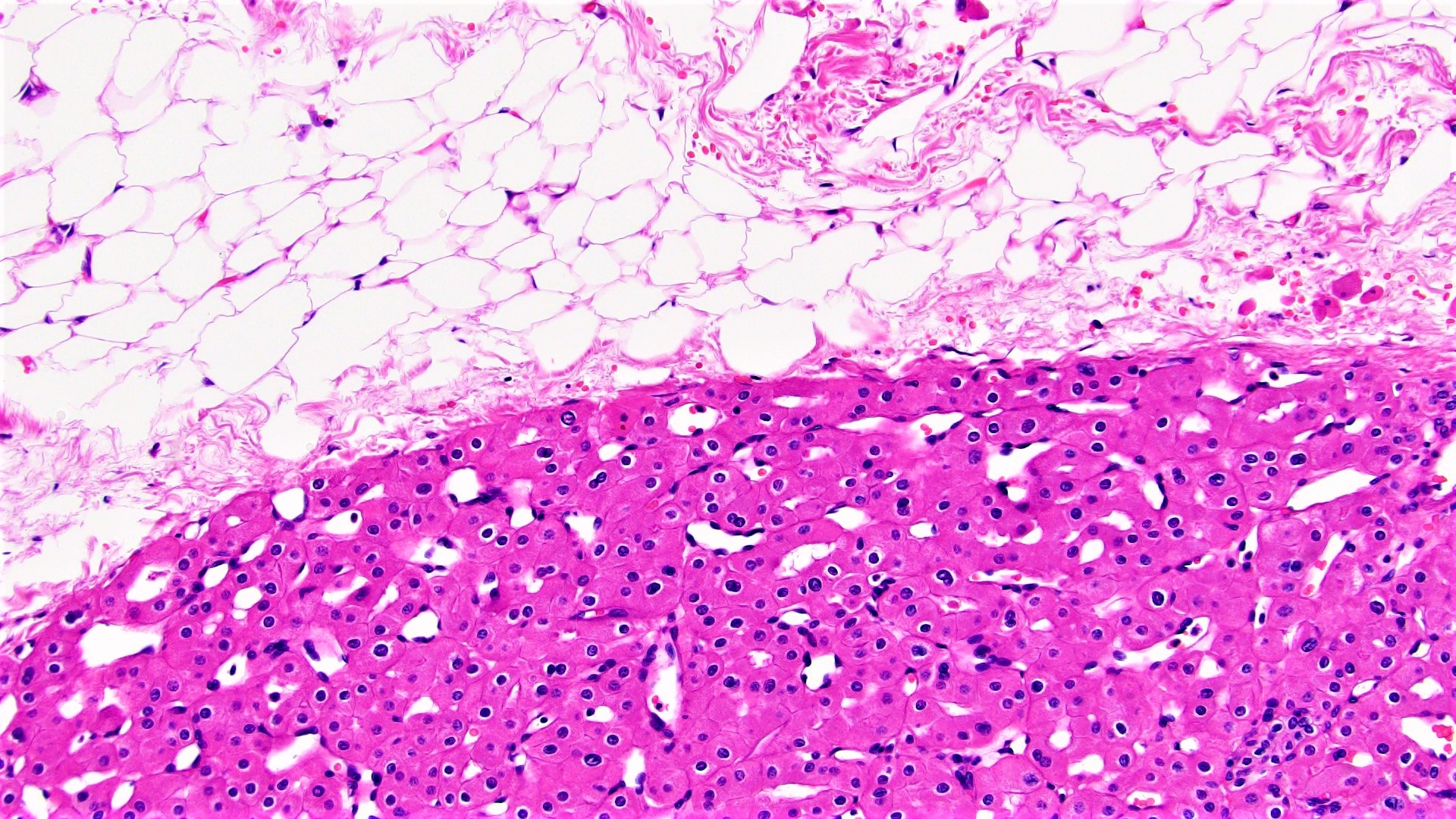
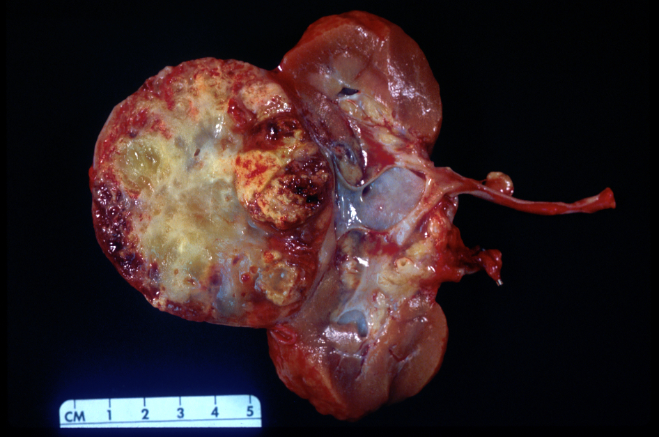
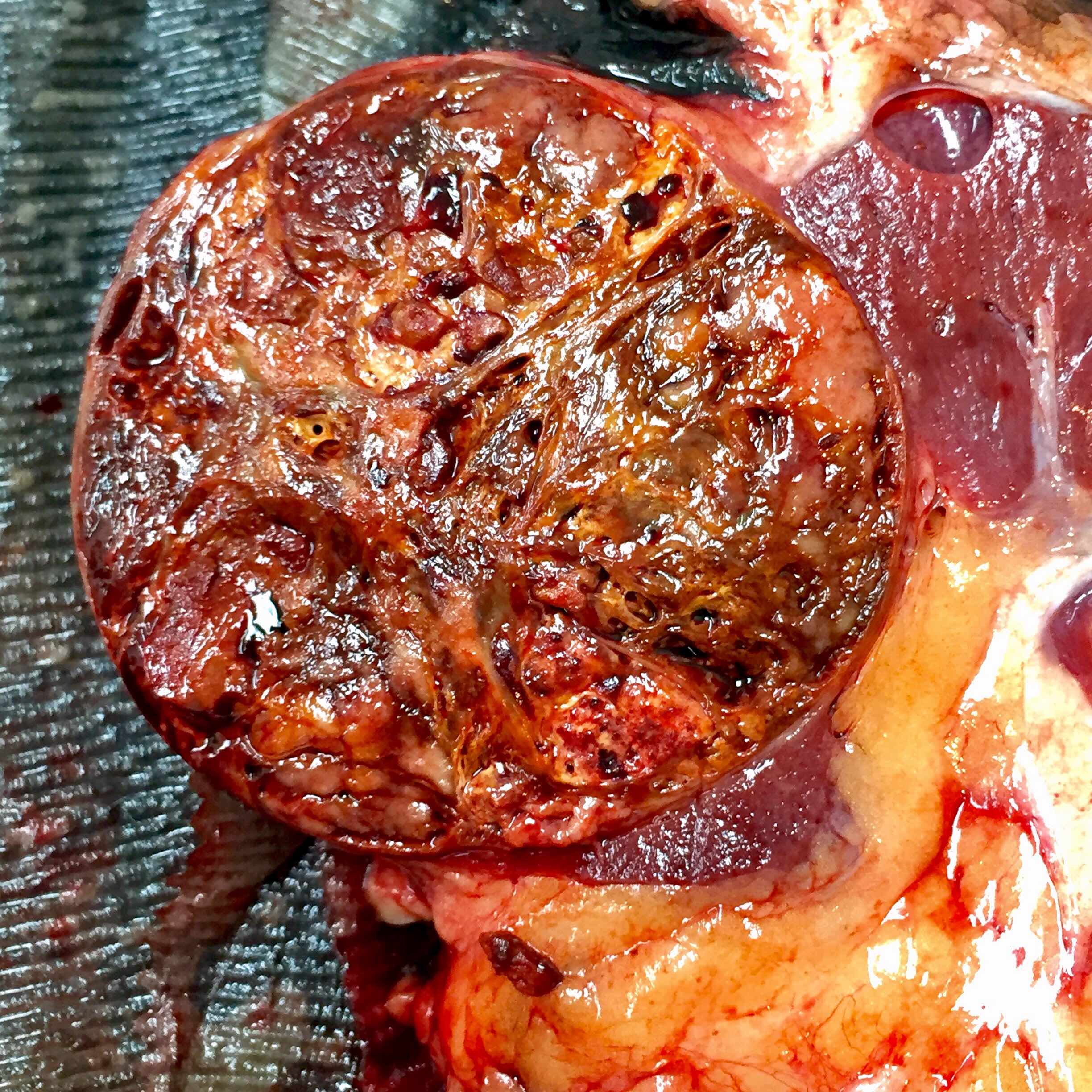
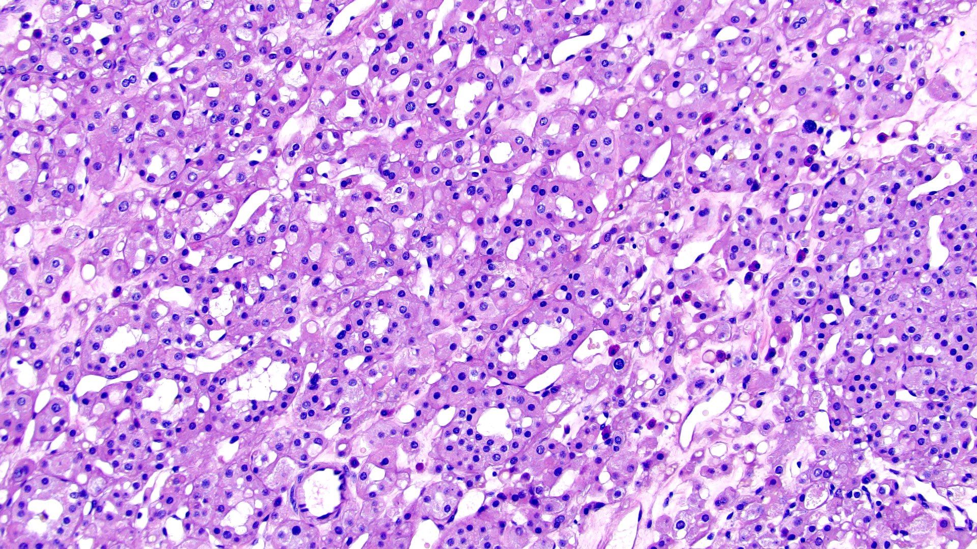
Clear cell RCC (pT1a)
Clear cell papillary RCC (pT1a)
ACDA RCC (pT1a)
Clear cell RCC (pT1b)
Chromophobe RCC (pT1b)
Papillary type 2 RCC (pT2a)
Chromophobe RCC (pT2a)
Papillary RCC (pT2b)
Clear cell RCC (pT3a)
Clear cell RCC (pT3b)
Clear cell RCC (pT4)
Contributed by Debra Zynger, M.D.

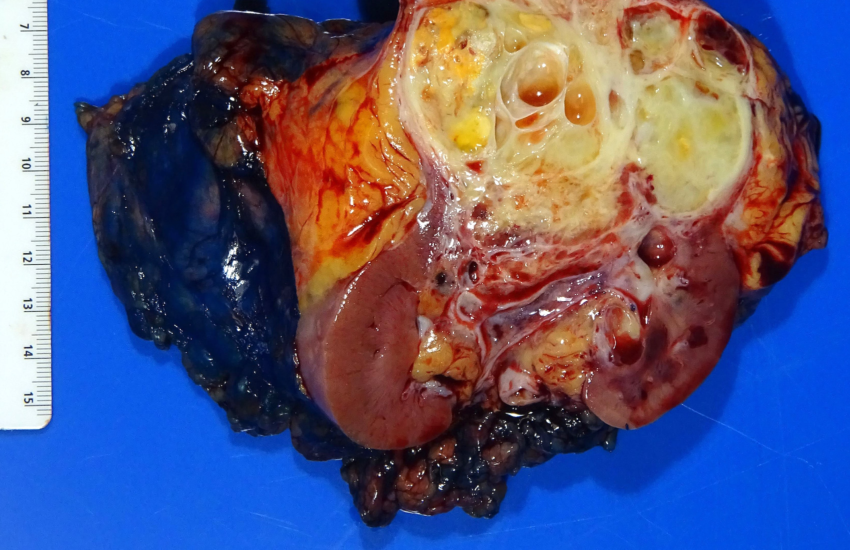
Renal sinus fat invasion (pT3a)
Perinephric fat invasion (pT3a)
Renal vein invasion (pT3a)
Pelvicaliceal invasion (pT3a)
Direct extension to adrenal (pT4)
Periaortic lymph node metastasis (pN1)
Bone metastasis (pM1)
Lung metastasis (pM1)
Ovary metastasis (pM1)
Stomach metastasis (pM1)
Contributed by Maria Tretiakova, M.D., Ph.D. and Debra L. Zynger, M.D.
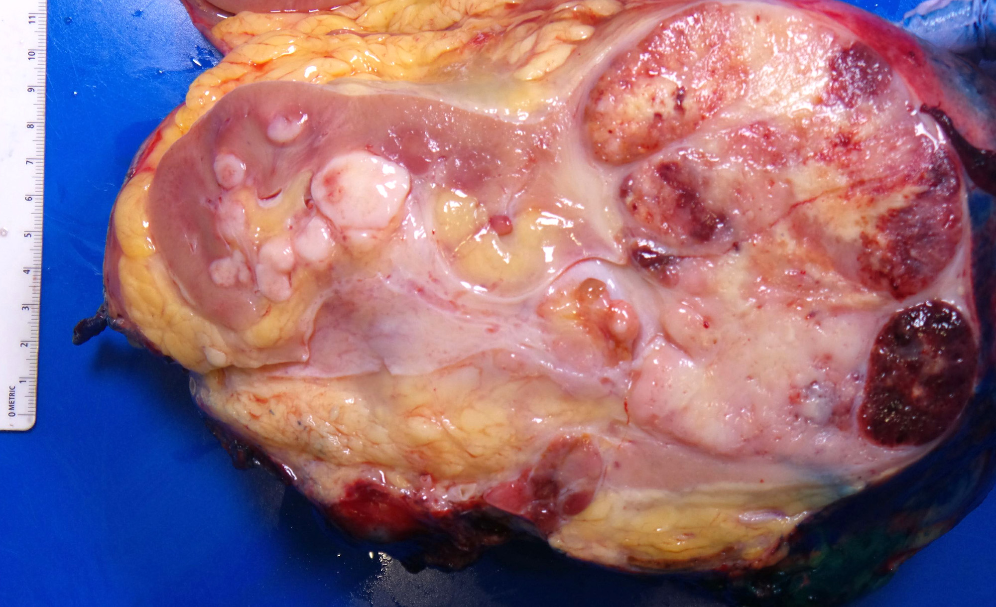
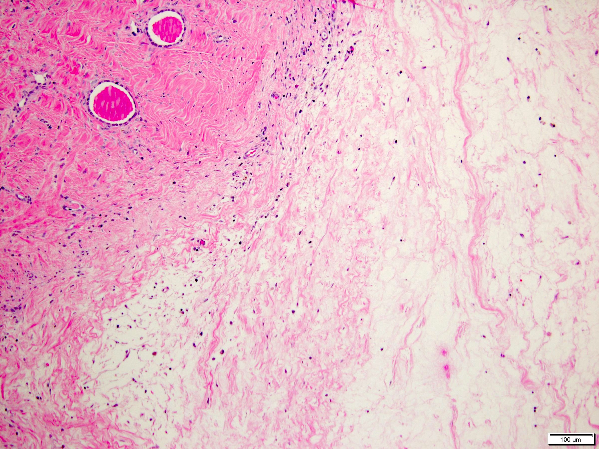
Solid tumor
Cystic tumor
Contributed by Maria Tretiakova, M.D., Ph.D.
Classic triad
Cell blistering
Psamomma bodies
TFE3 rRCC mimicking CCPRCT
Papillary and alveolar growth pattern
CCPRCT
pRCC
TFEB renal cell carcinoma
TFE3 positive IHC
MiT family translocation renal cell carcinoma by Rajal Shah, M.D.
Contributed by Maria Tretiakova, M.D., Ph.D. and Debra L. Zynger, M.D.
Solid tumor
Cystic tumor
Contributed by Maria Tretiakova, M.D., Ph.D.
Classic triad
Cell blistering
Psamomma bodies
TFE3 rRCC mimicking CCPRCT
Papillary and alveolar growth pattern
CCPRCT
pRCC
TFEB renal cell carcinoma
TFE3 positive IHC
MiT family translocation renal cell carcinoma by Rajal Shah, M.D.
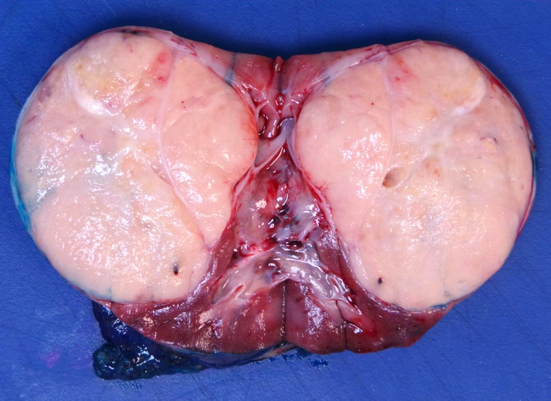
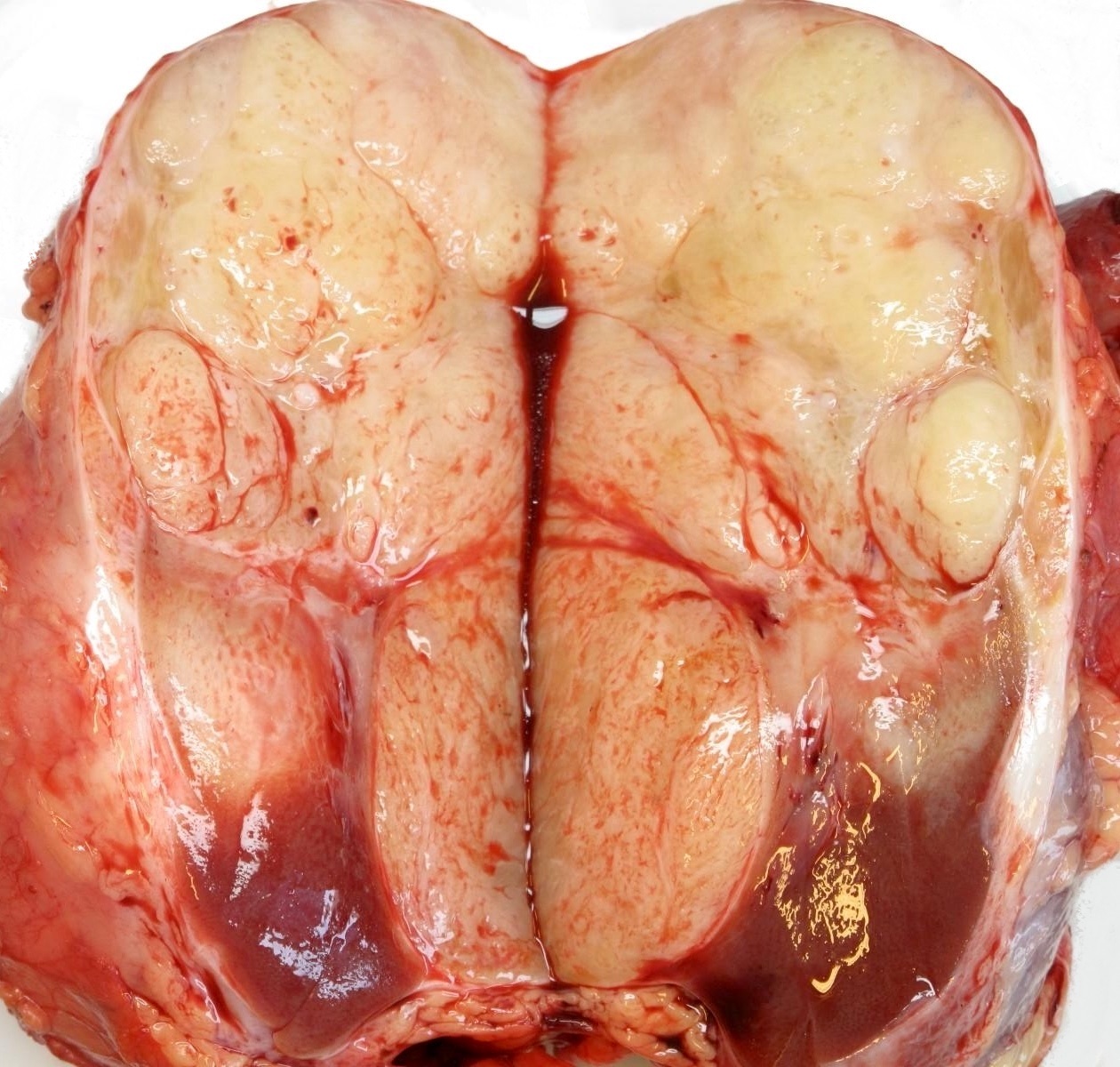
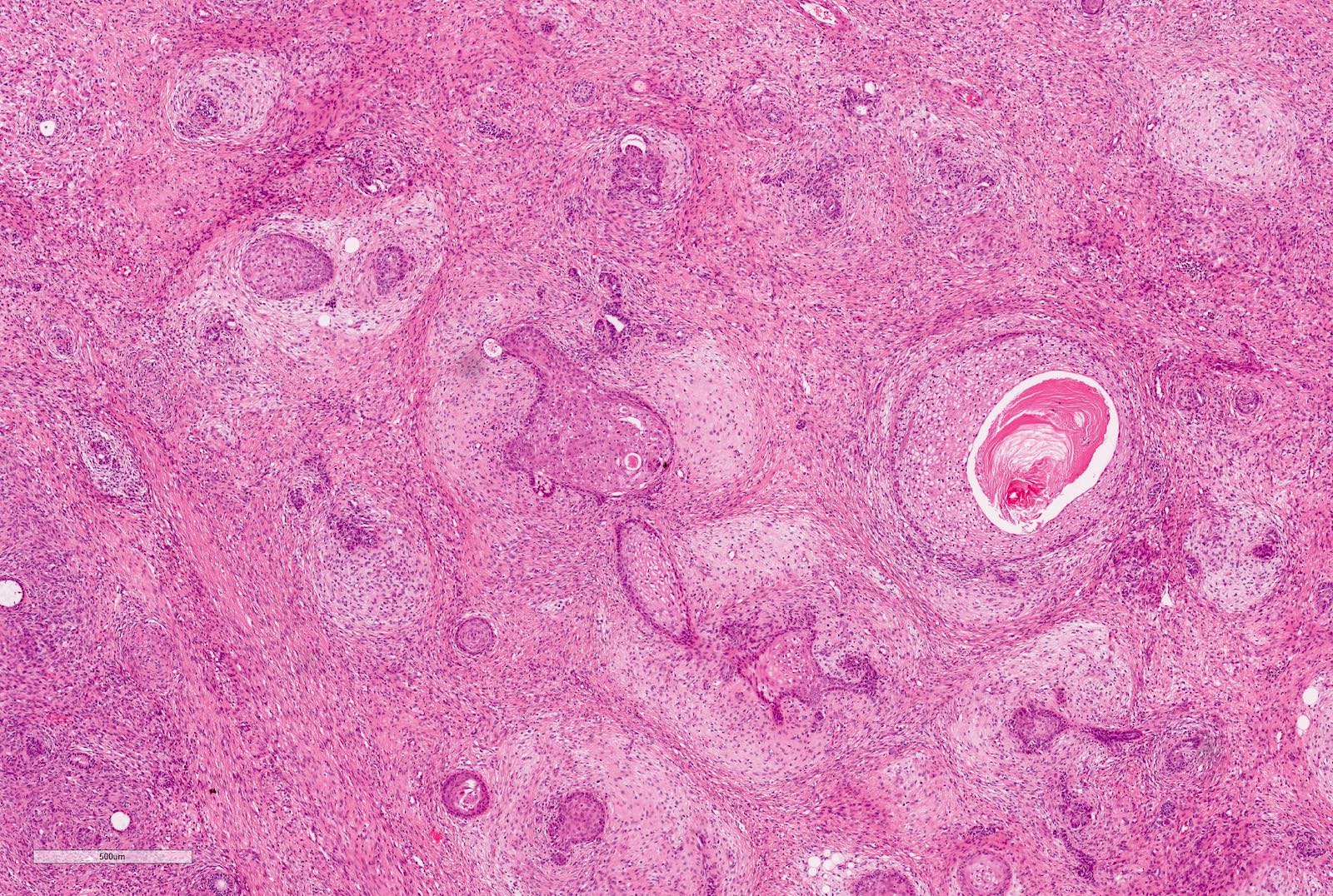
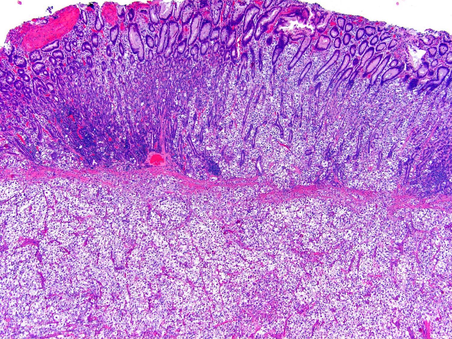
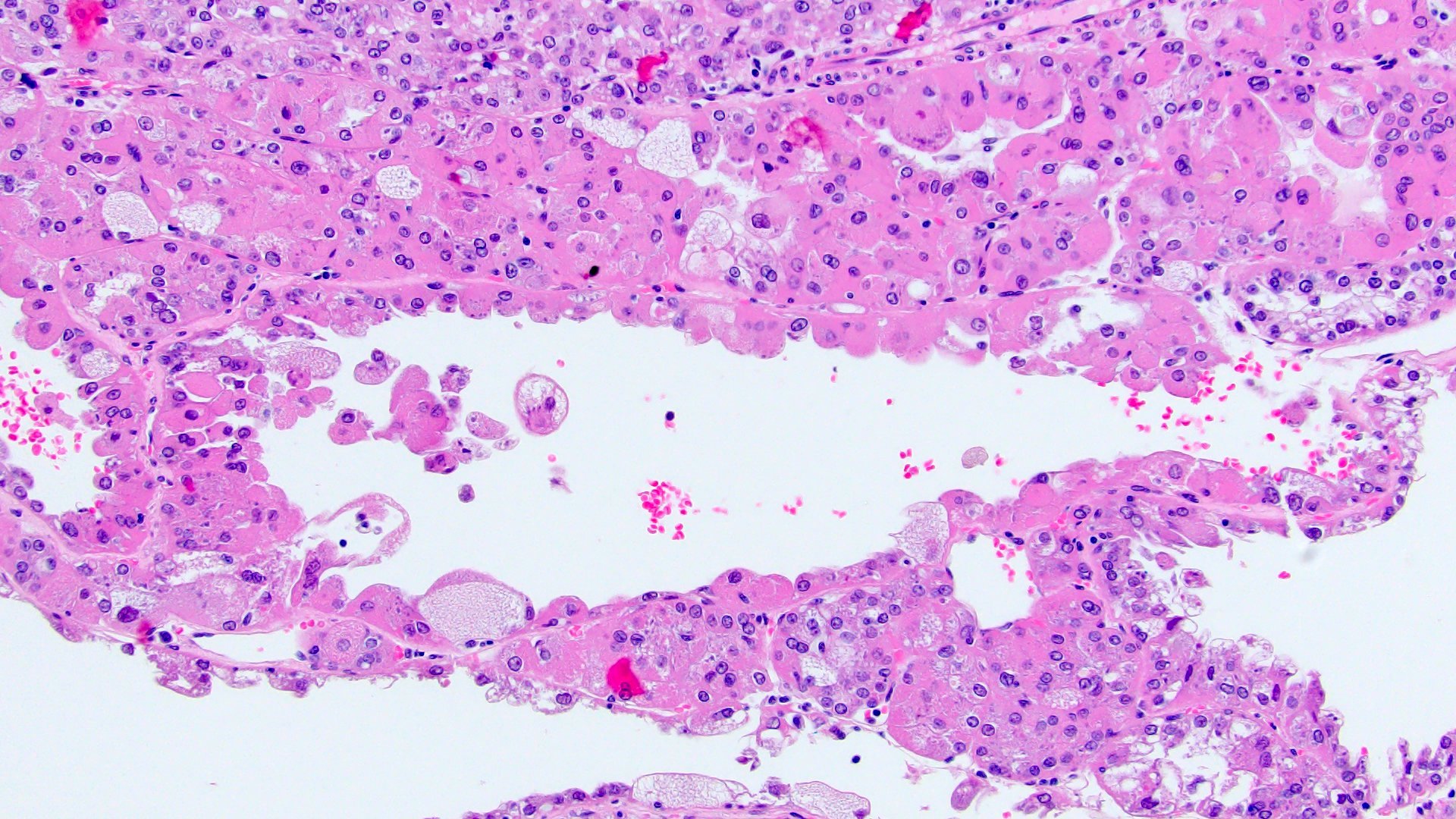
Contributed by Debra Zynger, M.D. (Case #484) and Daniel Anderson, M.D.

Creamy, soft mass

Partial nephrectomy
Contributed by Maria Tretiakova, M.D., Ph.D., Sabrina Sopha, M.D. and Debra Zynger, M.D. (Case #484)

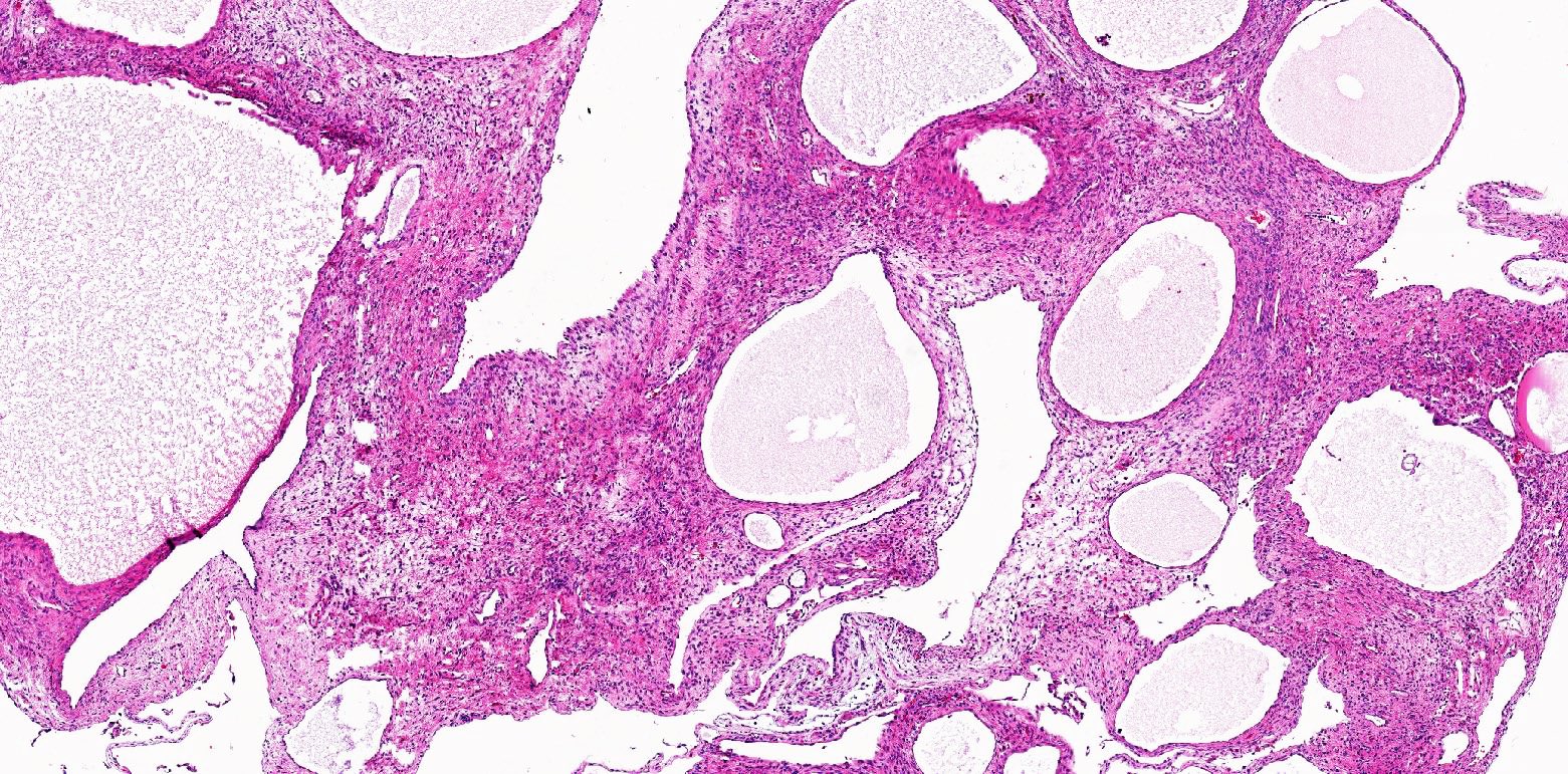
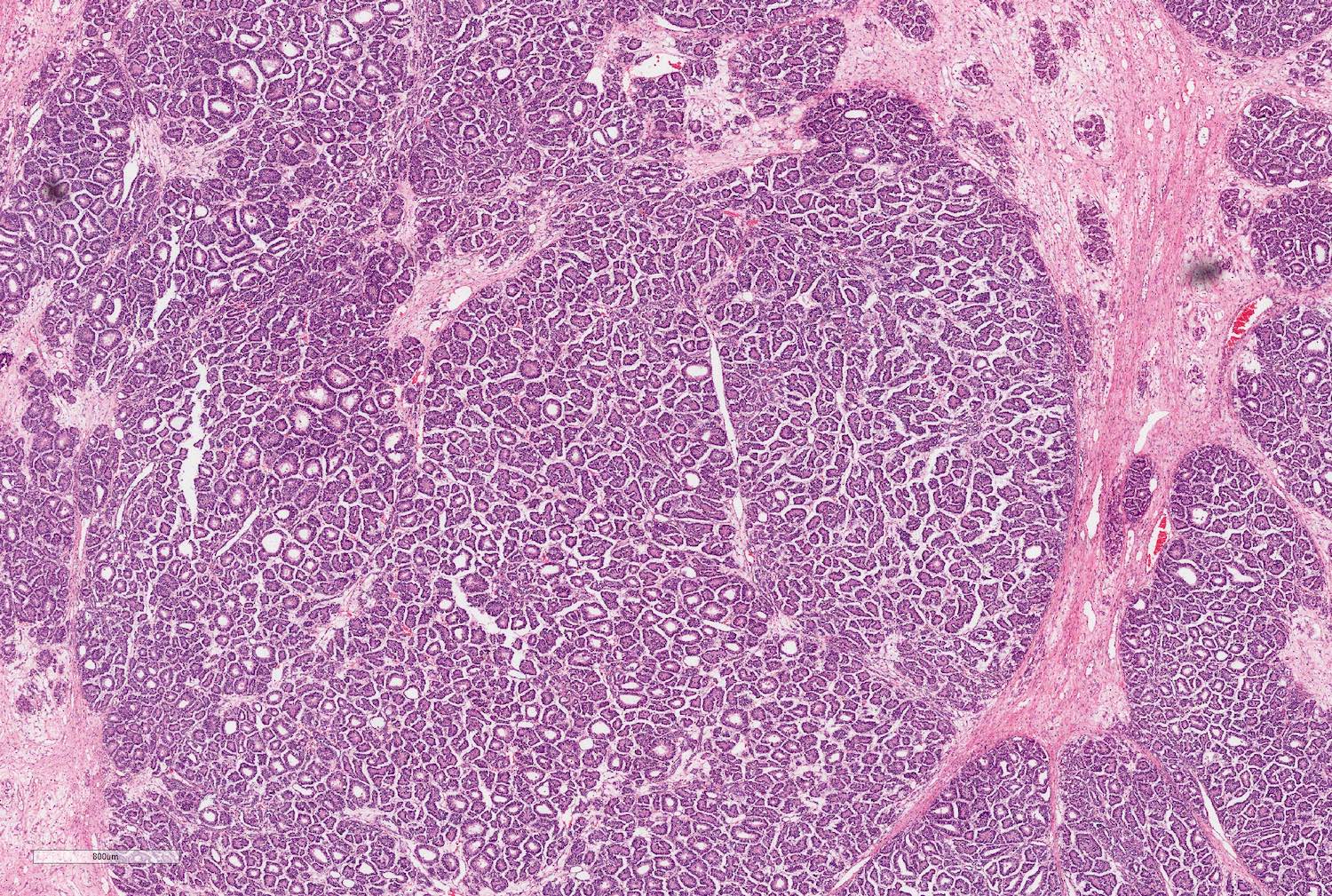
Tubules and variably sized cysts lined by cuboidal epithelium
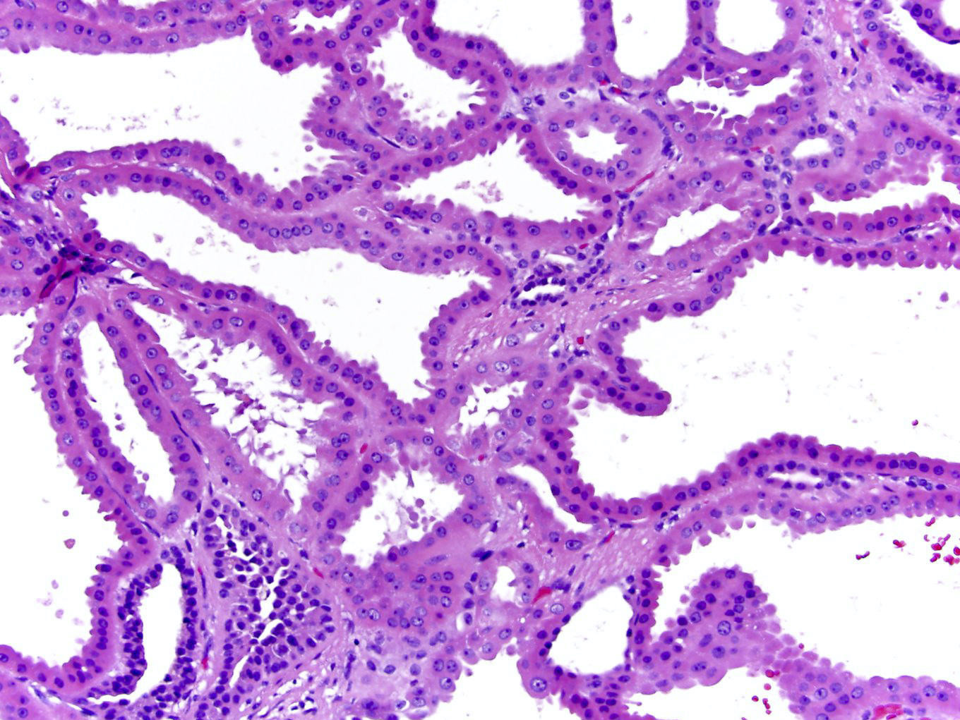
Hobnailing and distinct nucleoli
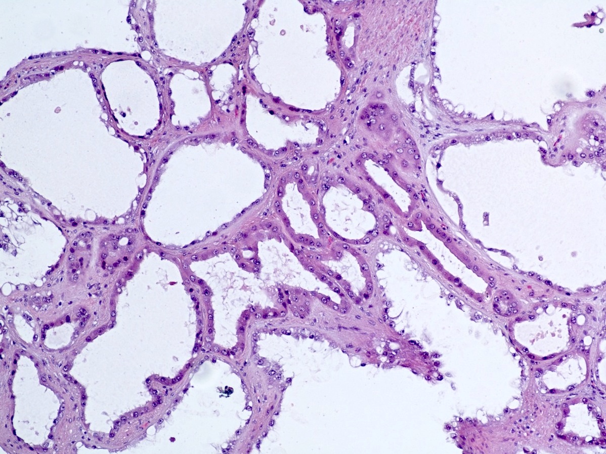
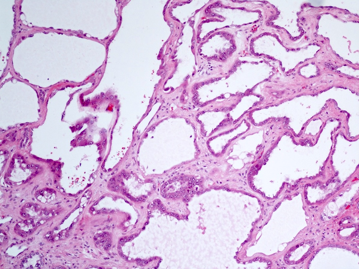
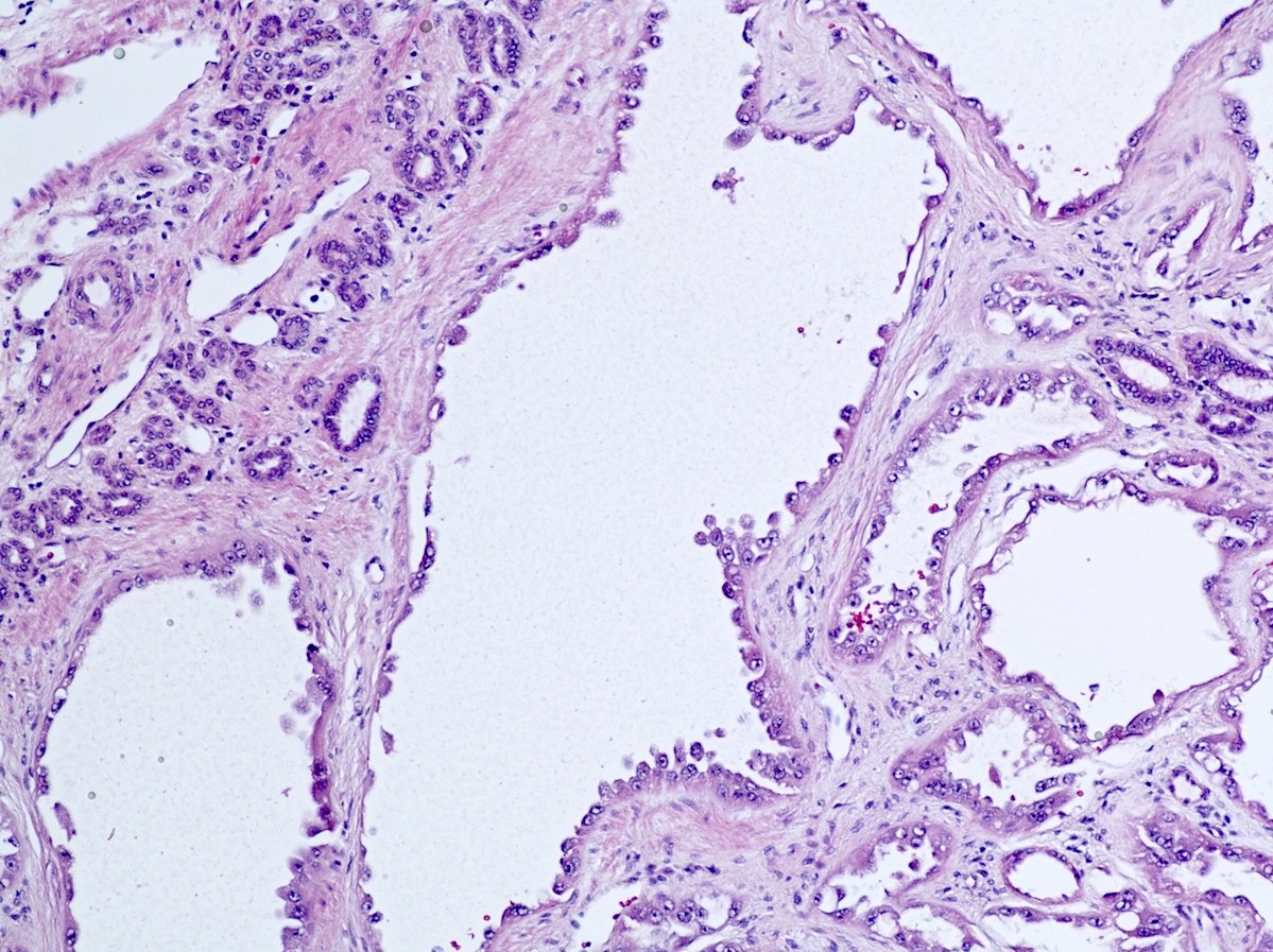

Cysts and tubules lined by cuboidal epithelium with scattered hobnailing
Numerous cysts
Hobnailed lining
Rigid cysts
Nucleoli
Small tubules

Cytoplasmic CD10, CK7 and EMA positivity
Fumarate hydratase
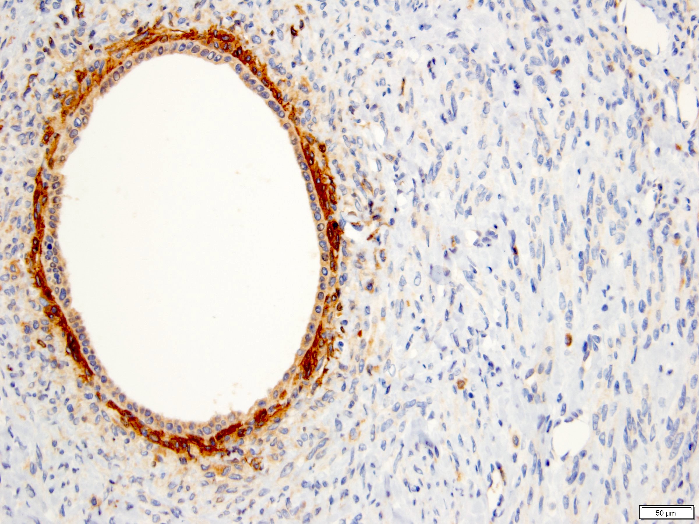
PR
Images hosted on other servers:

Sheets of cells with well defined borders
Images hosted on other servers:

Type I and type II cells
Images hosted on other servers:

Loss of chromosome 9 and gain of chromosome 17

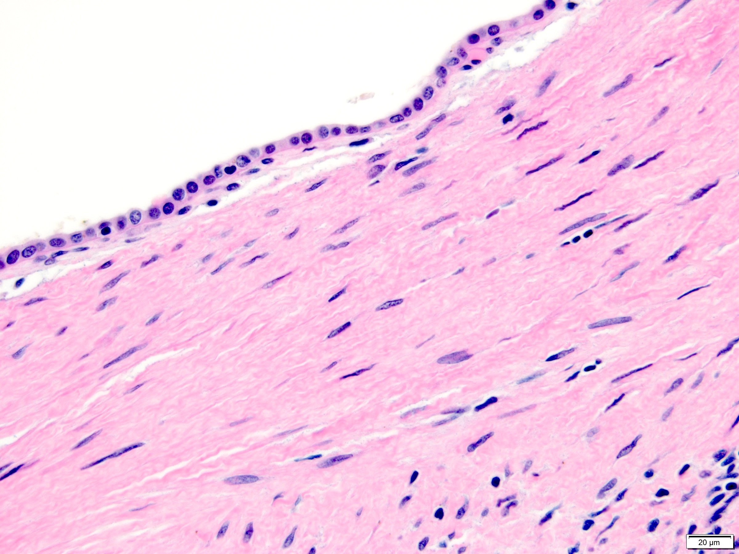
Loss of the Y chromosome in the lining epithelial cells
Tubulocystic renal cell carcinoma
by Dr. Rajal Shah
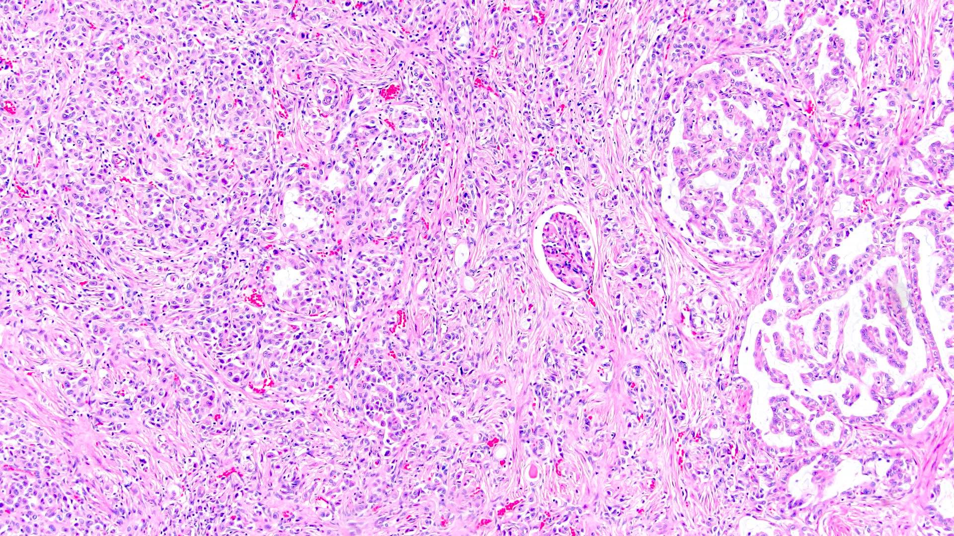
Contributed by Nicole K. Andeen, M.D. and Maria Tretiakova, M.D., Ph.D.

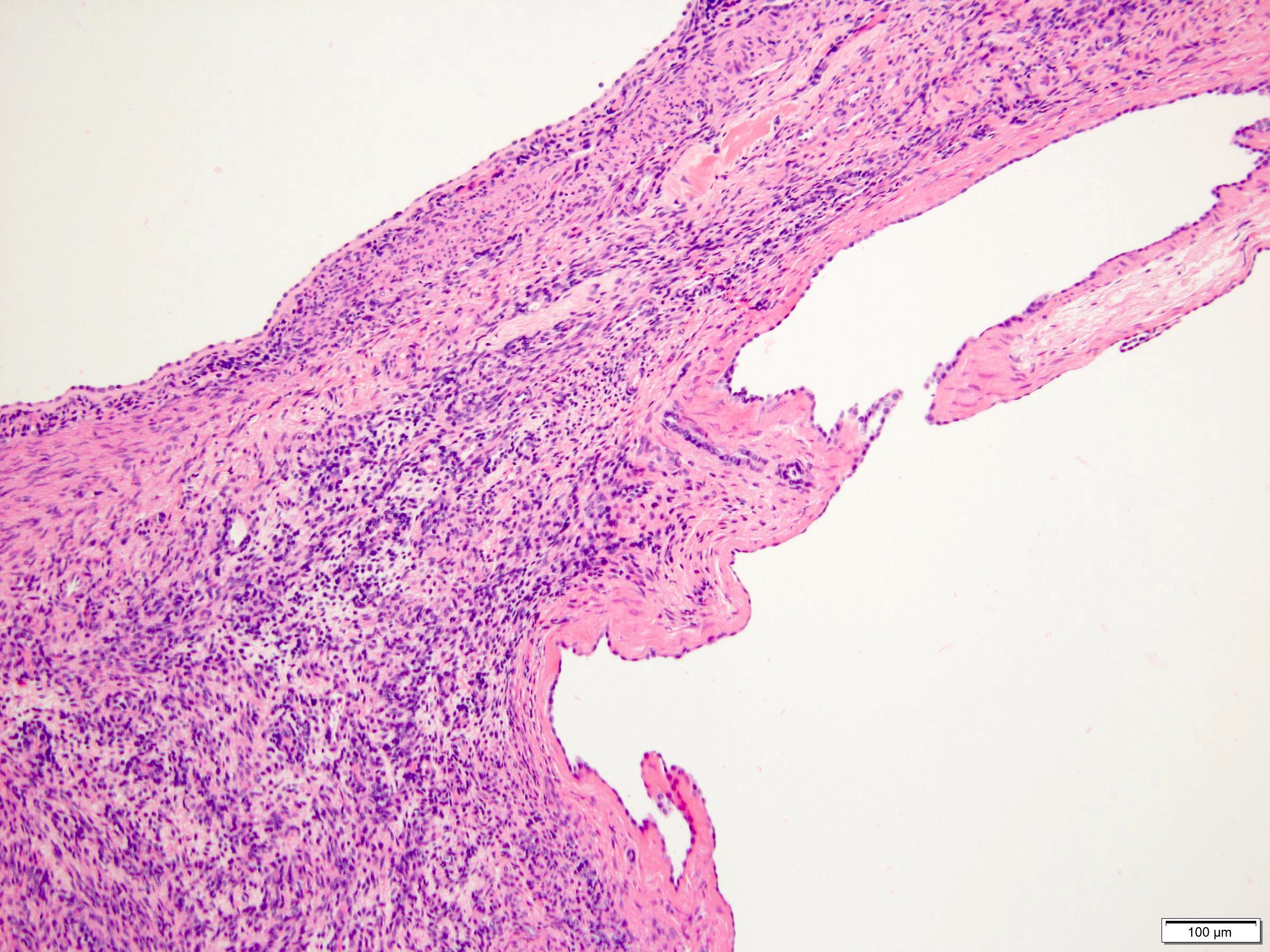
Unclassified RCC
Contributed by Nicole K. Andeen, M.D. and Maria Tretiakova, M.D., Ph.D.
Unclassified RCC CDC-like
Unclassified RCC
Initially unclassified RCC (chromophobe)
Initially unclassified RCC (clear cell)
ISUP presents: reporting of MRI guided prostate biopsies
by Dr. Jennifer Gordetsky
ISUP presents: maximizing the impact of renal mass biopsy
by Dr. Sara Wobker
ISUP presents: emerging WHO entities in renal neoplasia
by Dr. Ondrej Hess
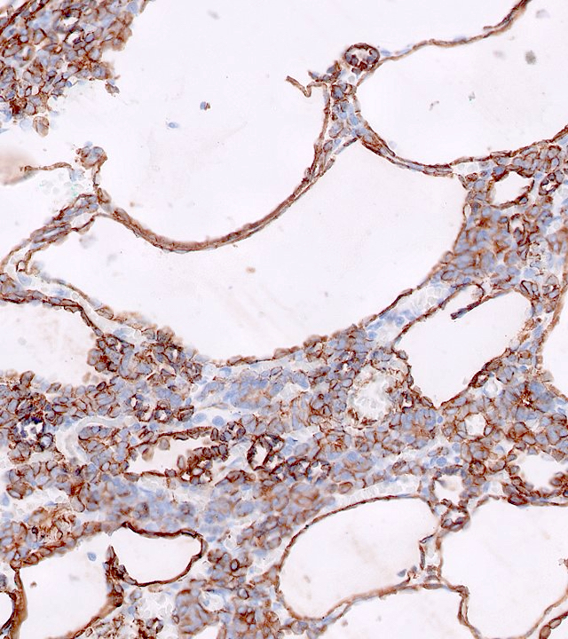
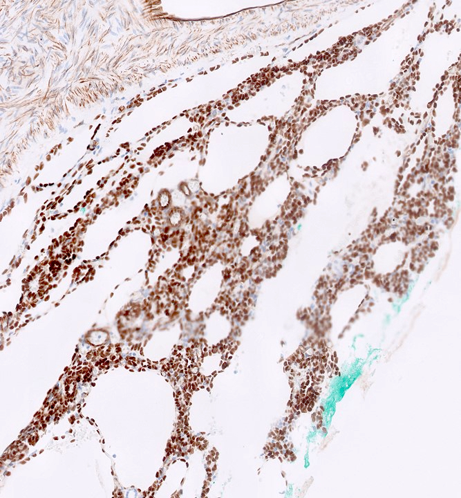
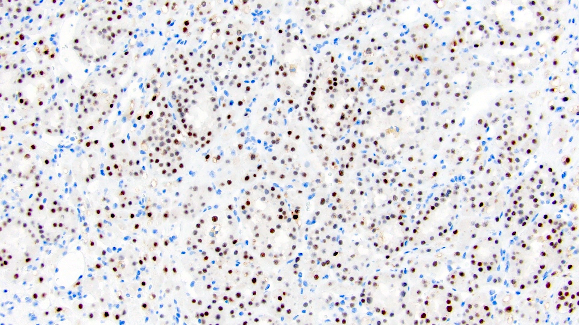
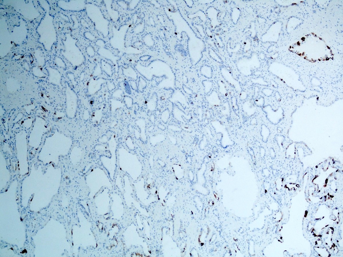
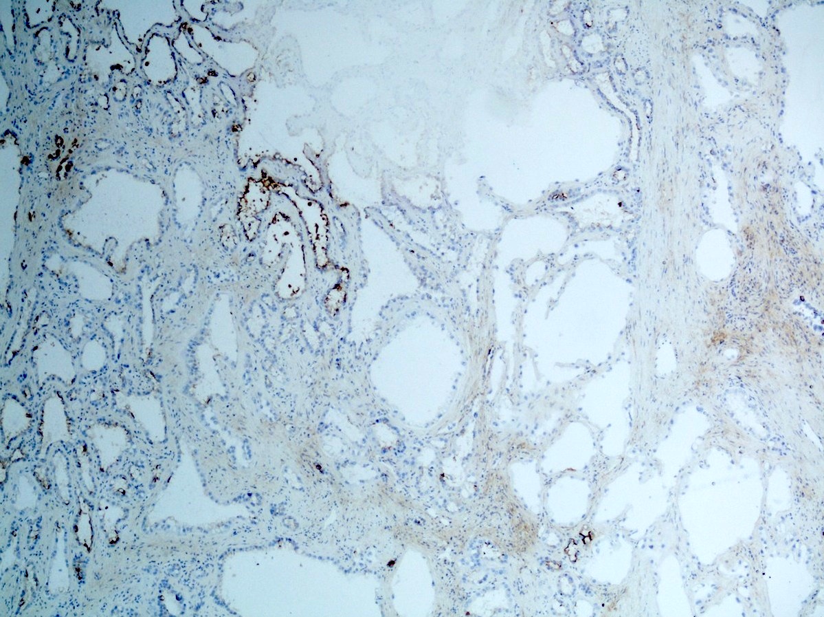
Contributed by Maria Tretiakova, M.D., Ph.D.
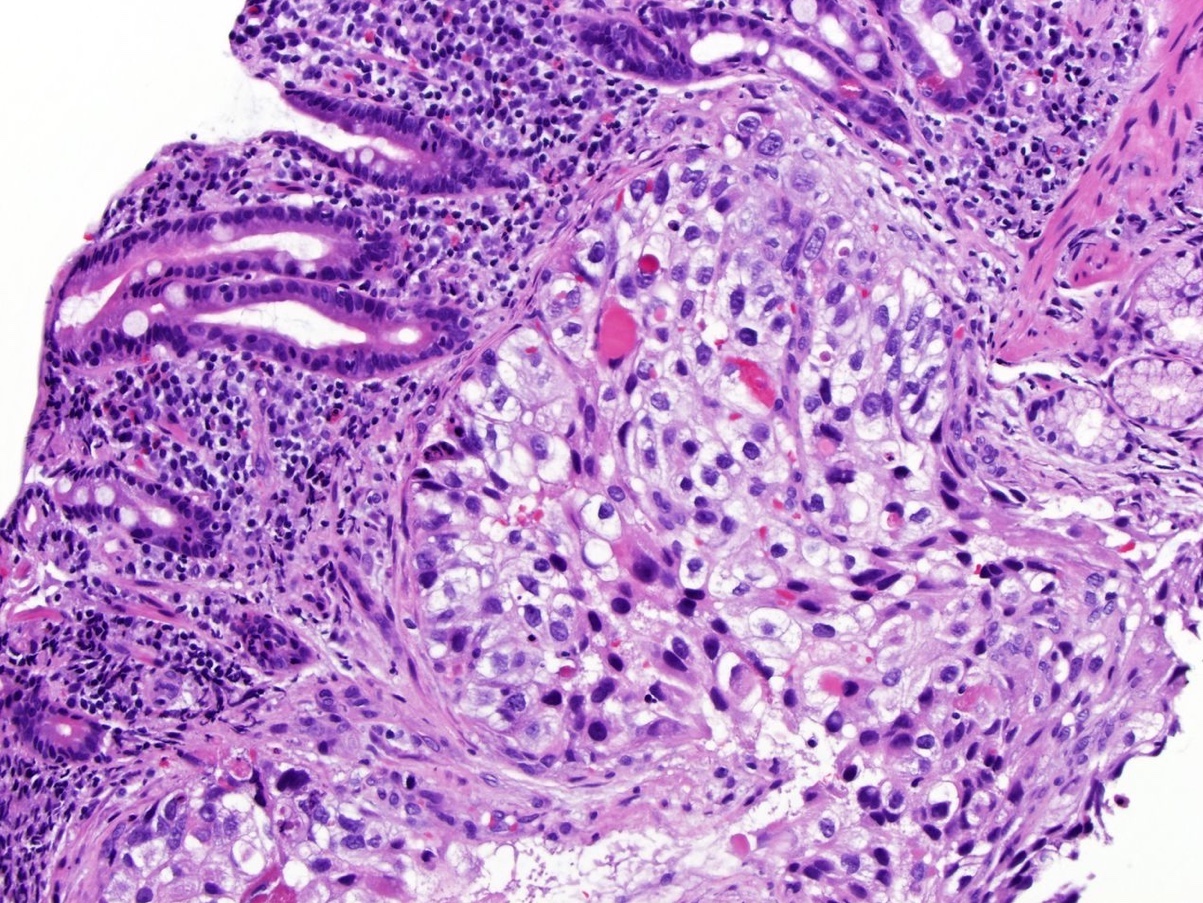
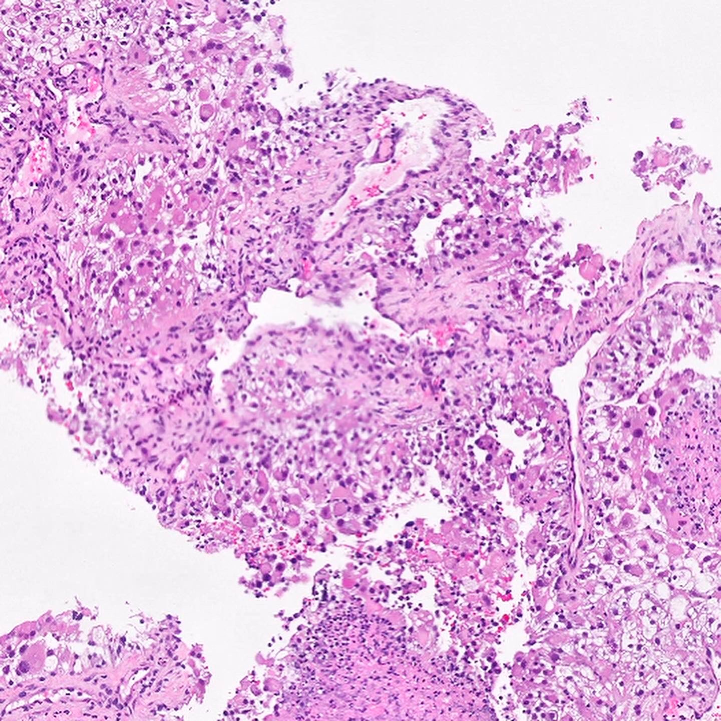
ALK rearranged renal cell carcinoma
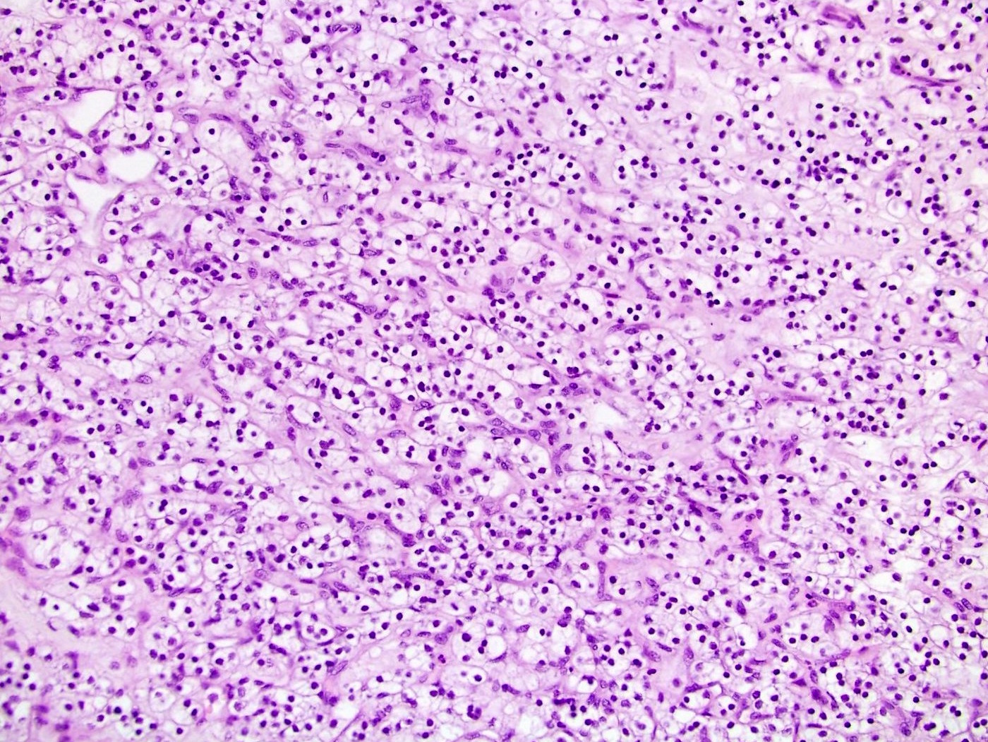
Eosinophilic solid and cystic renal cell carcinoma
ELOC (formerly TCEB1) mutated renal cell carcinoma
Fumarate hydratase deficient renal cell carcinoma
Macronucleoli
Low grade oncocytic tumor
Images hosted on other servers:

Overall and disease specific survival by histology
Images hosted on other servers:

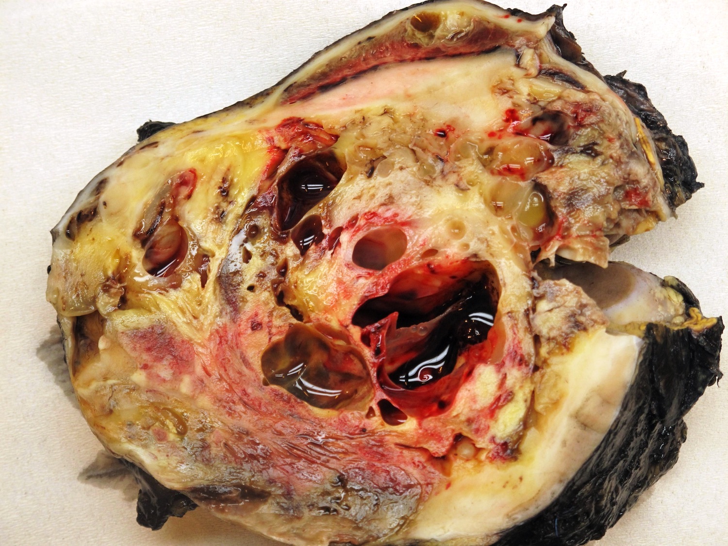
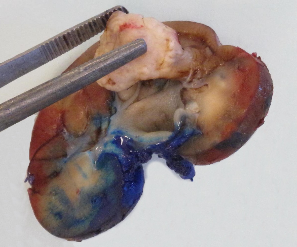
Right renal mass

Enhancing right renal mass

Tumor extending into inferior vena cava

Cystic tumor

Slightly hyperdense tumor

Metastases on DOTATATE scan


CT and MRI showing left renal mass in a horseshoe kidney
Contributed by Jennifer Jeung, M.D. (Case #204)
Tan, soft, well encapsulated mass in lower pole
Images hosted on other servers:

Tumor in horseshoe kidney

Renal mass with hemorrhage

Necrotic renal mass

Tumor involving sinus fat

Cystic renal mass
Contributed by Maria Tretiakova, M.D., Ph.D., Leandro Freitas, M.D., Ana Grizotto, M.D., Athanase Billis, M.D.,
the Genitourinary Pathology Society (Case #521) and Sleiman Khalil, M.D.
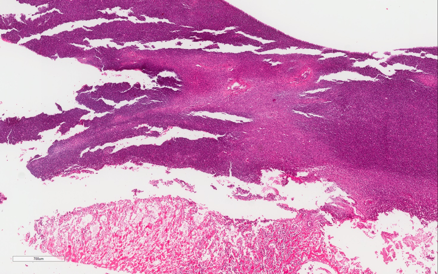
Well demarcated tumor

Nests and pseudoglands
Monotonous tumor cells
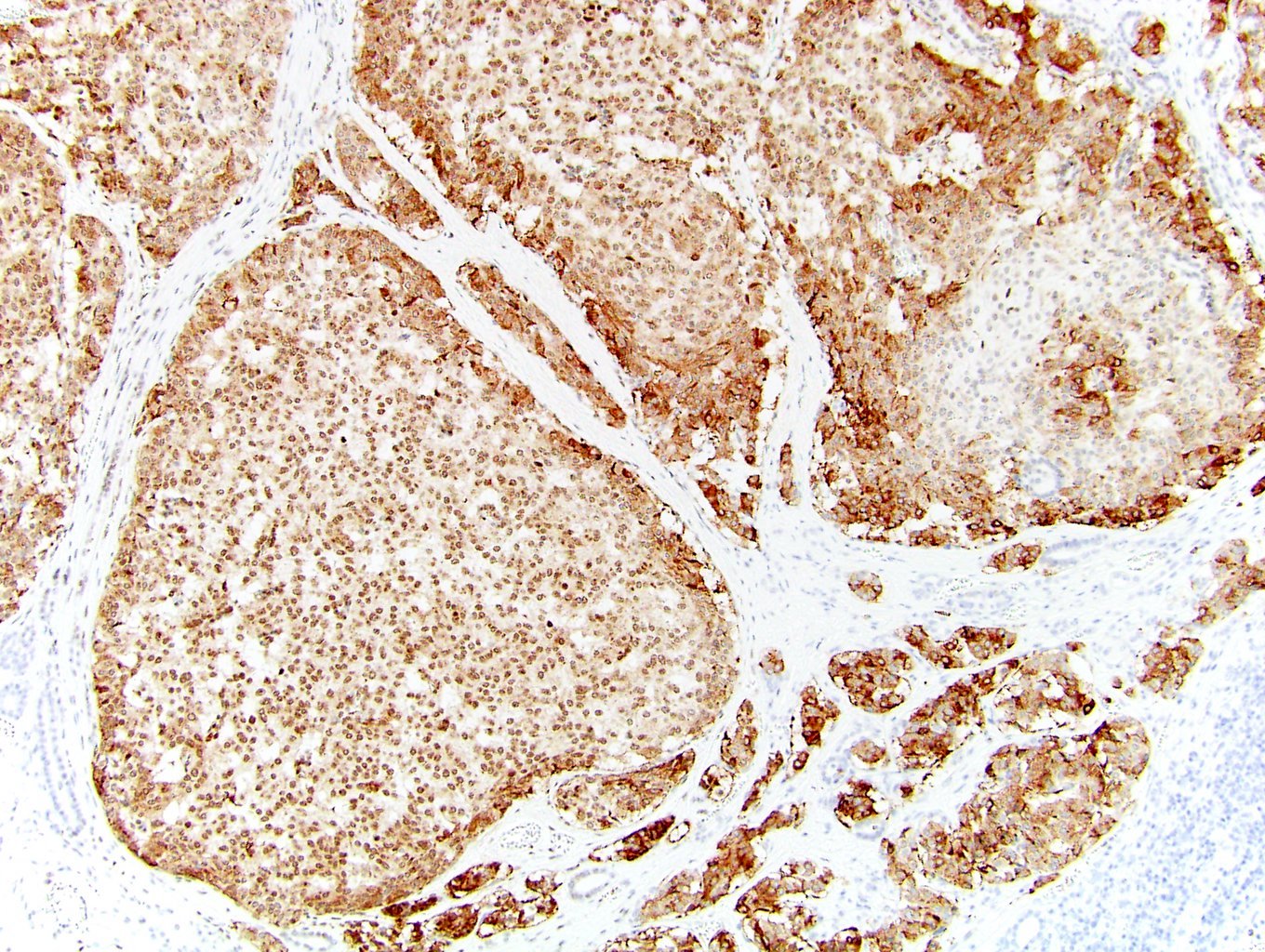
Classic appearance
Prominent stroma
Areas of solid growth
Gland-like lumina

Positive synaptophysin
Positive chromogranin

Positive CD99
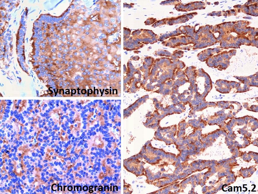
Synaptophysin, chromogranin, CAM5.2
Images hosted on other servers:

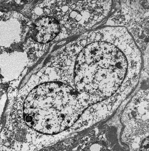
Monotonous cells

Positive synaptophysin stain

Granular cytoplasm
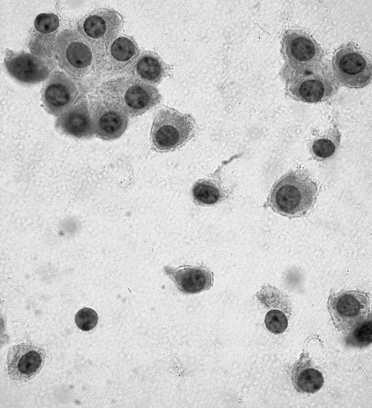
Contributed by Jennifer Jeung, M.D. (Case #204)
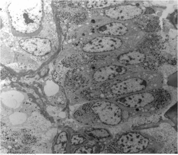
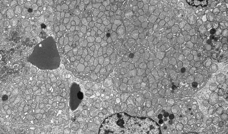
TEM of tumor cells

Prominent neurosecretory granules
Amin: 2022

Cheng: 2019
Eble: 2022
Fatima: 2019
IARC: 2022

Jennette: 2023
Jiang: 2023
Lara: 2015
Pritchard-Jones: 2015
Tickoo: 2015
VandenBussche: 2022

Wobker: 2021

Yang: 2020
Zhou: 2022
Zhou: 2022
Find related Pathology books: GU/adrenal, renal, cytopathology
