Superpage - Images

Age related changes
Diagrams / tables

Contributed by Dragos C. Luca, M.D.

Age related normal values in bone marrow

Gross images

Images hosted on other servers:

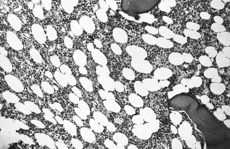
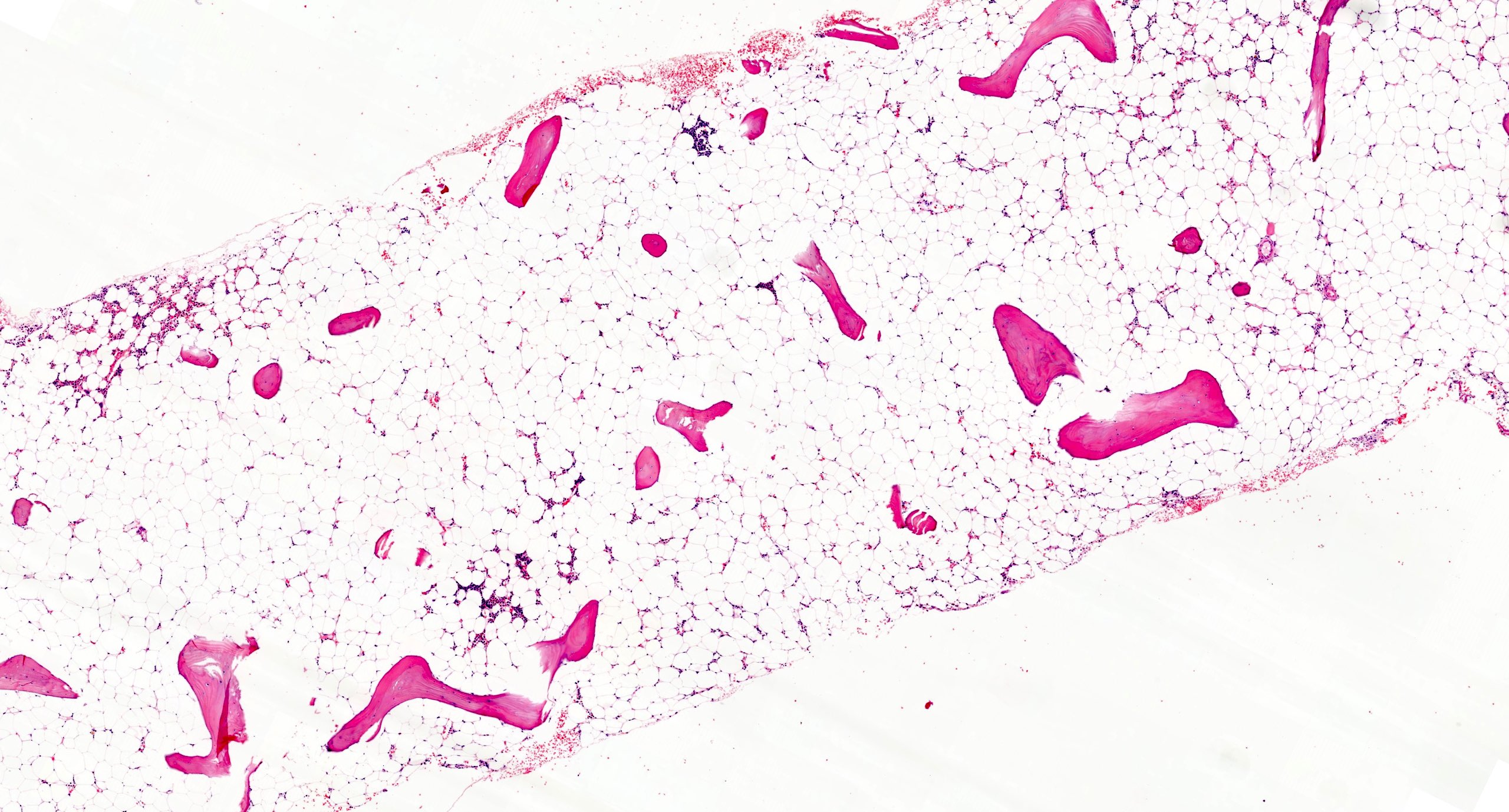
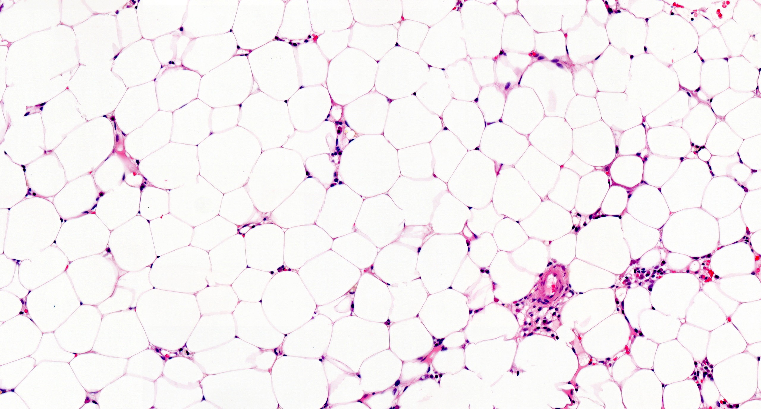
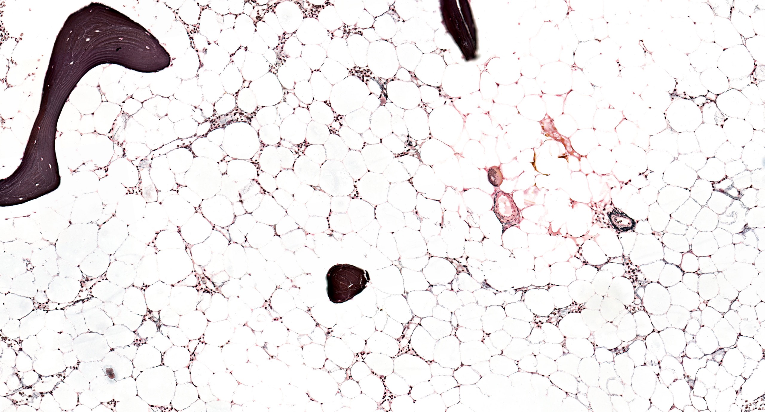
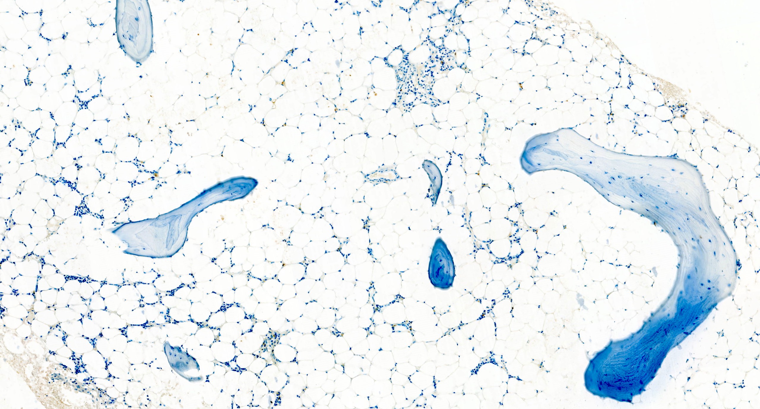
Fatty marrow

Microscopic (histologic) images

AFIP images

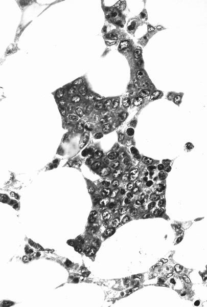
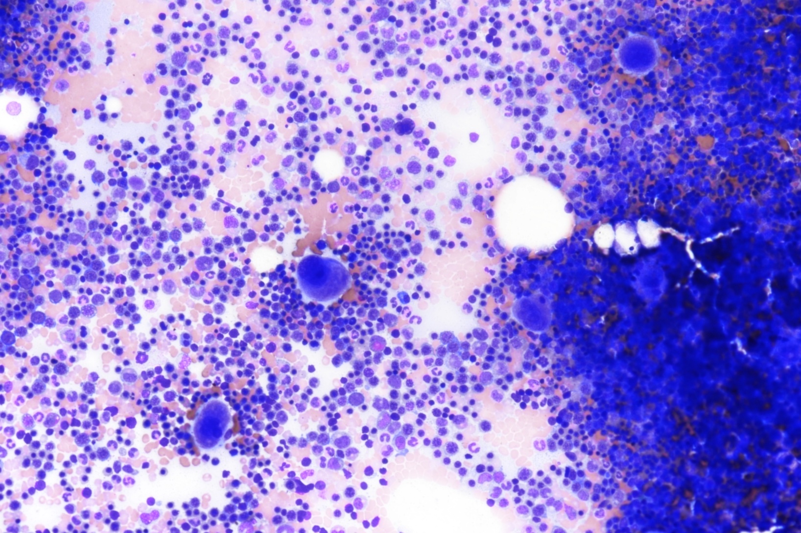
6 month old child has highly cellular marrow (normal for age)

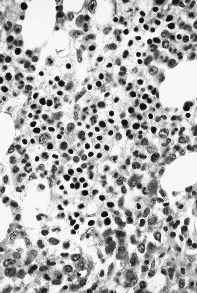
Adult marrow (ages 30 - 69 years)


Images hosted on other servers:

Adult marrow


Alcohol abuse
Microscopic (histologic) images

Images hosted on other servers:

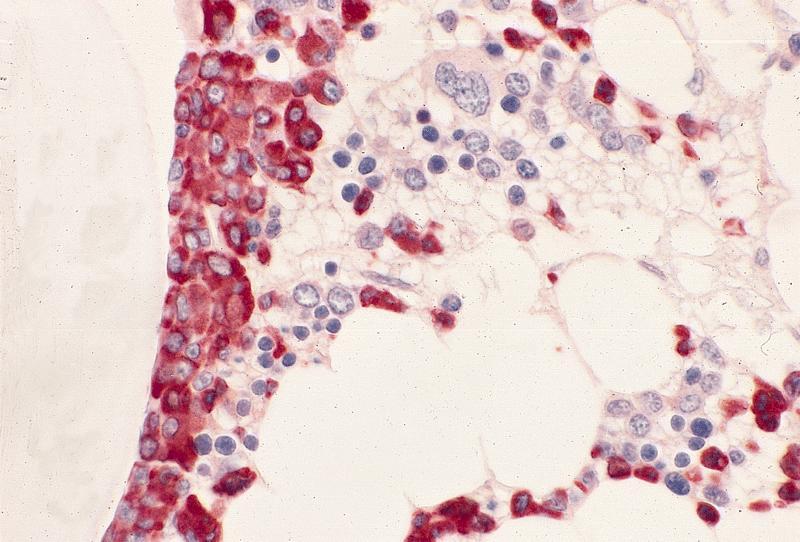
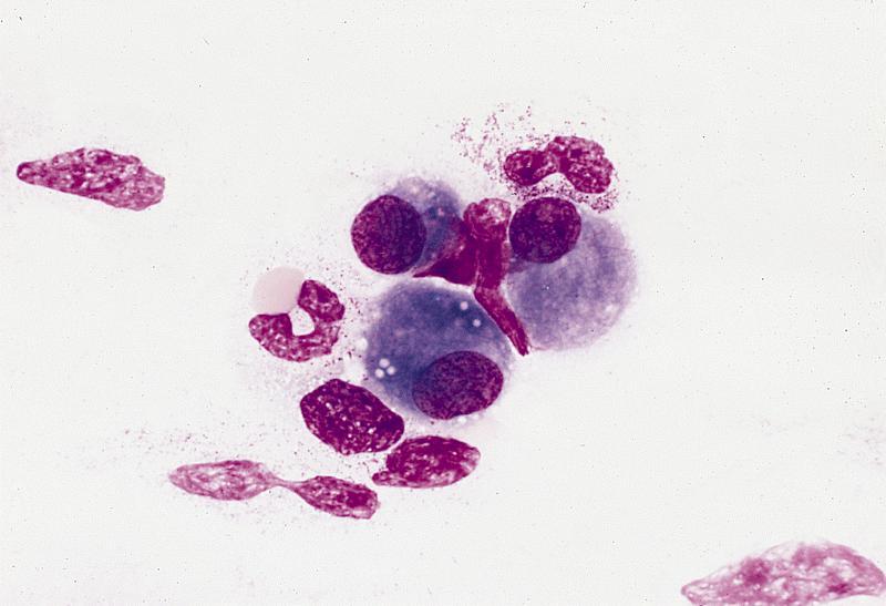
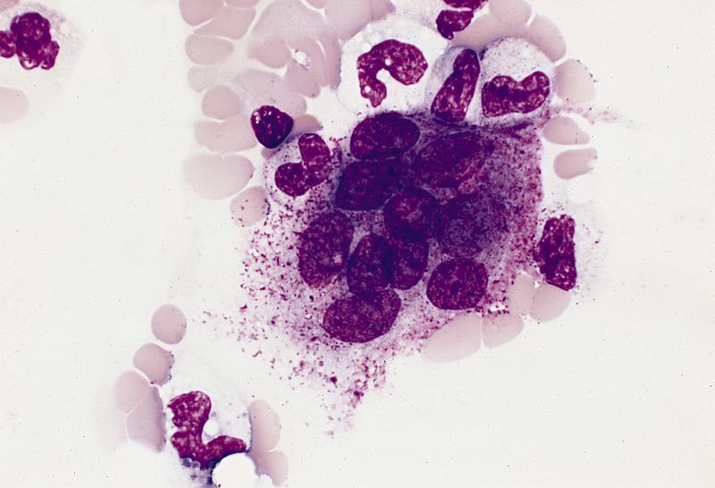
Marked megaloblastic features

Markedly hypercellular

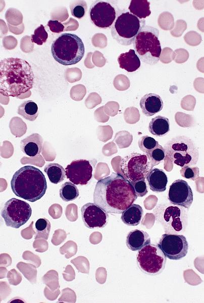
Bone marrow: intracytoplasmic vacuoless

Multiple coarses granules

Plasma cell: iron granules forming plaque

Hemosiderin granules in normoblast

Peripheral smear images

Images hosted on other servers:

Peripheral blood erythrocytes

Vacuolation of peripheral blood cells

Electron microscopy images

Images hosted on other servers:

Marrow plasma cell has dense aggregate of iron

Iron laden organelle
in upper cell may
correspond to
microscopic plaque


Aplastic anemia (AA)
Microscopic (histologic) images

Contributed by Valentina Sangiorgio, M.D.

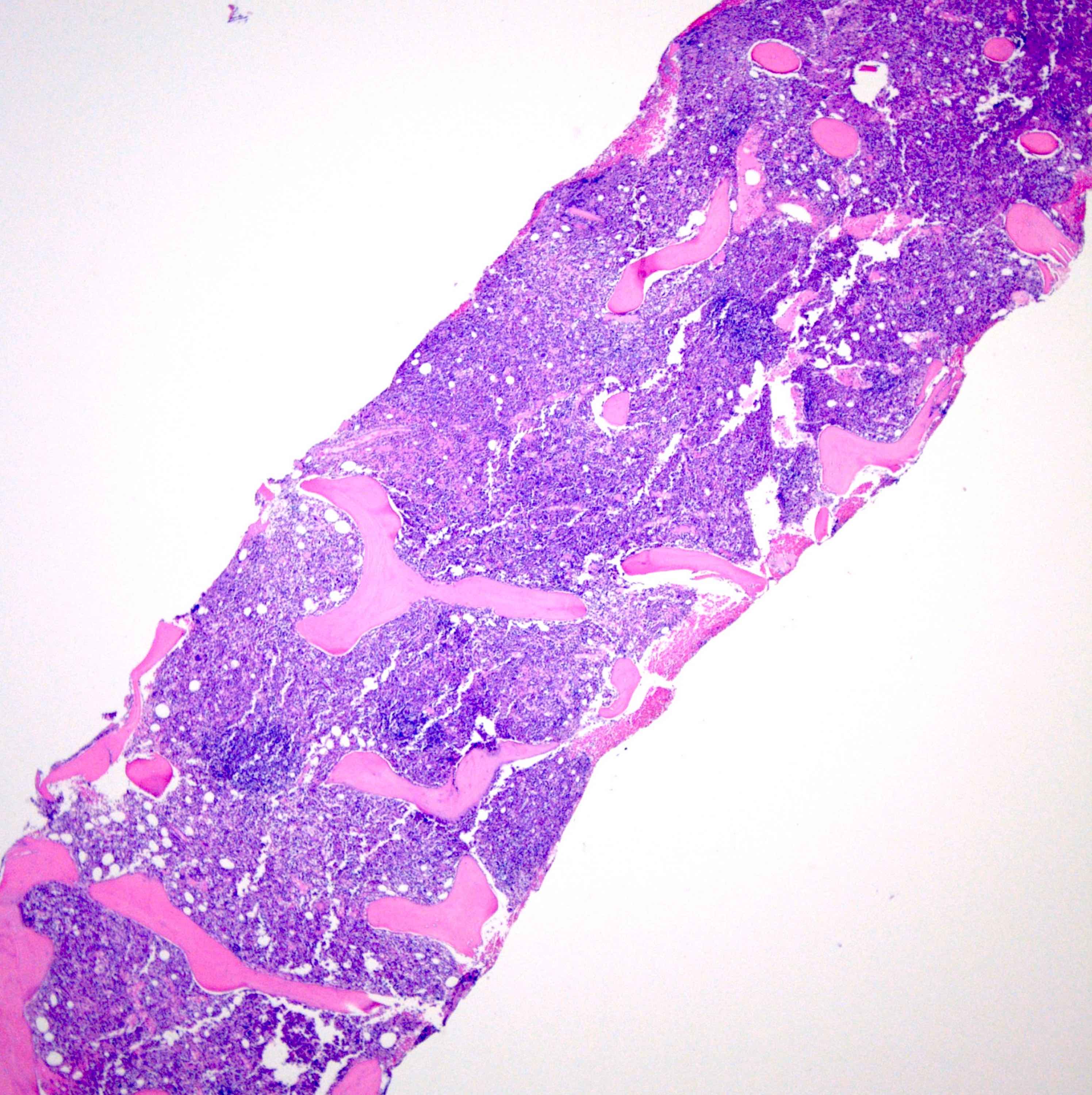
Hypocellular bone marrow

Absence of fibrosis

Virtual absence of myeloid cells


Isolated residual erythroid islands

Virtual absence of megakaryocytes

Normal CD34 count

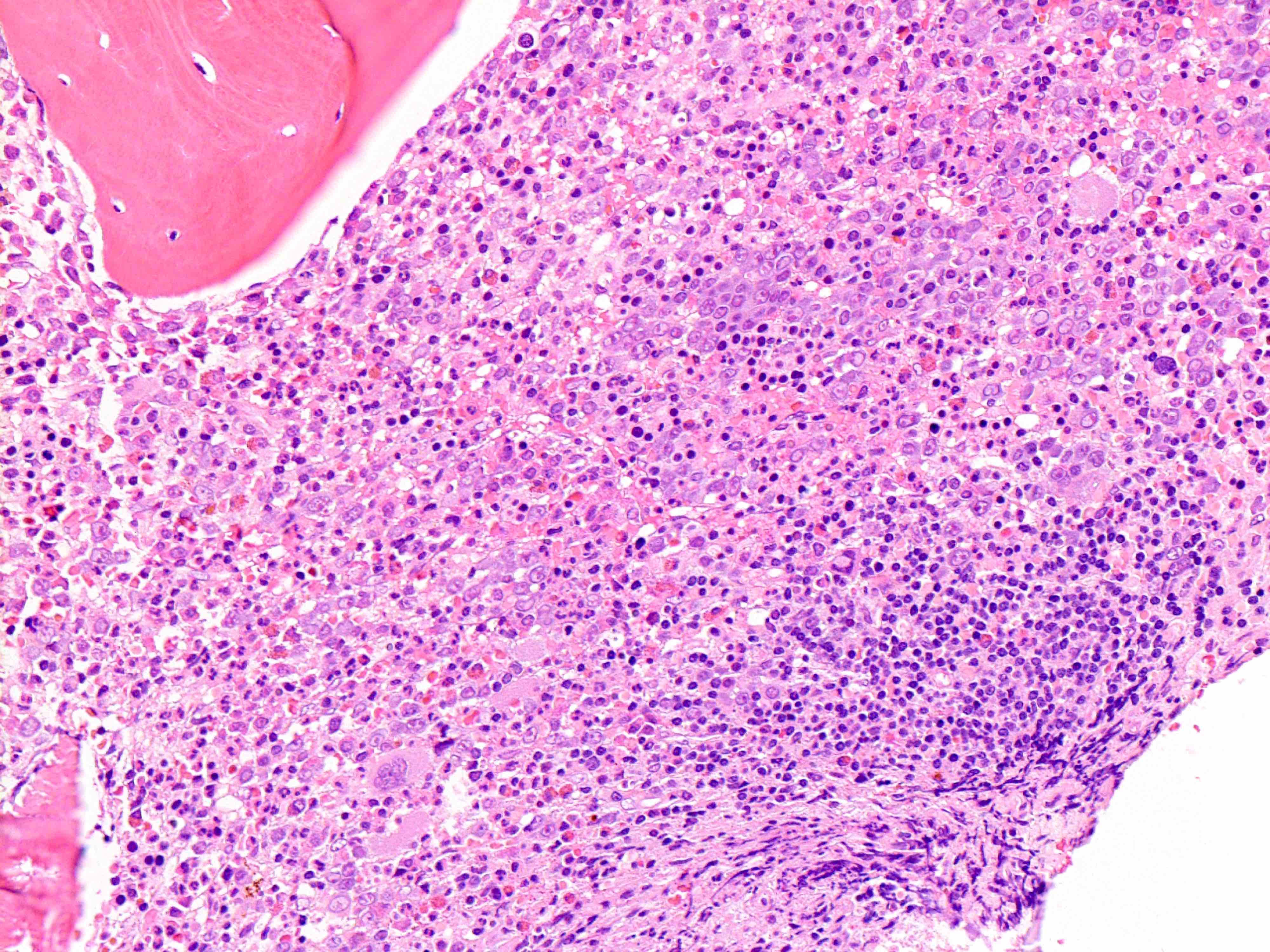
Mast cell hyperplasia

Flow cytometry images

Contributed by Valentina Sangiorgio, M.D.

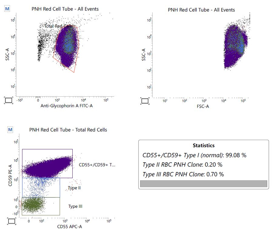
PNH red cell tube


Arsenic toxicity
Clinical images

Images hosted on other servers:

Pigment changes and
palmoplantar hyperkeratoses
are characteristic of
chronic arsenic exposure

Arsenic keratosis -
so called "raindrops
on a dusty road"

Skin cancer

Microscopic (histologic) images

AFIP images

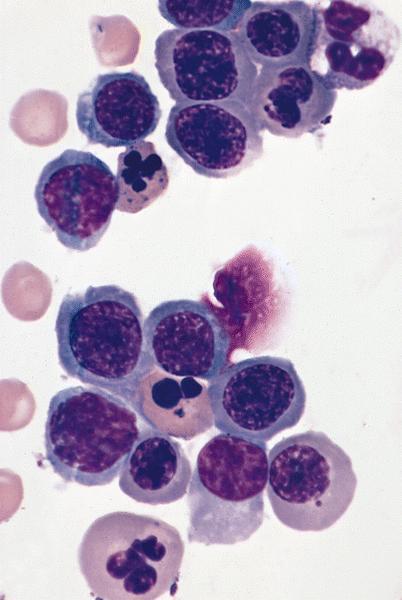
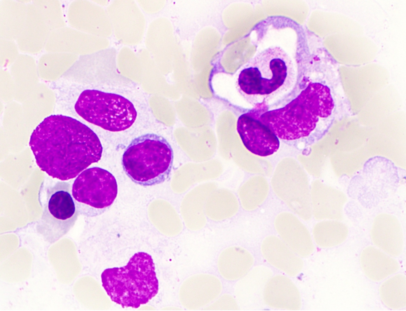
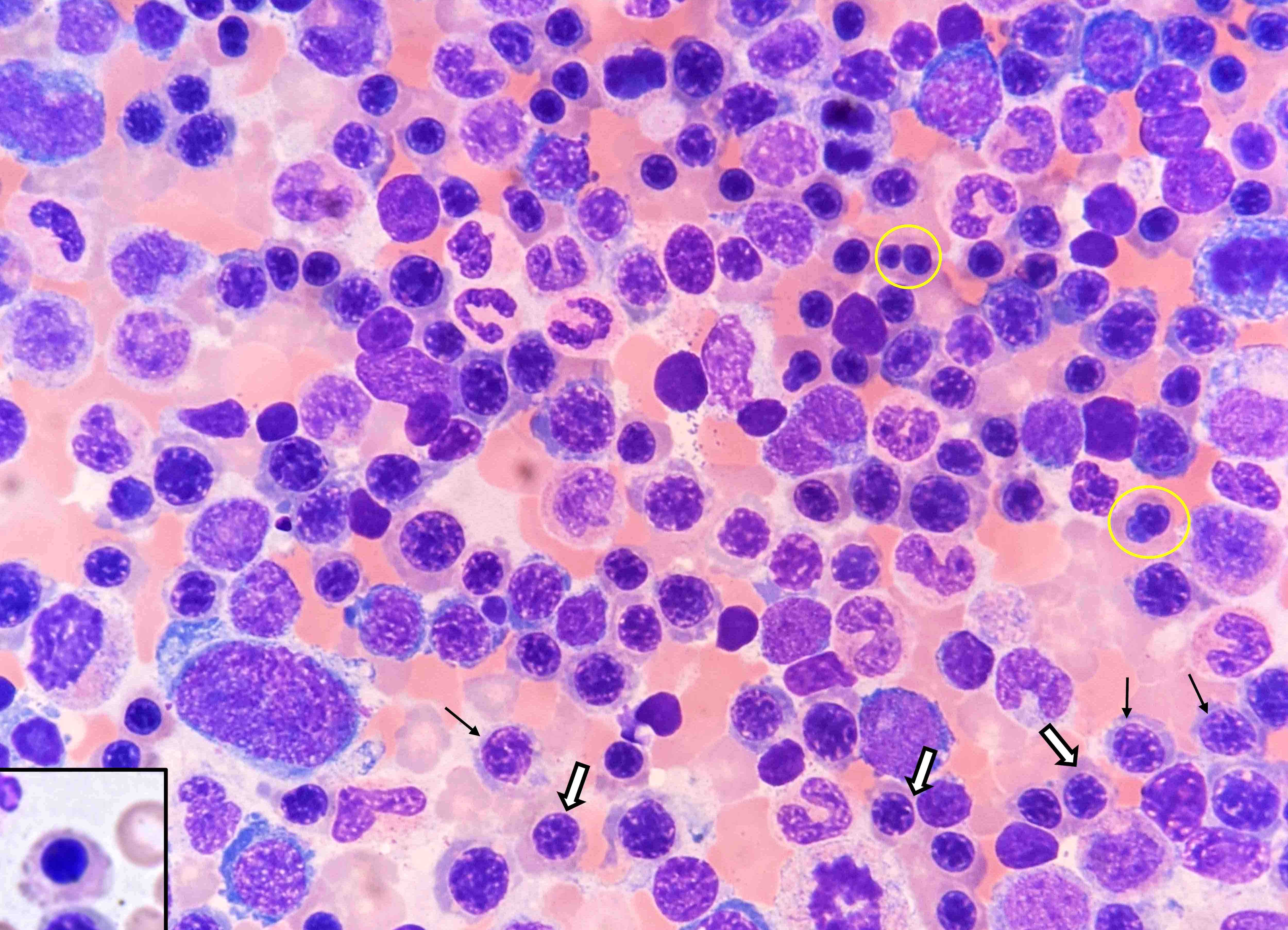
Bone marrow: erythroid
precursors have marked
dysplastic changes including
nuclear lobulation and karyorrhexis

Peripheral smear images

AFIP images

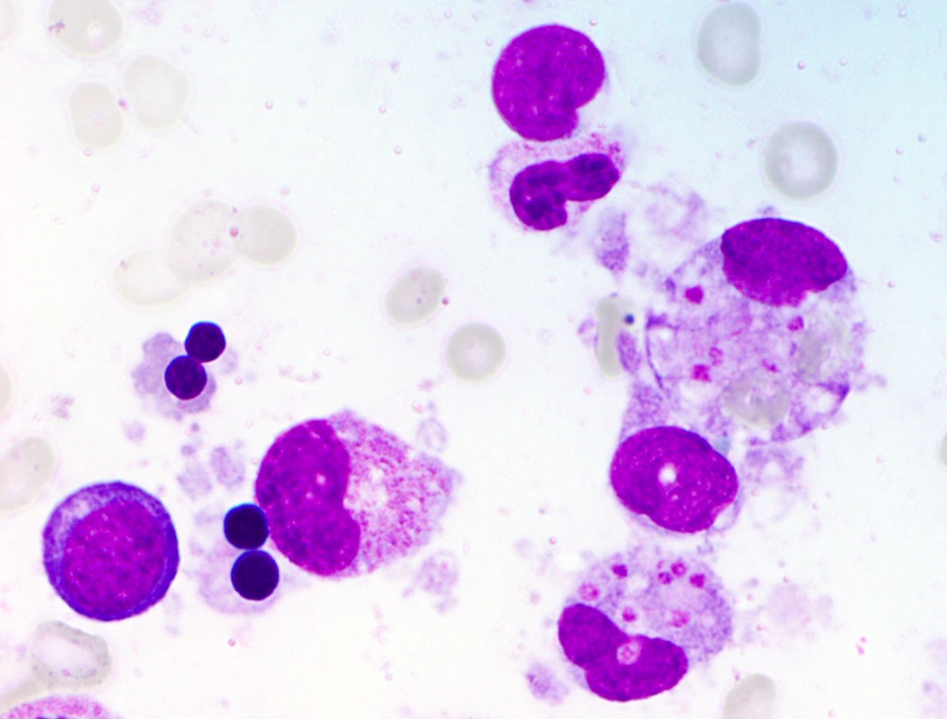
Erythroid precursor with
slightly lobulated
megaloblastoid nucleus

Electron microscopy images

Images hosted on other servers:

Abnormal erythroblasts


Autoimmune myelofibrosis (AIMF)
Microscopic (histologic) images

Contributed by Ria Vergara-Lluri, M.D.
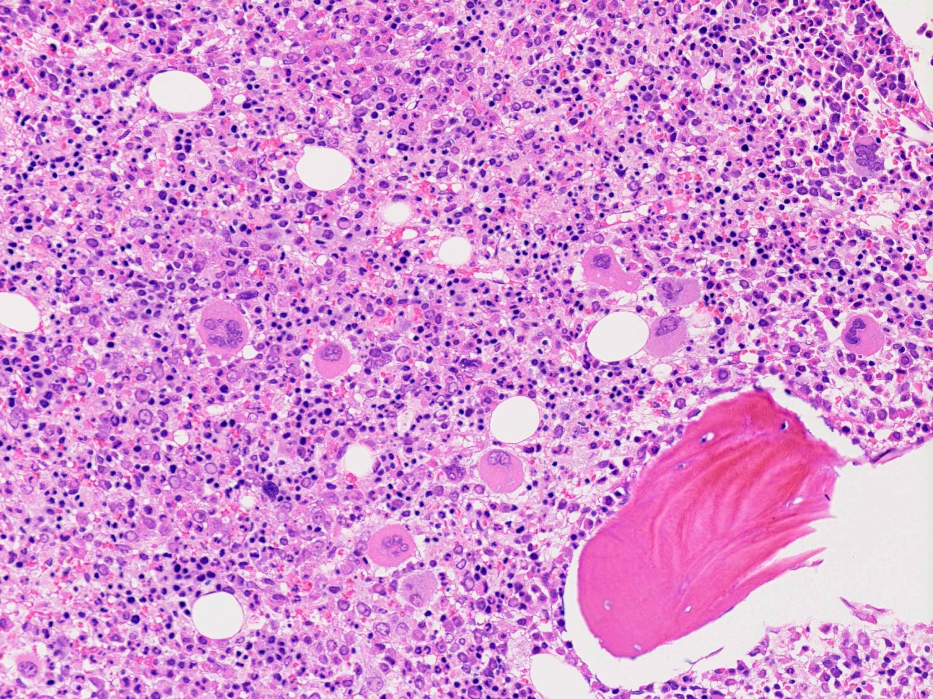
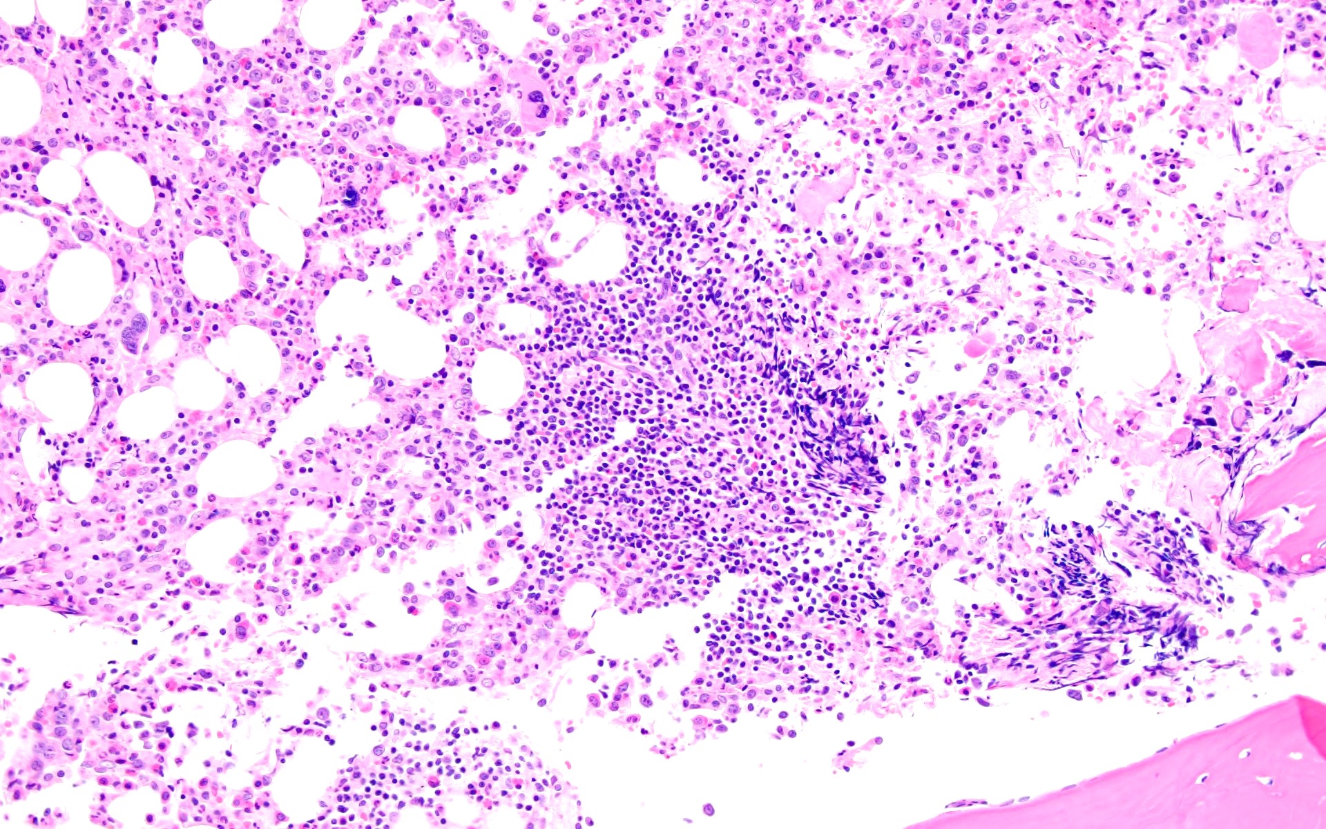
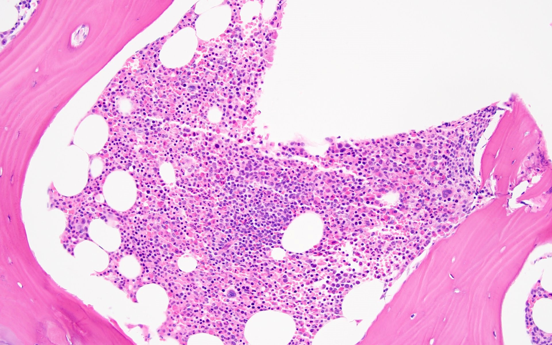
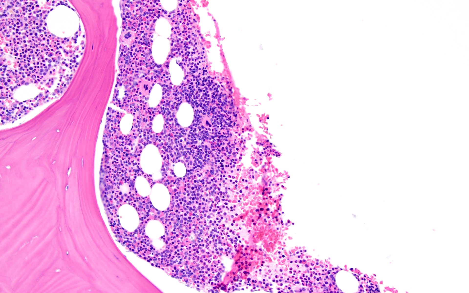
Markedly hypercellular bone marrow

Markedly hypercellular bone marrow

Erythroid hyperplasia

Erythroid hyperplasia

Megakaryocytic hyperplasia without atypia

Megakaryocytic hyperplasia without atypia

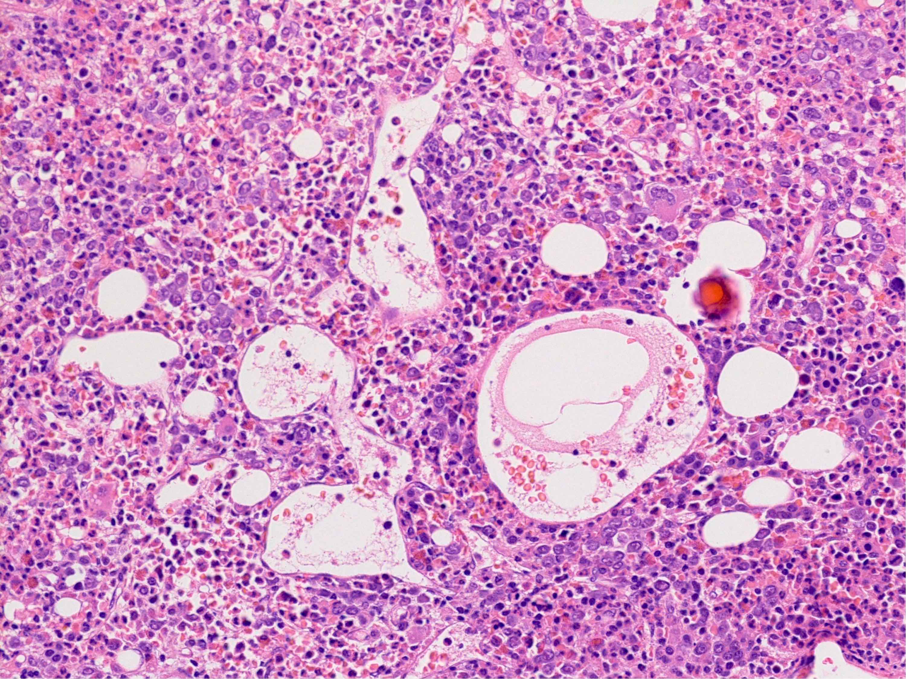
Sinusoidal dilation

Sinusoidal dilation

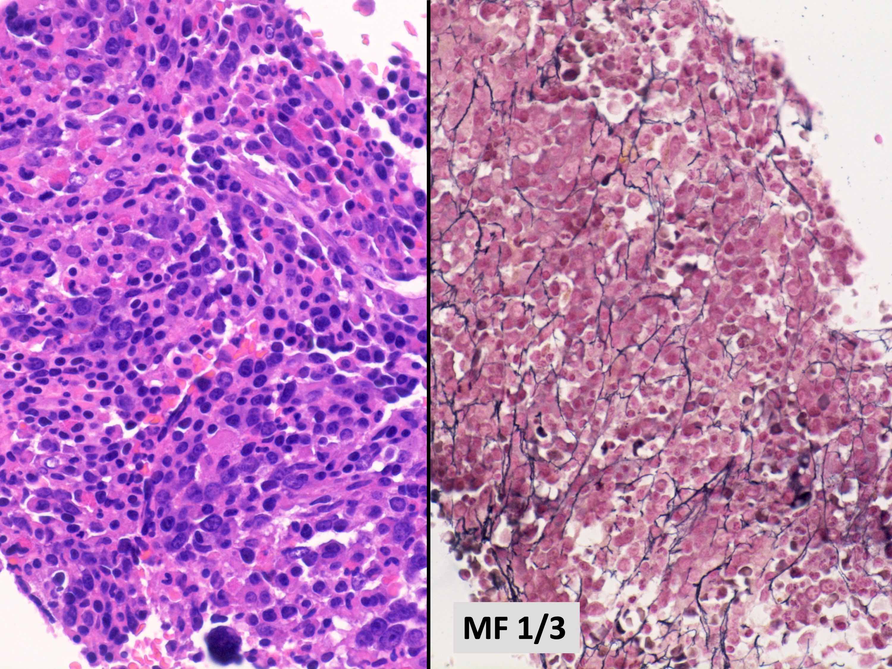
MF grade 1

MF grade 1

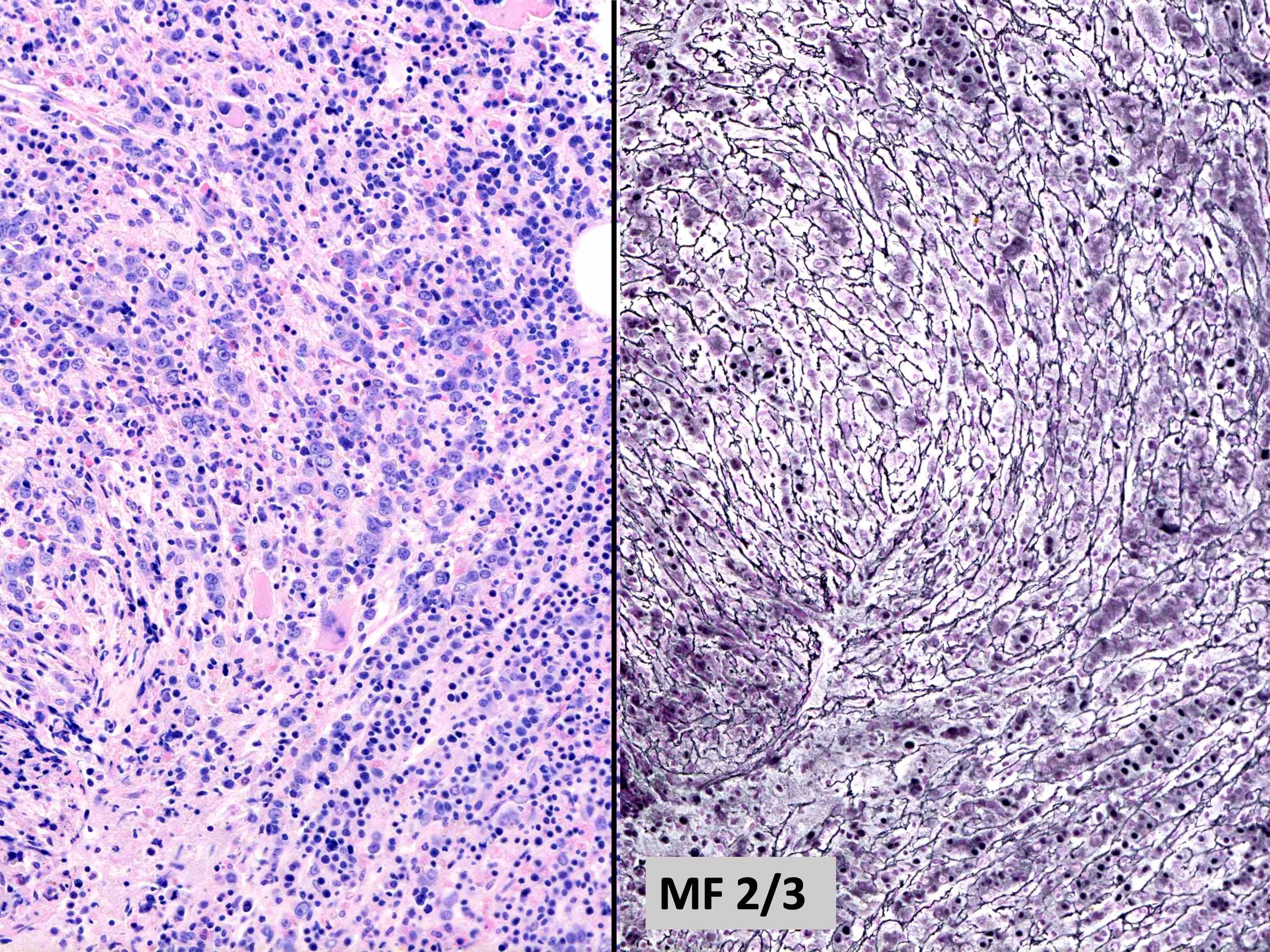
MF grade 2

MF grade 2


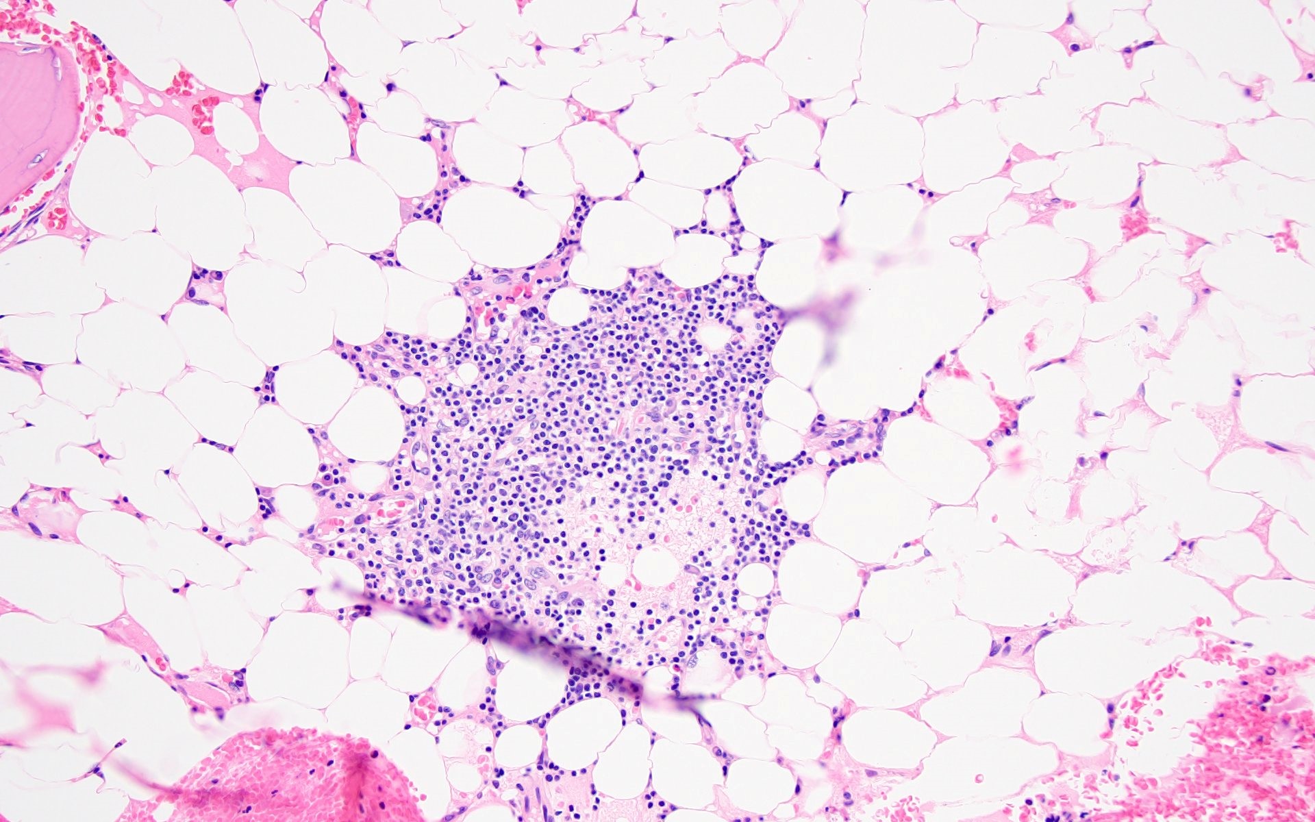
Benign lymphoid aggregate

Benign lymphoid aggregate

Benign lymphoid aggregate immunostains Benign lymphoid aggregate immunostains

Benign lymphoid aggregate immunostains

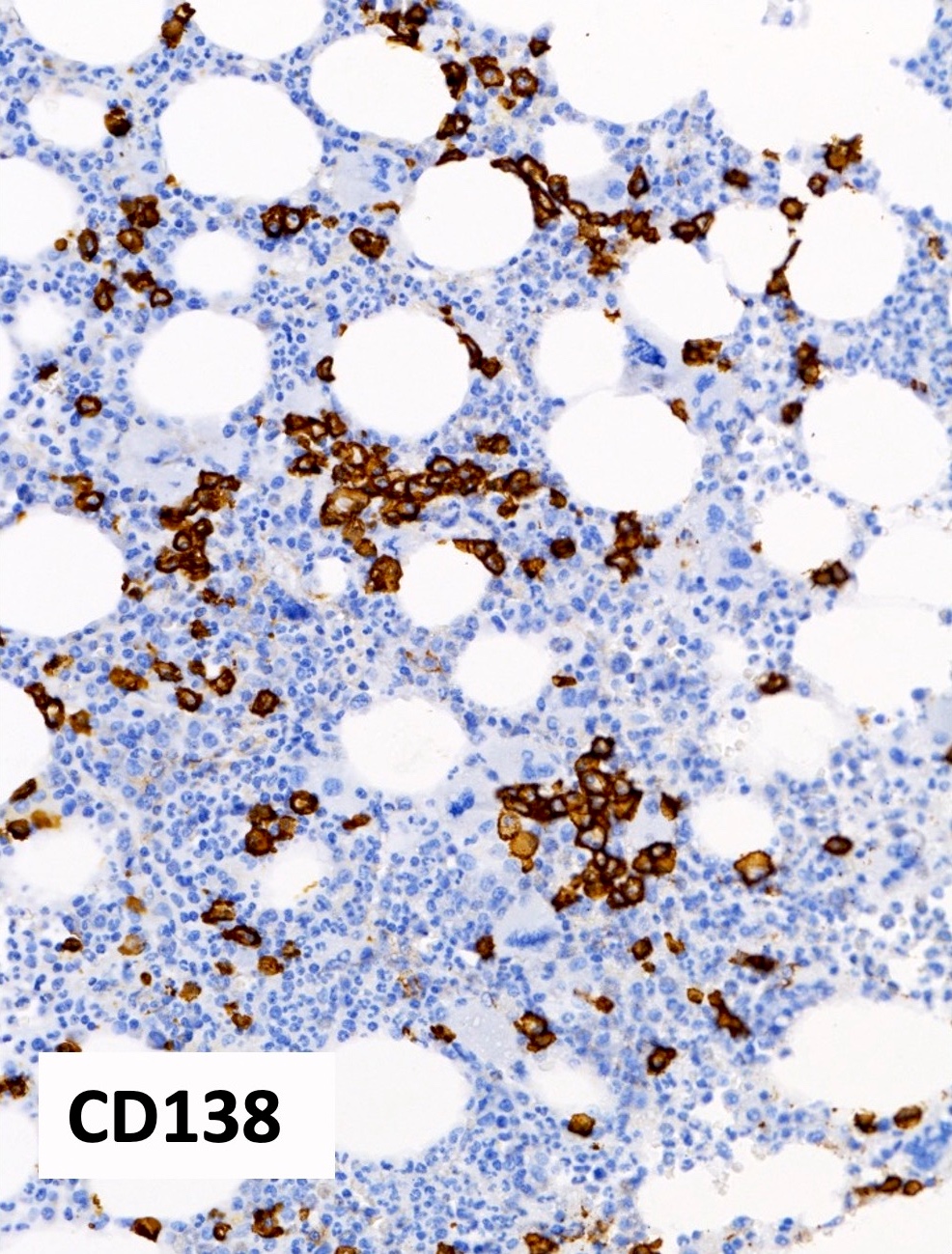
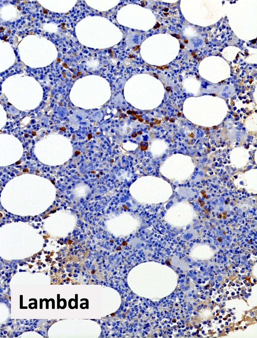
Polytypic plasmacytosis Polytypic plasmacytosis Polytypic plasmacytosis

Polytypic plasmacytosis

Peripheral smear images

Contributed by Ria Vergara-Lluri, M.D.
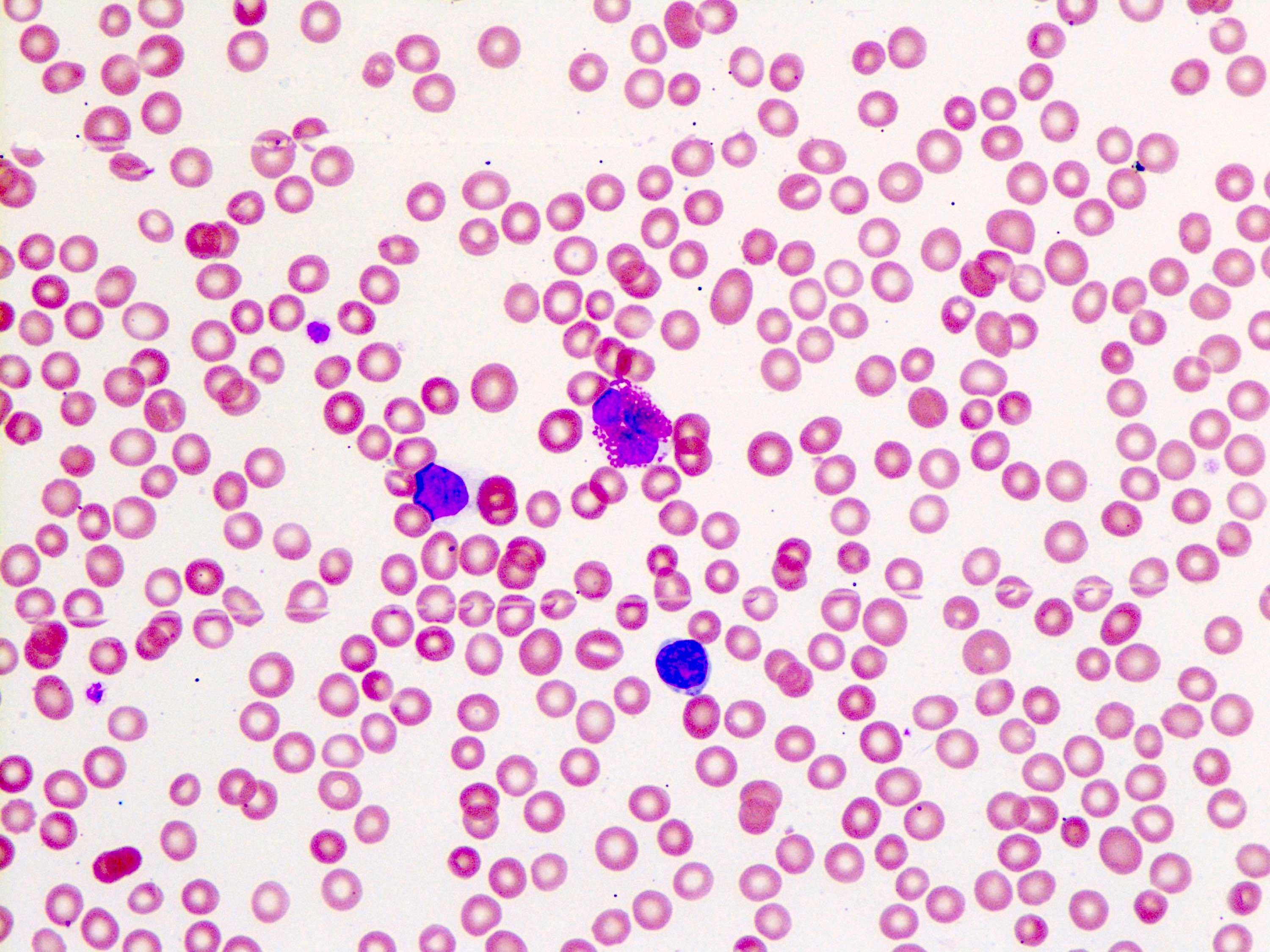
Peripheral blood smear

Peripheral blood smear


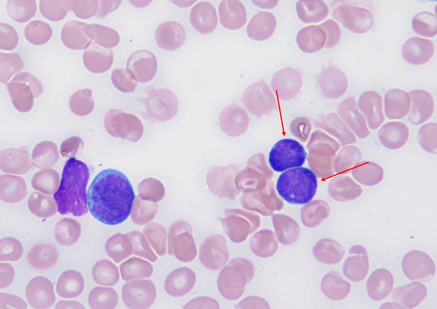
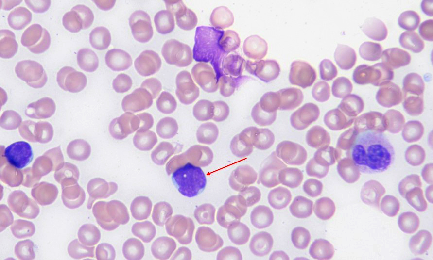
Basophils
Microscopic (histologic) images

Images hosted on other servers:
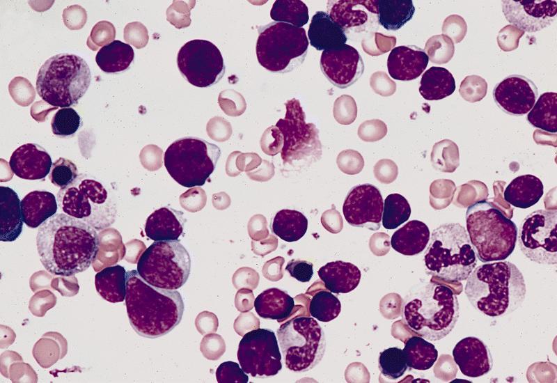
Basophilic myelocyte

Basophilic myelocyte

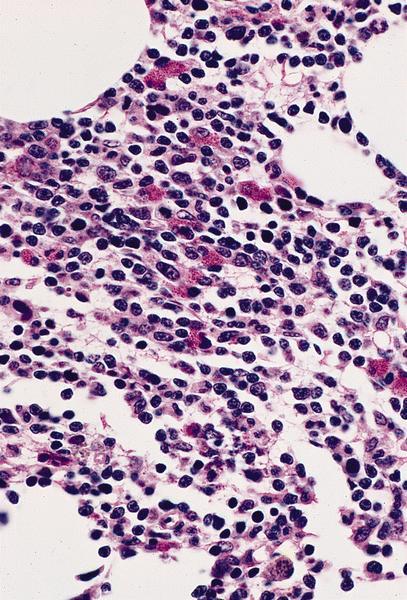
Basophilic metamyelocyte

Basophilic metamyelocyte

Peripheral smear images

Images hosted on other servers:
Basophil

Basophil

Electron microscopy images

Images hosted on other servers:

Basophil


Biopsy and aspirate smear
Diagrams / tables

Images hosted on other servers:

Schematics

Clinical images

Images hosted on other servers:

Blood clot

Microscopic (histologic) images

Images hosted on other servers:

Poor technique

Smear: good and poor technique

Epoxy resin embedded bone marrow biopsies

Molecular / cytogenetics images

Images hosted on other servers:

FISH from tissue imprints


Bone marrow transplantation
Microscopic (histologic) images

AFIP images

Promyelocytes and myelocytes in upper field

Foci of erythropoiesis


Chédiak-Higashi syndrome
Peripheral smear images

Images hosted on other servers:

Giant inclusion bodies


CMV
Microscopic (histologic) images

Images hosted on other servers:

Site unknown (not bone marrow)


Copper deficiency
Microscopic (histologic) images

Images hosted on other servers:

Extensive vacuolation of erythroid precursors

Vacuoles in erythroid precursors

Vacuoles in immature granulocytes

Vacuoles in myelocyte and basophilic normoblast


Cytoplasmic vacuoles in two pronormoblasts

Sideroblasts (Prussian blue)

Pelger-Huët nuclear anomaly

Peripheral smear images

Images hosted on other servers:

Dimorphic RBCs

More pronounced
anisopoikilocytosis


Diamond-Blackfan anemia
Diagrams / tables

Images hosted on other servers:
Missing Image

Hypothetic model for abortive
ribosome assembly and p53
activation relevant to DBA


Dyskeratosis congenita
Diagrams / tables

Images hosted on other servers:

Defects in cellular proliferation
and differentiation that give rise
to dyskeratosis congenita

Clinical images

Images hosted on other servers:

Reticular skin pigmentation

Reticular pigmentation

Dorsum of tongue

Ulceroproliferative growth

Clinical data of the DKC proband in the first pedigree


Plantar surface of hands

Palmar surface of hands

Hyperpigmentation and dystrophic nails

Epiphora and ectropion

Microscopic (histologic) images

Images hosted on other servers:

Histopathological features of dyskeratosis congenita

Lymph node: metastatic squamous cell cac

Dorsum of tongue: squamous cell carcinoma

Dorsum of tongue:
richly vascular
with mild chronic
inflammatory infiltrate


Embryonic development
Diagrams / tables

Images hosted on other servers:

Blood islands in embryo

Microscopic (histologic) images

Images hosted on other servers:

Yolk sac at 18 days

Fetal liver at 17 weeks

Fetal bone marrow at 19 weeks


Eosinophils
Microscopic (histologic) images

Images hosted on other servers:


Eosinophilic metamyelocyte

Eosinophilic metamyelocyte

Developing eosinophils

Developing eosinophils


AML-M4eo:
Various eosinophil precursors Various eosinophil precursors Various eosinophil precursors Various eosinophil precursors

Various eosinophil precursors


Hypereosinophilic syndrome:
Bone marrow aspirate

Bone marrow aspirate

Bone marrow biopsy

Bone marrow biopsy

Peripheral smear images

Images hosted on other servers:

Hypereosinophilic syndrome:
Peripheral blood

Peripheral blood


Peripheral blood:
Eosinophils Eosinophils Eosinophils

Eosinophils

Electron microscopy images

Images hosted on other servers:

Normal eosinophil with crystalloid granules


Erythroid maturation (erythropoiesis)
Diagrams / tables

Images hosted on other servers:

Erythropoiesis

Microscopic (histologic) images

AFIP images

Erythroblast island

Proerythroblast is large cell at
upper left of center; there are
also basophilic, polychromatophilic
and orthochromatic normoblasts


Images hosted on other servers:

Orthochromatic normoblast

Proerythroblast


Polychromatophilic normoblast

Basophilic normoblast


Reticulocyte

Basophilic and polychromatophilic normoblasts

Electron microscopy images

Images hosted on other servers:

Normal erythrocyte

Reticulocyte


Fanconi anemia
Diagrams / tables

Images hosted on other servers:

Diagnostic algorithm

Clinical images

Images hosted on other servers:

Clinodactyly with brachymesophalangia

Microscopic (histologic) images

Images hosted on other servers:

Hypocellular spicule with fatty infiltration

Aspirate smear findings

Molecular / cytogenetics images

Images hosted on other servers:

Chromosome breakage test

Representative karyotypes


Gelatinous transformation
Microscopic (histologic) images

Images hosted on other servers:

Prominent gelatinous transformation

Serous atrophy

Various images


General
Diagrams / tables

AFIP images

Hematopoietic cell differentiation


Images hosted on other servers:

Hematopoietic cell differentiation

Normal bone marrow differential in adults

Microscopic (histologic) images

Images hosted on other servers:

Bone marrow biopsy:

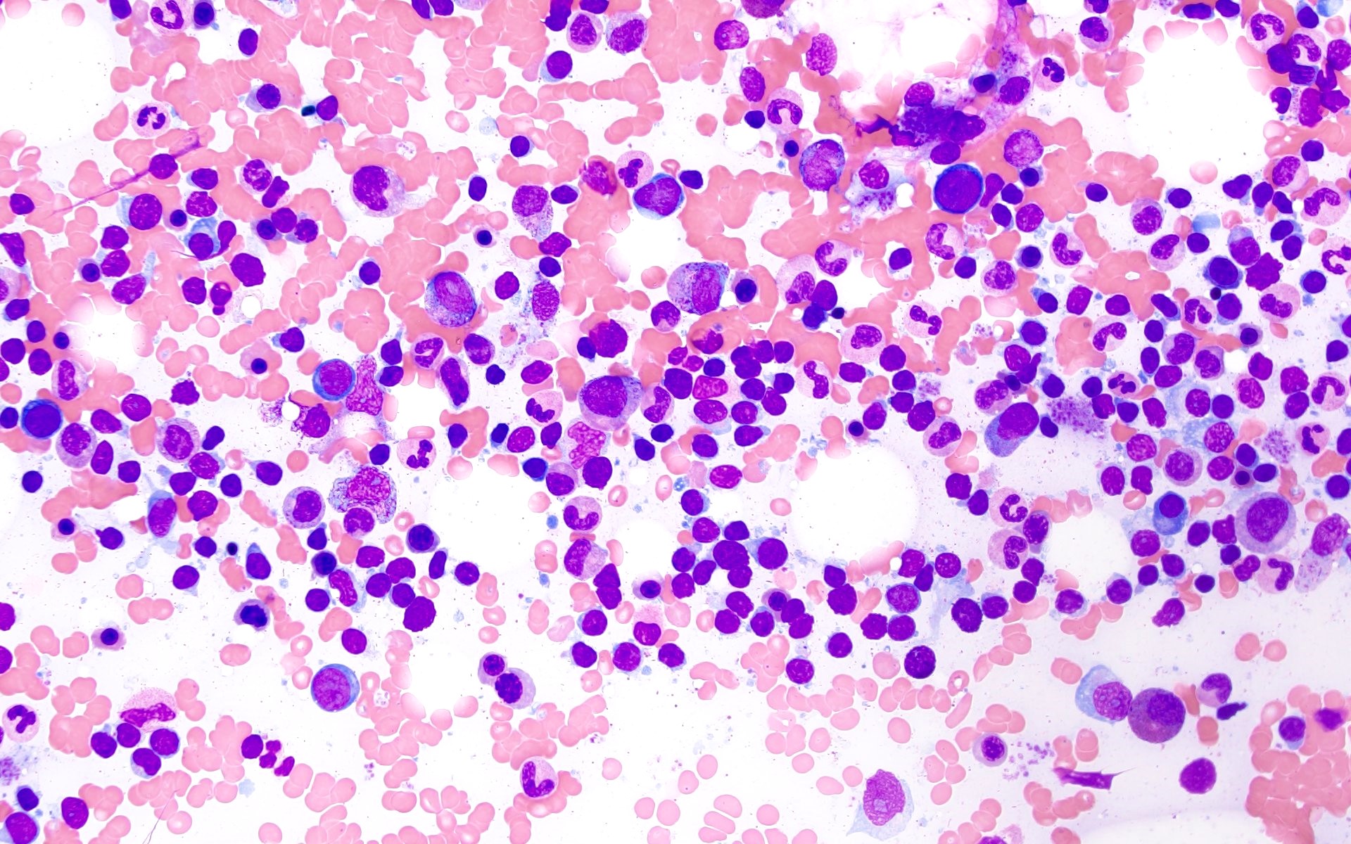
Normal marrow


Bone marrow aspirate smear:

Normal


Granulomatous inflammation
Microscopic (histologic) images

Contributed by Natasha Savage, M.D. and Yulan Jin, M.D., Ph.D.
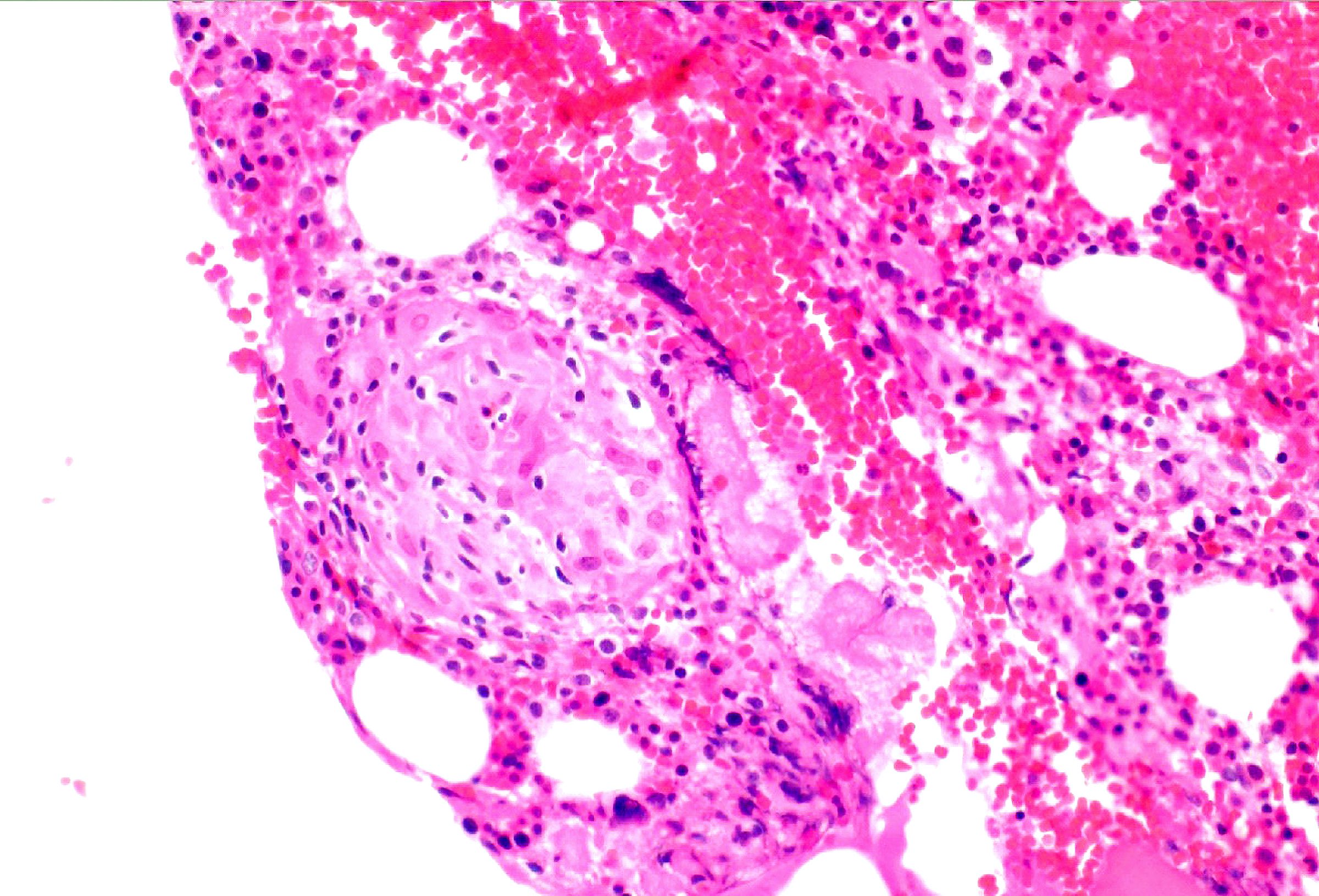
Nonnecrotizing granuloma

Nonnecrotizing granuloma

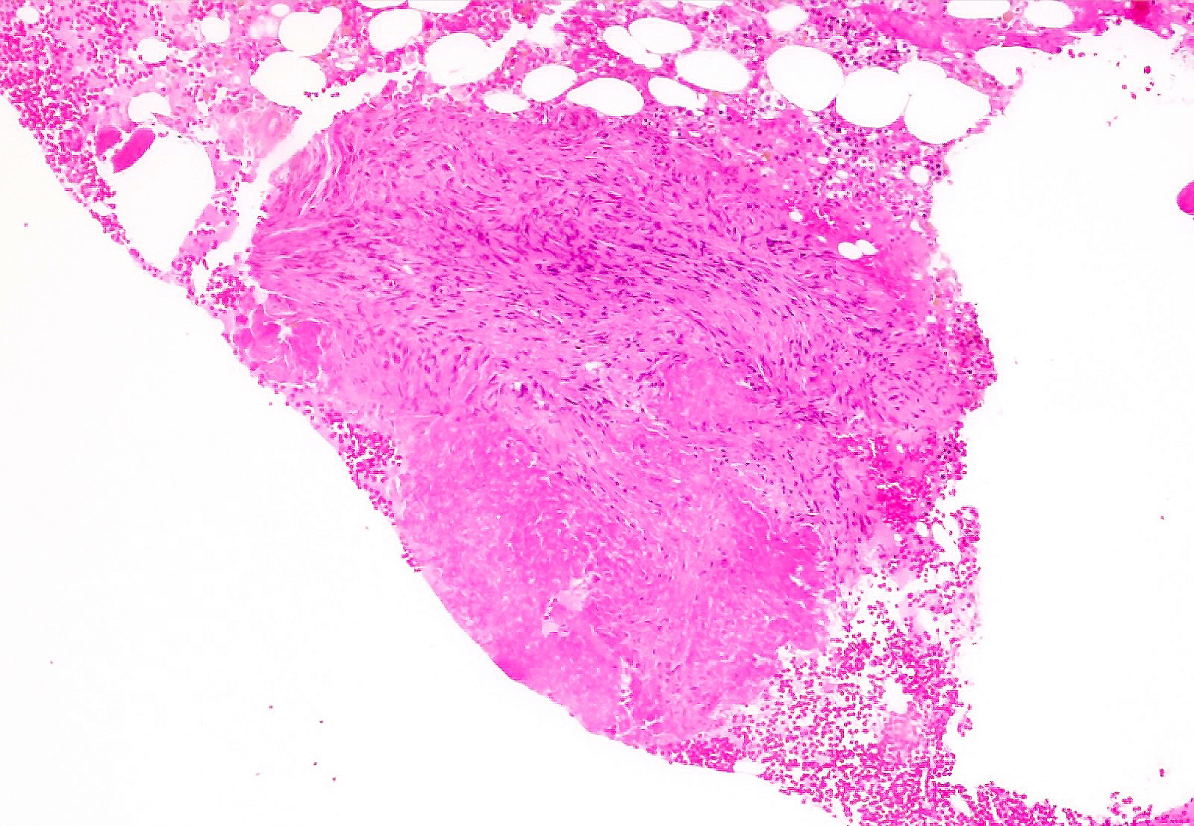
Necrotizing granuloma

Necrotizing granuloma

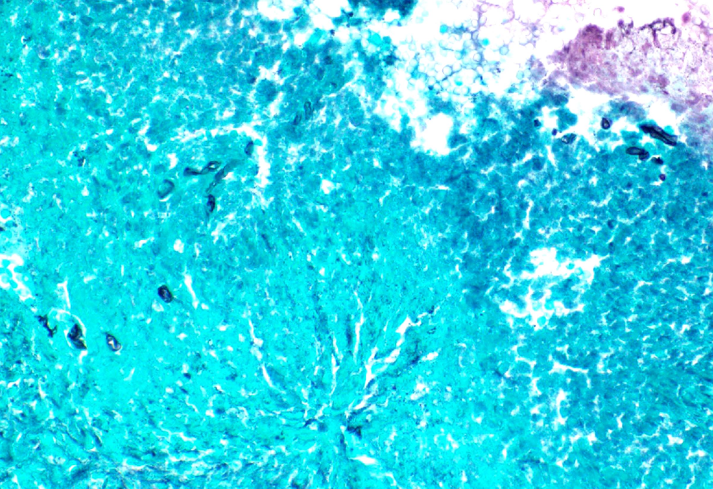
GMS stain

GMS stain


Hematogones
Diagrams / tables

Images hosted on other servers:
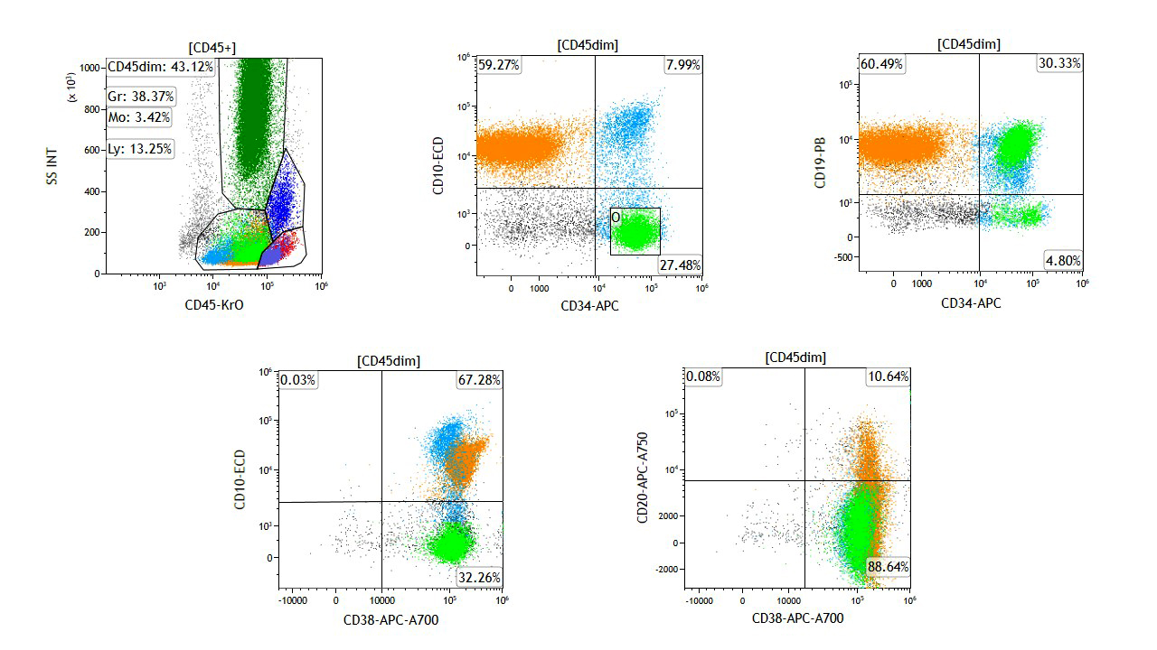
Normal hematogone maturation by flow cytometry

Normal hematogone maturation

Microscopic (histologic) images

Contributed by Giby V. George, M.B.B.S. and AFIP
Bone marrow aspirate hematogones Bone marrow aspirate hematogones Bone marrow aspirate hematogones

Bone marrow aspirate hematogones

Bone marrow aspirate hematogones Bone marrow aspirate hematogones Bone marrow aspirate hematogones

Bone marrow aspirate hematogones


Sparse, lightly basophilic cytoplasm

Sparse, lightly basophilic cytoplasm

Increased hematogones evenly distributed

Increased hematogones evenly distributed

Clumped nuclear chromatin

Clumped nuclear chromatin

Flow cytometry images

Contributed by Giby V. George, M.B.B.S.
Hematogone maturation in the background of B ALL

Hematogone maturation in the background of B ALL


Images hosted on other servers:
Normal hematogone maturation by flow cytometry Normal hematogone maturation by flow cytometry

Normal hematogone maturation by flow cytometry

Videos

ICCS: B cells, features of immaturity

Ace My Path: B cell maturation


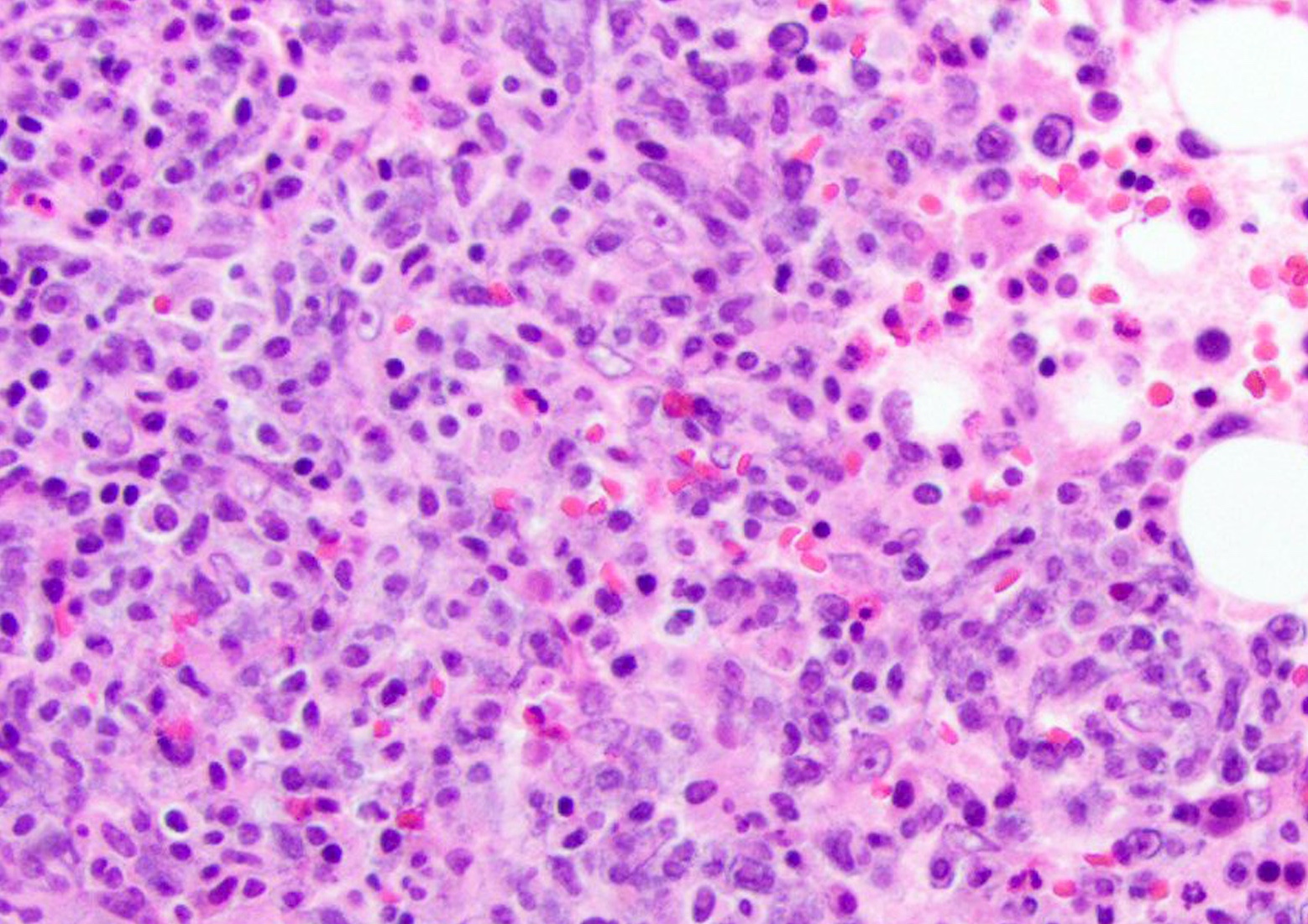
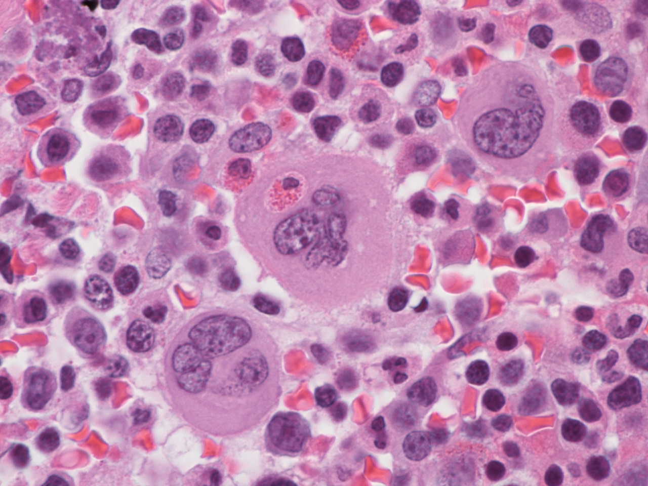
Hemophagocytic lymphohistiocytosis
Microscopic (histologic) images

Contributed by Sherif A. Rezk, M.D. and AFIP

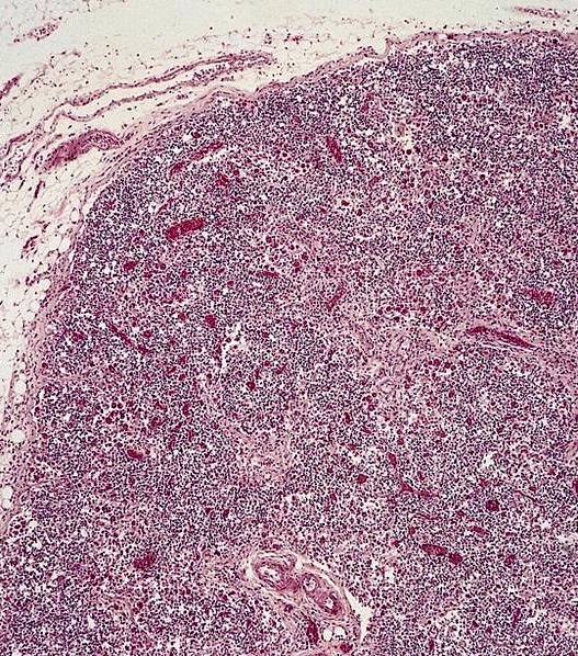
Bone marrow
hemophagocytosis

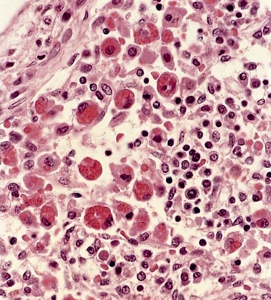
Reactive hemophagocytosis in lymph node


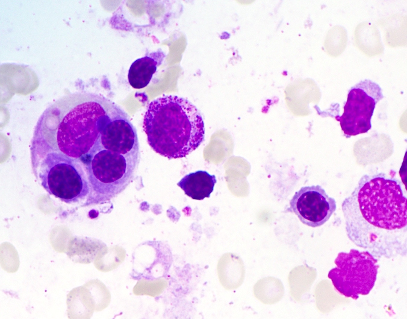
Erythrophagocytosis

Neutrophil
hemophagocytosis

Platelet
hemophagocytosis

Bone marrow in
reactive
hemophagocytic
syndrome


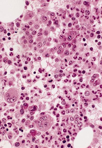
Hypercellularity
Microscopic (histologic) images

Images hosted on other servers:

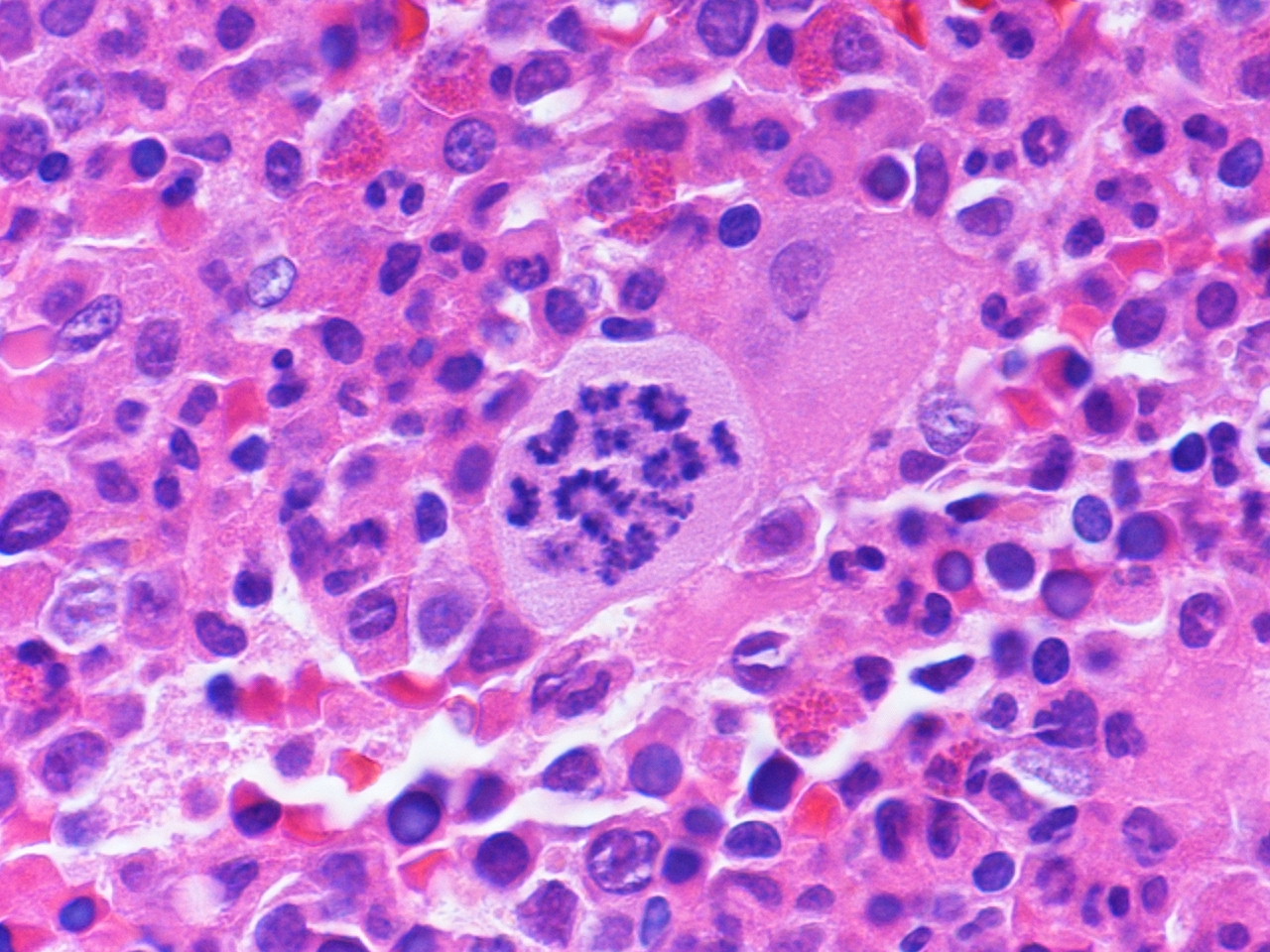
Megaloblastic anemia


Iron in nonneoplastic marrow
Microscopic (histologic) images

Images hosted on other servers:

Bone marrow fragment showing iron deposits

Iron

Normal iron stores

Increased iron stores

Electron microscopy images

Images hosted on other servers:

Clumped and free ferritin

Erythroblast contain ferritin

Macrophage and erythroblast cytoplasm

Erythroblasts with ferritin


Clumped, vesticle and free ferritin

Erythroblast with a clump of ferritin

Iron in mitochondria

Erythroblast containing mitochondrial iron


Lymphocyte maturation
Peripheral smear images

Images hosted on other servers:
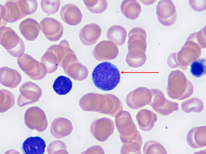
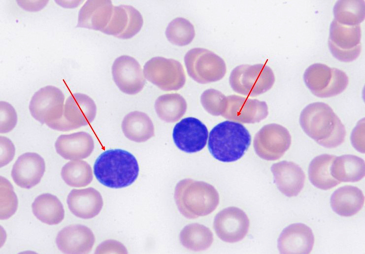
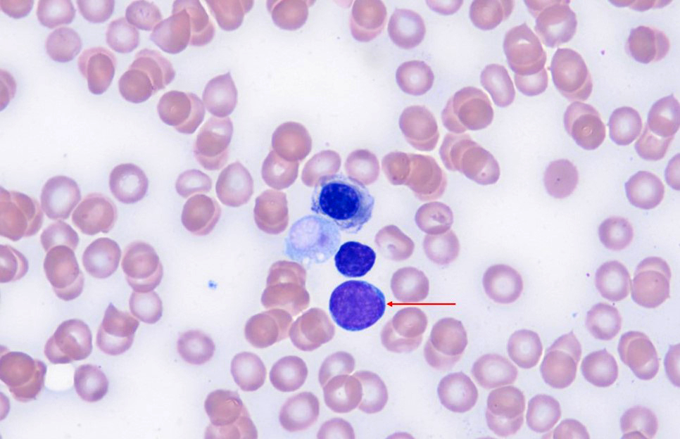
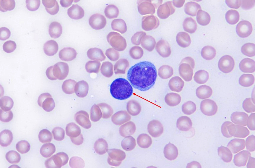
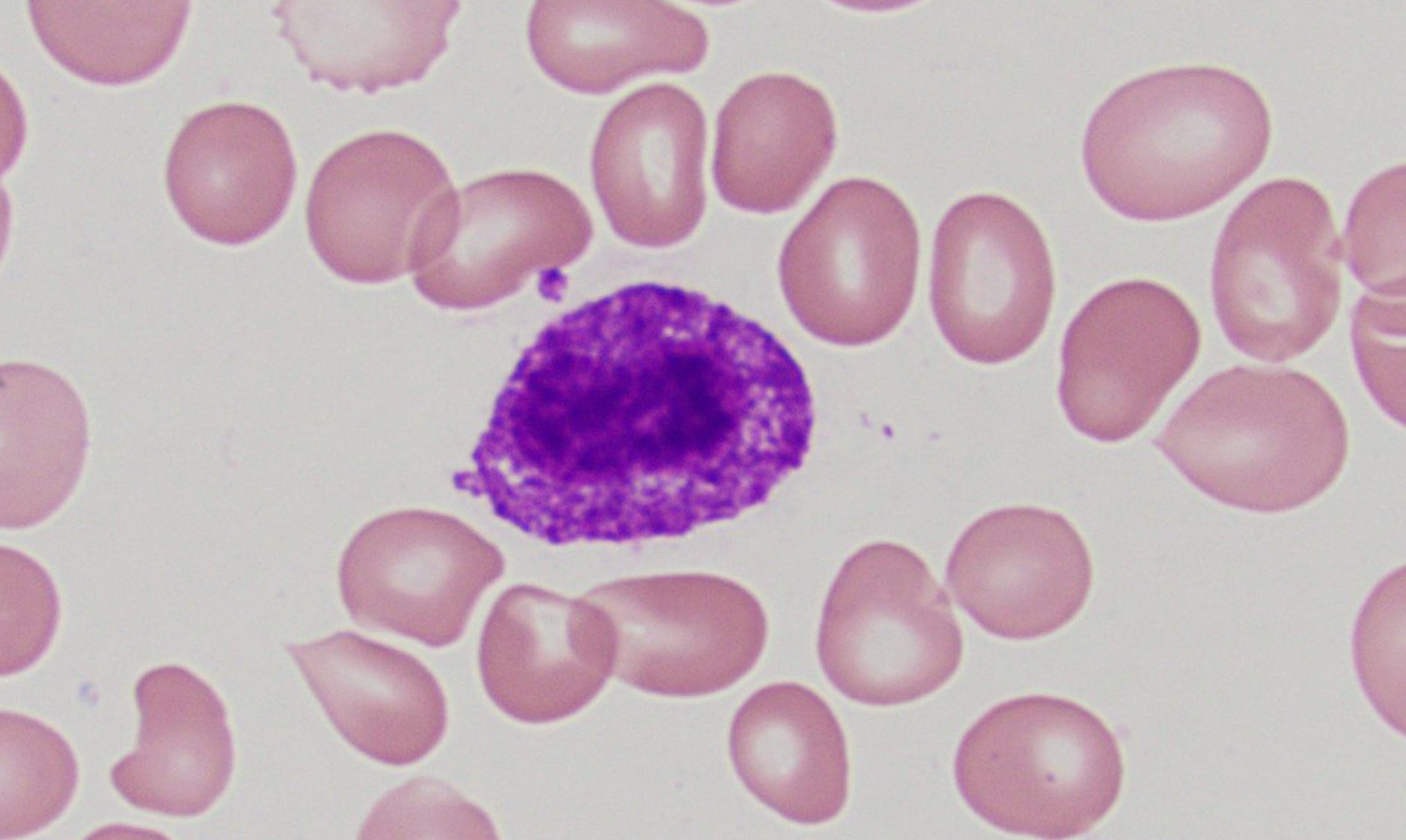
Normal lymphocytes

Normal lymphocytes

Electron microscopy images

Images hosted on other servers:

Small lymphocyte

With cytoplasmic projections


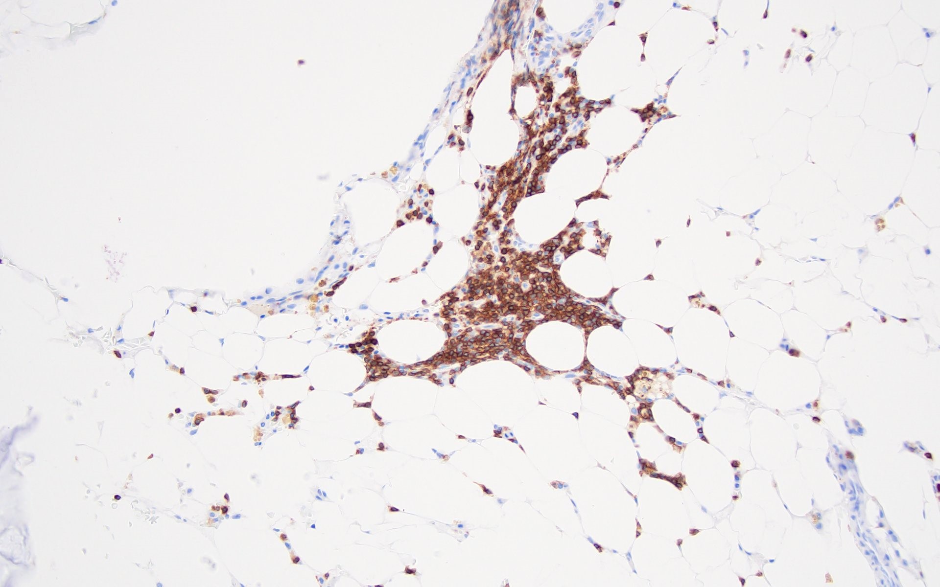
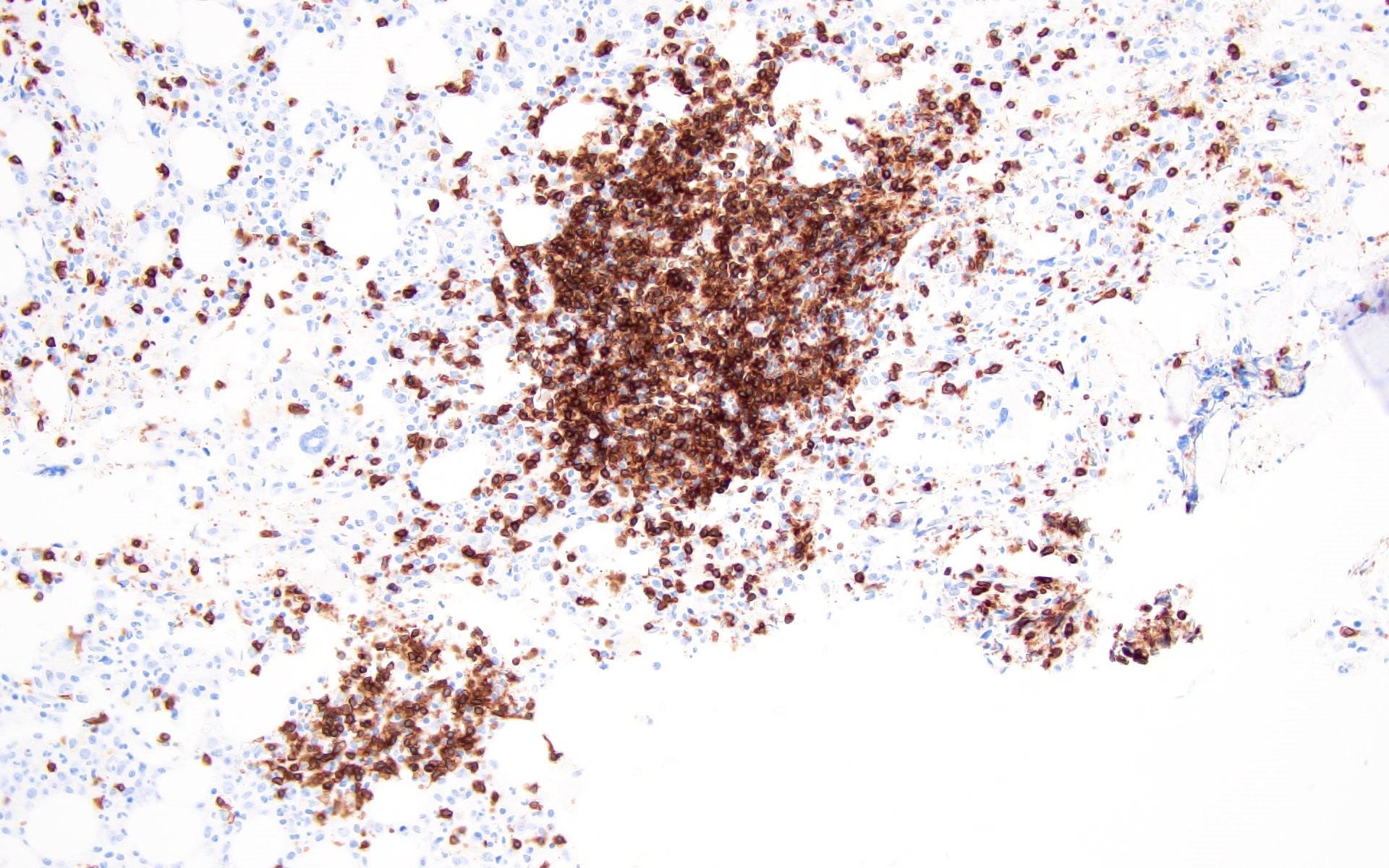
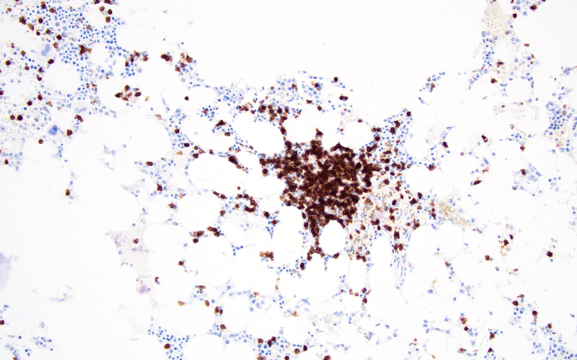
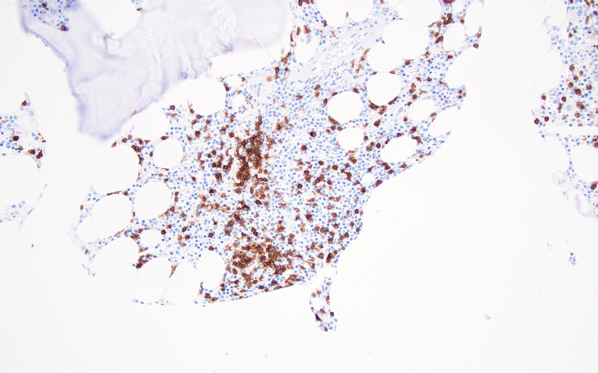
Lymphoid aggregates (benign)
Microscopic (histologic) images

Contributed by Barina Aqil, M.D.
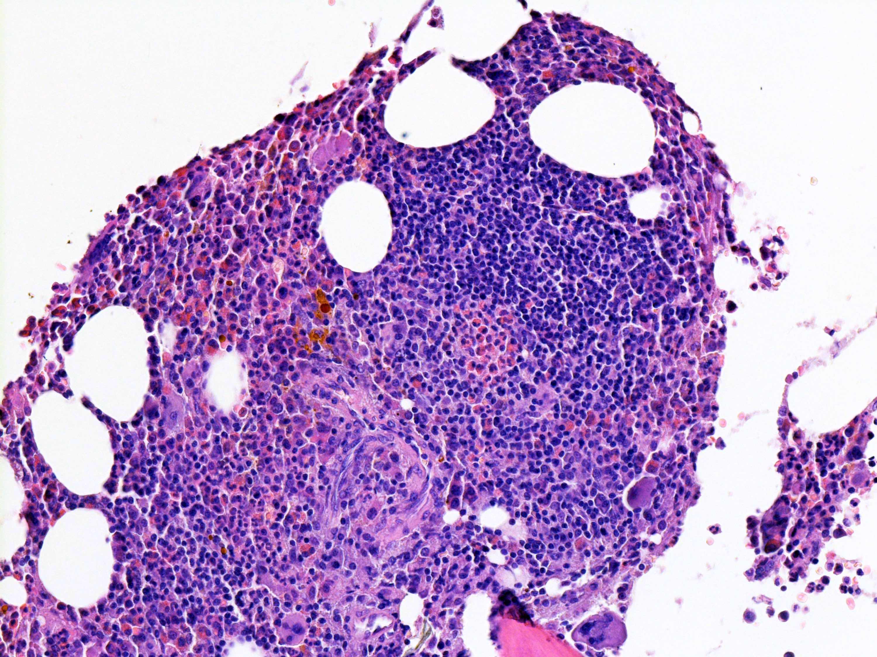
Pattern 1

Pattern 1

Pattern 2

Pattern 2

Pattern 3

Pattern 3

Pattern 4

Pattern 4

Pattern 5

Pattern 5


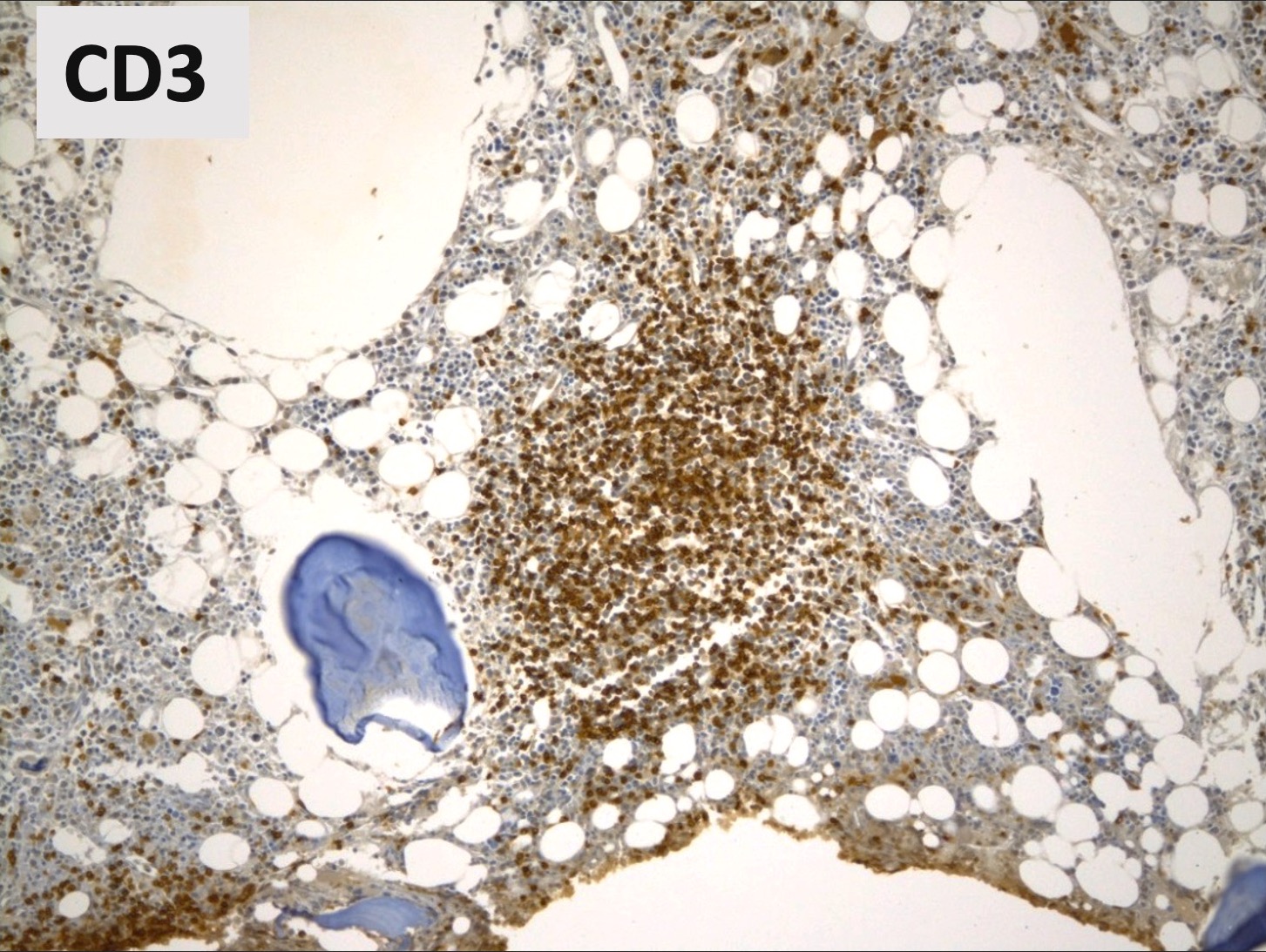
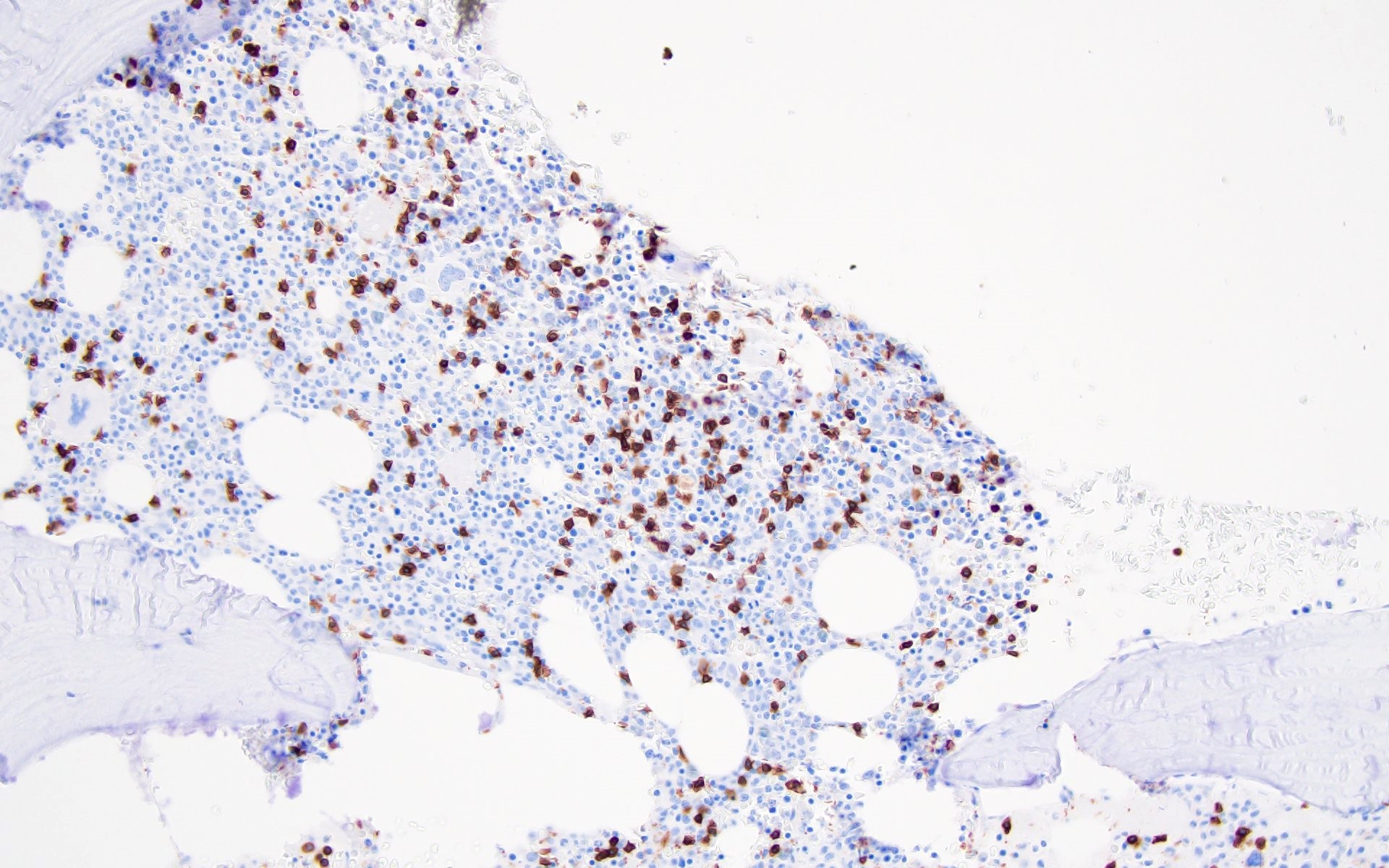
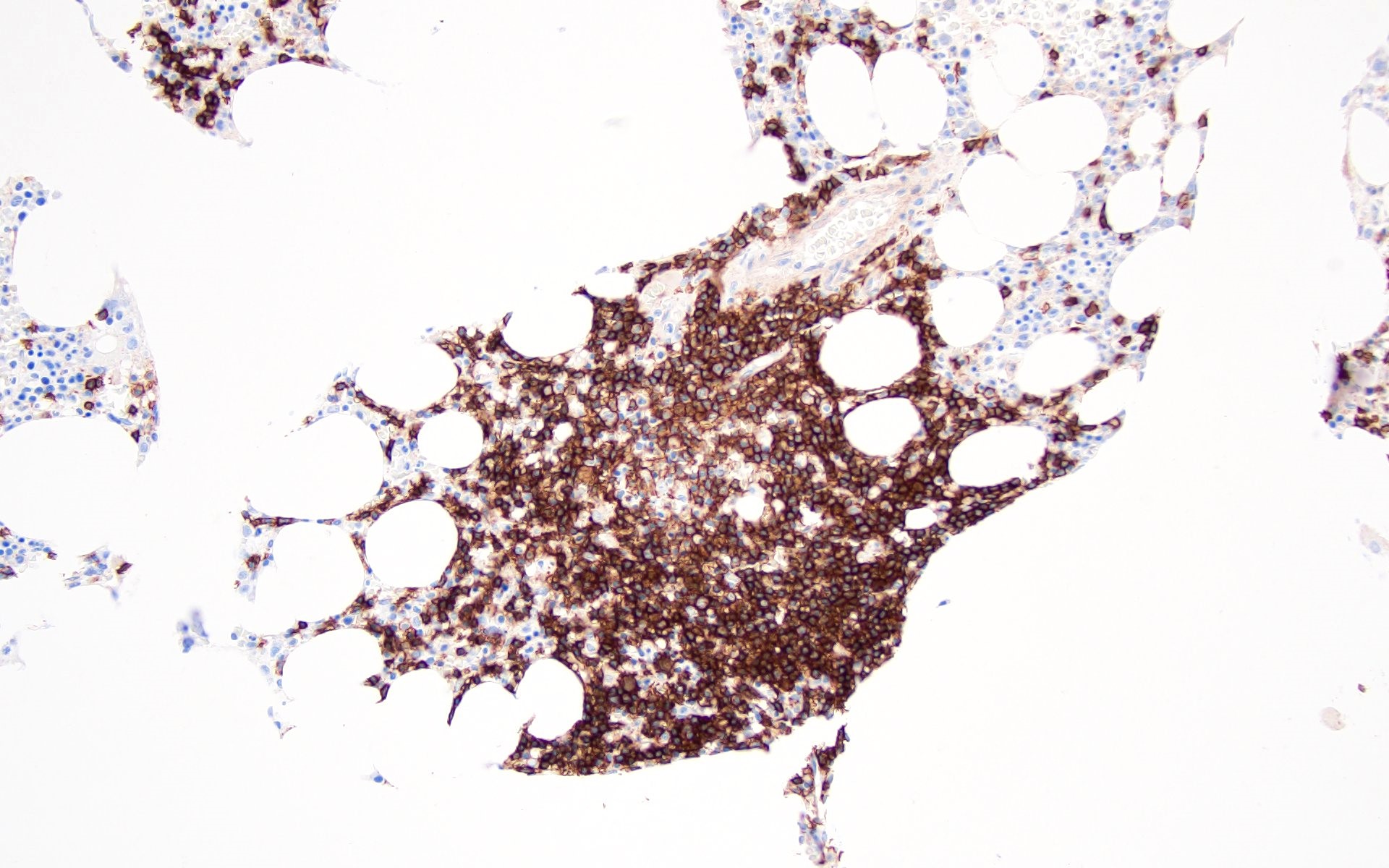
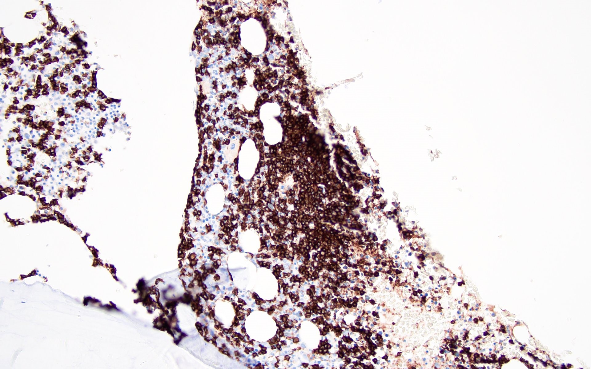
Pattern 1 CD3

Pattern 1 (CD3)

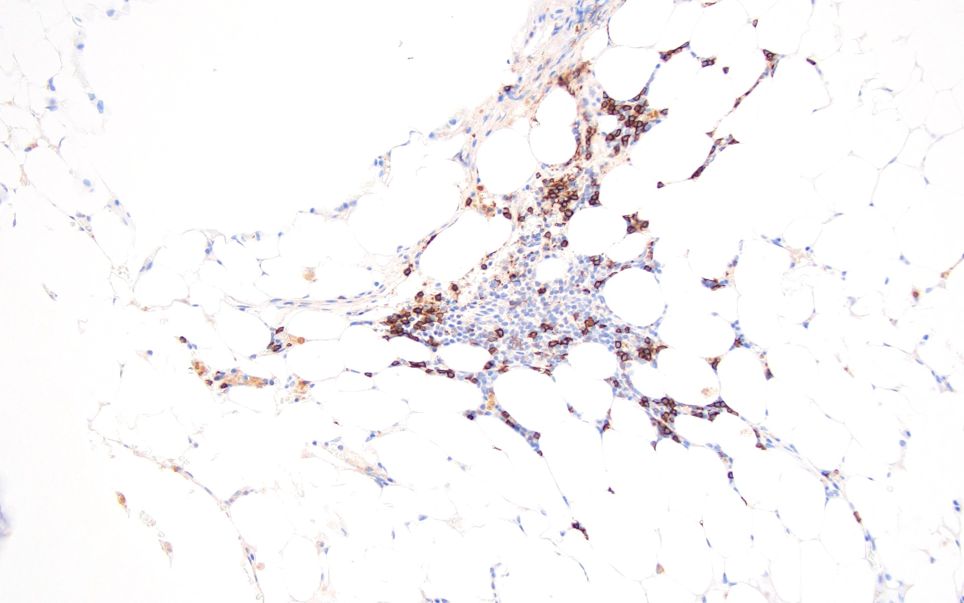
Pattern 2 (CD3)

Pattern 2 (CD3)

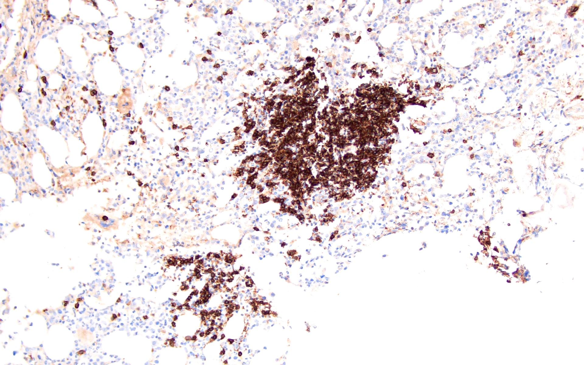
Pattern 3 (CD3)

Pattern 3 (CD3)

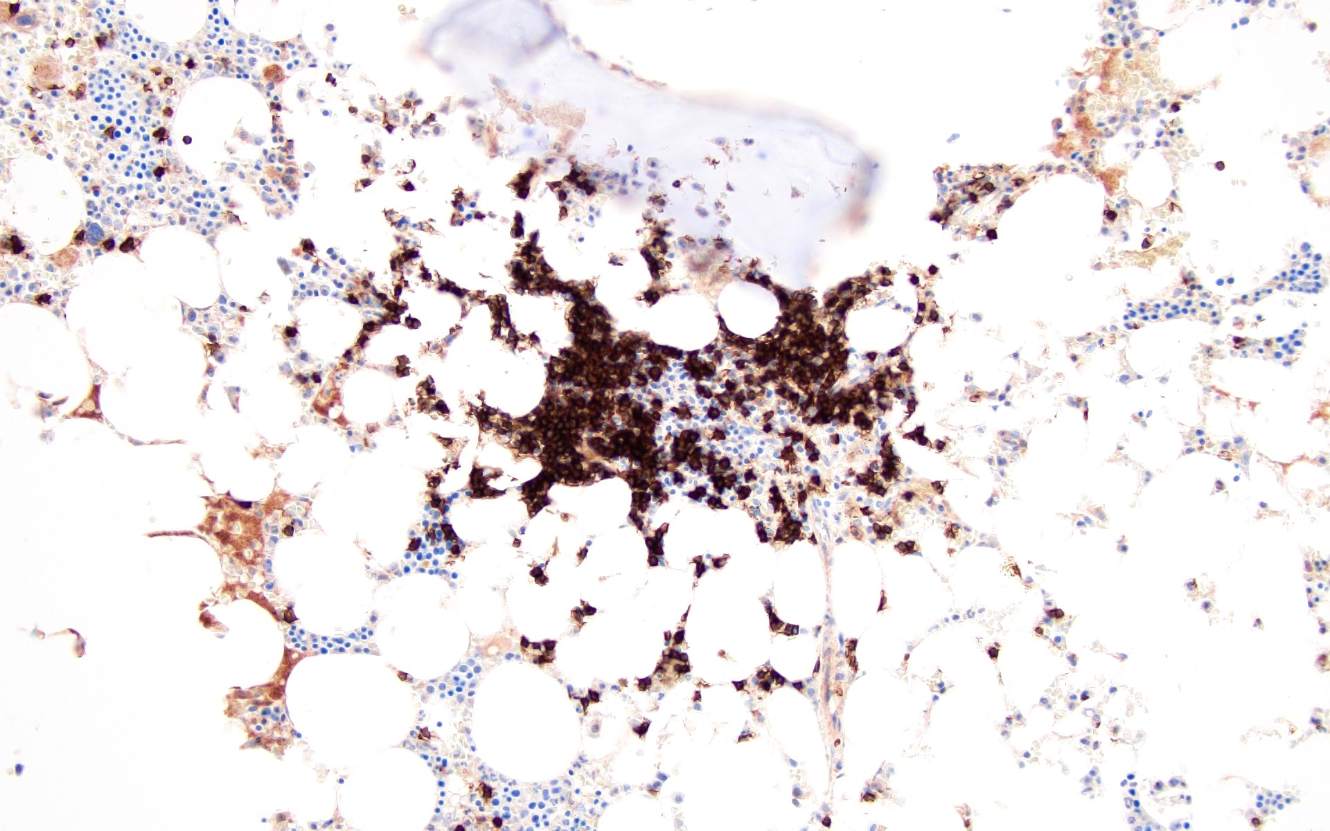
Pattern 4 (CD3)

Pattern 4 (CD3)

Pattern 5 (CD3)

Pattern 5 (CD3)


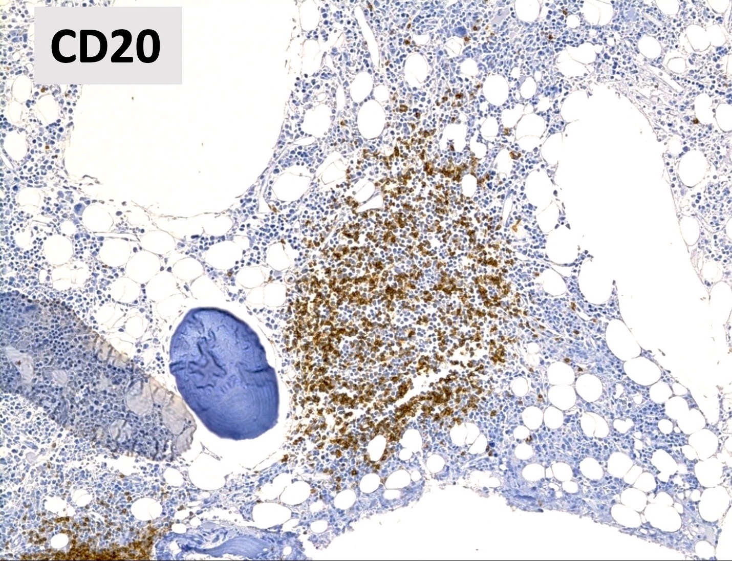
Pattern 1 (CD20)

Pattern 1 (CD20)

Pattern 2 (CD20)

Pattern 2 (CD20)

Pattern 3 (CD20)

Pattern 3 (CD20)

Pattern 4 (CD20)

Pattern 4 (CD20)

Pattern 5 (CD20)

Pattern 5 (CD20)

Cytology images

Contributed by Barina Aqil, M.D.
Sheet of small lymphocytes

Sheet of small lymphocytes


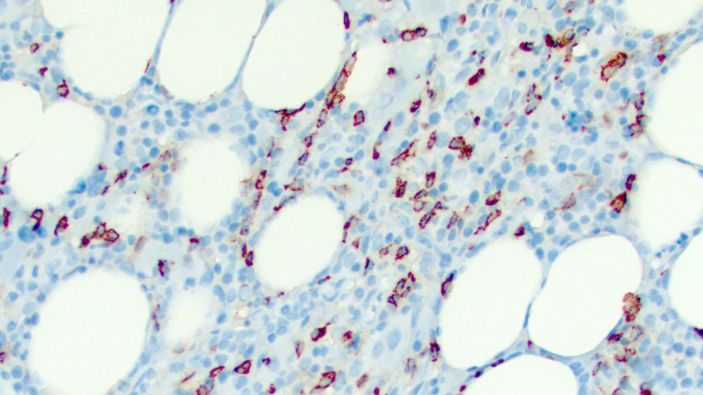
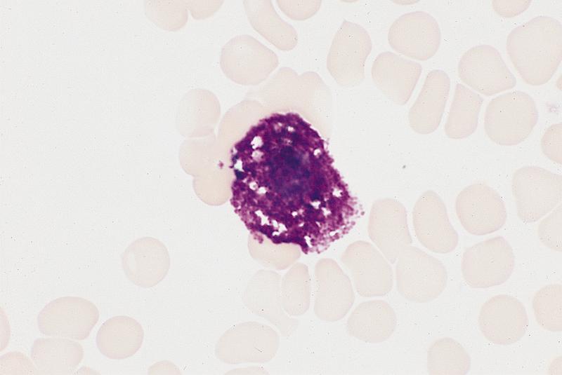
Mast cells
Microscopic (histologic) images

Contributed by Ramya Gadde, M.D. and AFIP
Mast cells

Scattered mast cells

H&E

Spindle shaped mast cells

Tryptase

Tryptase

Scattered mast cells with basophilic granules

Scattered mast cells with basophilic granules

Electron microscopy images

Images hosted on other servers:

Degranulating mast cell

Mast cells with abundant granules


Megakaryocytes
Diagrams / tables

Images hosted on other servers:

Megakaryopoiesis pathways

Proplatelet formation

Microscopic (histologic) images

Contributed by Dragos C. Luca, M.D. and AFIP images

Emperipolesis

Endomitosis

Megakaryocytes


Images hosted on other servers:

Megakaryoblasts

Megakaryocytes


Emperipolesis

CD62P+ megakaryocyte

Platelet formation

Electron microscopy images

Images hosted on other servers:

Platelets


Megaloblastic anemia
Diagrams / tables

Contributed by Julie Teruya-Feldstein, M.D. and Swati Bhardwaj, M.B.B.S., M.D.
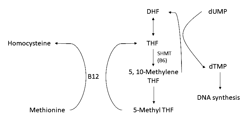
Intracellular metabolism of folate

Intracellular metabolism of folate

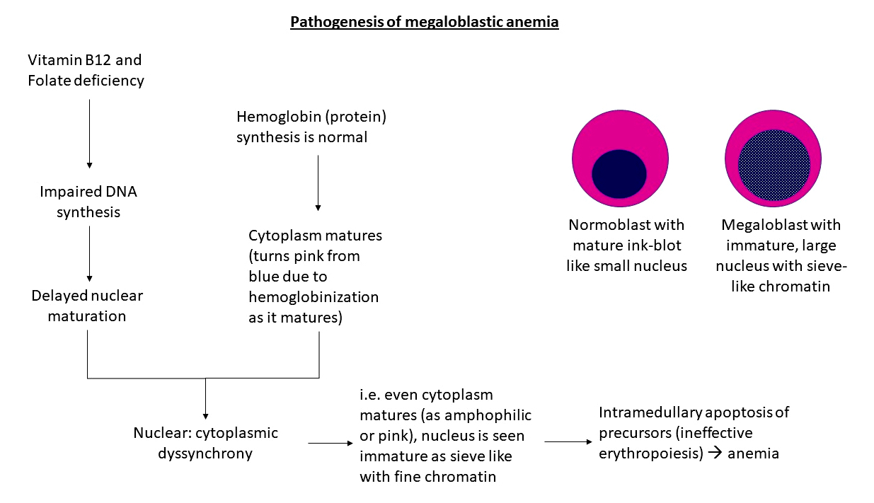
Pathophysiology of megaloblastic anemia

Pathophysiology of megaloblastic anemia


Images hosted on other servers:

Thymidine and methionine synthesis

Hemogram

Clinical images

Images hosted on other servers:
Glossitis, smooth tongue

Glossitis, smooth tongue

Microscopic (histologic) images

Contributed by Julie Teruya-Feldstein, M.D. and Swati Bhardwaj, M.B.B.S., M.D.
Erythroid hyperplasia

Erythroid hyperplasia

Megaloblasts and erythroid nuclear dysplasia

Megaloblasts and erythroid nuclear dysplasia

Erythroid hyperplasia and nuclear abnormalities

Erythroid hyperplasia and nuclear abnormalities

Frequent mitoses and neutrophil hypersegmentation

Frequent mitoses and neutrophil hypersegmentation

Hypersegmented neutrophil


Hypersegmented neutrophil


Peripheral smear images

Images hosted on other servers:

Macroovalocytosis /
round macrocytes
in liver disease

Macrocytosis with a lymphocyte

Videos

Megaloblastoid erythroids, basophils, mast cells in a bone marrow smear


Monocytes
Microscopic (histologic) images

AFIP images

Promonocyte


Bone marrow in acute monocytic leukemia (AML M4):

Monoblasts are larger cells with abundant
cytoplasm and round nuclei and
promonocytes are two cells with folded and
creased nuclei next to monoblast in center


Images hosted on other servers:

Monocyte

Electron microscopy images

Images hosted on other servers:

Monocyte - TEM and SEM


Necrosis
Microscopic (histologic) images

Images hosted on other servers:
Necrotic and nonnecrotic areas Necrotic and nonnecrotic areas

Necrotic and nonnecrotic areas

Disruption of normal bone marrow architecture

Disruption of normal bone marrow architecture

Antiphospholipid syndrome

Antiphospholipid syndrome

Nuclear features associated with necrosis

Nuclear features associated with necrosis

Ghost-like hematopoietic cells

Ghost-like hematopoietic cells


Neutrophil maturation
Microscopic (histologic) images

AFIP images

Myelocytes along endosteum of bone trabeculae: H&E and myeloperoxidase stain

Perivascular immature granulocytes


Images hosted on other servers:

Type I myeloblast

Type II myeloblast

Type III myeloblast

Blast (upper) and promyelocyte (lower)


Myeloblast (type not specified)

Promyelocytes: with Auer rods


Promyelocytes

3 myelocytes + 2 metamyelocytes (right and lower)


Neutrophilic myelocytes

Myelocyte and bands

Metamyelocytes

Electron microscopy images

Images hosted on other servers:

Neutrophil

Details of granules


Osteoblasts
Diagrams / tables

Images hosted on other servers:

Interaction of osteoblasts and hematopoietic stem cells

Microscopic (histologic) images

AFIP images

Osteoblasts in marrow smear


Images hosted on other servers:

Osteoblasts

Electron microscopy images

Images hosted on other servers:

Colorized SEM


Osteoclasts
Diagrams / tables

Images hosted on other servers:

Cross section

Bone resorptive cycle

Microscopic (histologic) images

AFIP images

Very large cell


Images hosted on other servers:

Osteoclasts

Electron microscopy images

Images hosted on other servers:

Osteoclast


Parvovirus (erythrovirus) B19
Microscopic (histologic) images

Images hosted on other servers:

50 year old man: rare pronormoblast forms

Large and megaloblastic pronormoblast forms


Giant cell with eosinophilic nuclear inclusion

B19+

B19+ cells in bone marrow biopsy

Electron microscopy images

Images hosted on other servers:

Native parvovirus B19


Pearson syndrome
Microscopic (histologic) images

Images hosted on other servers:

Aspirate smear shows few spicules

Pronormoblast with vacuoles

Occasional ringed sideroblasts (Prussian blue)


Plasma cells
Microscopic (histologic) images

Images hosted on other servers:

Plasma cells

Plasma cells in myeloma

Electron microscopy images

Images hosted on other servers:

Plasma cell without labels


Plasmacytosis
Microscopic (histologic) images

Images hosted on other servers:

Lymph node biopsy

Differentiation of mature plasma cells

Peripheral smear images

Images hosted on other servers:

Stages of plasma cell differentiation

Flow cytometry images

Images hosted on other servers:

Plasma cells in bone marrow


Pure red cell aplasia
Microscopic (histologic) images

Images hosted on other servers:

No erythroid precursors


Q fever
Radiology images

Images hosted on other servers:

Q fever meningoencephalitis

Q fever pneumonia

Microscopic (histologic) images

Images hosted on other servers:

Fibrin ring granuloma with central fatty vacuoles

Liver: fibrin ring granuloma


Liver: doughnut granuloma

Cardiac valve

Liver: doughnut granuloma (arrow)

Cell lines infected with C. burnetii

Electron microscopy images

Images hosted on other servers:

Within vacuole of host cell


Sea blue histiocytosis (syndrome)
Microscopic (histologic) images

Images hosted on other servers:

Chronic myelogenous leukemia

Refractory anemia with excess blasts

Refractory anemia with excess blasts in transformation

Electron microscopy images

Images hosted on other servers:

Hyperlipidemia: cytoplasmic electron dense granules

"Finger print" arrangement of membranes


Shwachman-Diamond syndrome
Microscopic (histologic) images

Images hosted on other servers:

Bone marrow aspirate

Decreased bone marrow cellularity


Thrombocytopenia absent radii (TAR) syndrome
Radiology images

Images hosted on other servers:

Absent radius


Uncommon storage diseases
Microscopic (histologic) images

Images hosted on other servers :

Fabry disease:

Kidney: podocytes are diffusely distended by intracytoplasmic vacuoles; scattered foam cells in interstitium

Kidney: PAS shows podocytes with foamy cell change


Lymph nodes: reactive hyperplasia

Electron microscopy images

Images hosted on other servers - cystinosis:

Normal platelets

Platelets contain variable dense granules

Platelet undergoing spontaneous release

Back to top
Recent Bone marrow nonneoplastic Pathology books

Cherry: 2022

Foucar: 2009

Foucar: 2017

Foucar: 2023

Hsi: 2017

Hudnall: 2019

IARC: 2017

Jaffe: 2016

Naeim: 2018

Wake: 2023

Wang: 2020


Find related Pathology books: hematopathology
Image 01 Image 02