Table of Contents
Definition / general | Essential features | Pathophysiology | Clinical features | Symptoms | Laboratory | Case reports | Treatment | Sample assessment & plan | Differential diagnosis | Board review style question #1 | Board review style answer #1 | Board review style question #2 | Board review style answer #2Cite this page: Chambers T, Virk M. Transfusion associated circulatory overload. PathologyOutlines.com website. https://www.pathologyoutlines.com/topic/transfusionmedcirculatoryoverload.html. Accessed April 2nd, 2025.
Definition / general
- Circulatory volume overload following transfusion
- Fluid accumulates in the lungs if the heart or kidneys are unable to compensate for the volume of the product transfused
Essential features
- Transfusion associated circulatory overload (TACO) is a form of cardiopulmonary edema due to the inability to tolerate the volume or rate of transfusion
- In patients with a history of heart failure, renal failure or evidence of positive fluid balance, carefully consider the need for transfusion
- Slowing the rate of transfusion or concurrent diuresis may prevent TACO in at risk patients
- TACO is the leading cause of death from transfusion in the US
Pathophysiology
- Transfusion increases intravascular volume
- If the heart is unable to increase cardiac output or the kidneys are unable to compensate for the increased volume, venous pressure will increase
- Increased pressure can force fluid from vasculature into the lungs
- Reference: Transfusion 2019;59:3617
Clinical features
- New onset or exacerbation of 3 or more of the following within 12 hours of the end of transfusion (CDC: National Healthcare Safety Network - Biovigilance Component Hemovigilance Module Surveillance Protocol [Accessed 12 April 2021])
- Acute respiratory distress (dyspnea, orthopnea, cough)
- Elevated B type natriuretic peptide (BNP)
- Elevated central venous pressure (CVP)
- Evidence of left heart failure
- Evidence of positive fluid balance
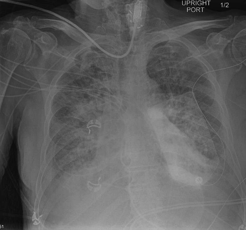
- Radiographic evidence of pulmonary edema
- Risk factors include (Am J Med 2013;126:357.e29):
- Positive fluid balance
- History of heart failure
- History of chronic renal failure
- Hemorrhagic shock
- Multiple blood products transfused
- TACO is a common transfusion reaction and is estimated to occur in 1 - 8% of transfused patients
- TACO caused the highest number of transfusion related fatalities (32%) in data reported to the FDA from 2014 to 2018 (FDA: Fatalities Reported to FDA Following Blood Collection and Transfusion [Accessed 12 April 2021])
- TACO is associated with increased morbidity measured by significantly increased hospital and ICU length of stay (Am J Med 2013;126:357.e29)
- Although TACO is the most frequent serious adverse event associated with blood transfusions, it is likely underrecognized and the incidence underestimated, especially in ICU patients and pediatrics (Crit Care Med 2019;47:849, Transfusion 2018;58:1037)
Symptoms
- Dyspnea, orthopnea, cough, headache, chest tightness, hypertension, tachycardia, hypoxia, widened pulse pressure, jugular venous distension
Laboratory
- Elevated circulating B type natriuretic peptide (BNP, formerly brain natriuretic peptide) or N-terminal-pro-BNP (NT-pro-BNP), a marker for congestive heart failure
- Post / pretransfusion NT‐proBNP ratio > 1.5 can aid in the diagnosis of TACO; posttransfusion levels of BNP < 300 or NT‐proBNP < 2000 pg/mL, drawn within 24 hours of the reaction, make TACO unlikely (Transfusion 2019;59:795)
Case reports
- 5 year old boy with neuroblastoma develops acute hypoxemia, tachypnea and tachycardia after red blood cell transfusion (Transfus Apher Sci 2017;56:445)
- 46 year old man develops acute dyspnea with hypoxemia following red blood cell transfusion (Indian J Crit Care Med 2014;18:396)
- 63 year old woman with diabetes develops acute respiratory distress following red blood cell transfusion (BMJ Case Rep 2020;13:e230426)
Treatment
- Stop transfusion
- Diuresis
- Supplemental oxygen
- Prevention:
- Avoid unnecessary transfusion (Transfusion 2014;54:2344)
- Reduce transfusion rate
- Diurese high risk patients before or during transfusion
Sample assessment & plan
- Assessment
- Given the patient’s history of heart failure, the presence of dyspnea and hypertension after transfusion, there is an increased concern for TACO. The chest Xray results after the transfusion show evidence of worsening pulmonary edema compared with a recent prior Xray. The patient's pre-transfusion BNP was 250 pg/ml and post-transfusion BNP is 400 pg/ml, further supporting the diagnosis of TACO. Clerical check of the transfused unit is correct and there is no visible evidence of hemolysis.
- Plan
- Carefully consider the need for transfusion, weighing it against the potential risks of transfusion and avoid unnecessary transfusions by adhering to restrictive thresholds for hemodynamically stable patients. Slowing the rate of transfusion or concurrent diuretic treatment may alleviate future incidents of TACO.
Differential diagnosis
- Transfusion related acute lung injury (TRALI) (Blood 2019;133:1840):
- Noncardiogenic pulmonary edema characterized by dyspnea, often accompanied by fever and hypotension
- Patients with TRALI do not have evidence of volume overload and do not respond to dieresis
- Transfusion associated dyspnea (TAD):
- Patients with TAD do not have evidence of volume overload and do not respond to diuresis
Board review style question #1
A 75 year old man with a history of heart failure presents to the hospital with a lower GI bleed. He requires a red blood cell transfusion for a hemoglobin of 7 g/dL. During the transfusion of the second red blood cell unit, he develops a cough, chest tightness, headache and hypertension. The transfusion is stopped, the patient is given supplemental oxygen with improvement of symptoms and a chest Xray is ordered (shown above). Which of the following is the most likely cause of his signs and symptoms?
- Acute hemolytic transfusion reaction
- Anaphylaxis
- Transfusion associated circulatory overload (TACO)
- Transfusion related acute lung injury (TRALI)
Board review style answer #1
C. Transfusion associated circulatory overload (TACO)
Transfusion associated circulatory overload is defined as the new onset or exacerbation of 3 or more of the following within 12 hours of cessation of transfusion:
Comment Here
Reference: Circulatory overload
Transfusion associated circulatory overload is defined as the new onset or exacerbation of 3 or more of the following within 12 hours of cessation of transfusion:
- Acute respiratory distress (dyspnea, orthopnea, cough)
- Elevated B type natriuretic peptide (BNP)
- Elevated central venous pressure (CVP)
- Evidence of left heart failure
- Evidence of positive fluid balance
- Radiographic evidence of pulmonary edema
Comment Here
Reference: Circulatory overload
Board review style question #2
Transfusion associated circulatory overload (TACO) may demonstrate which of the following features?
- Development of flank pain and dark colored urine
- Development of hypotension, dyspnea and angioedema
- Elevated B type natriuretic peptide (BNP) or NT-pro-BNP
- Presence of HLA antibodies in the transfused blood component
Board review style answer #2
C. Elevated B type natriuretic peptide (BNP) or NT-pro-BNP
Elevated circulating B type natriuretic peptide (BNP, formerly brain natriuretic peptide) or N-terminal-pro-BNP (NT-pro-BNP), markers for congestive heart failure, can indicate circulatory overload. Demonstration of anti-human leukocyte antigens or anti-human neutrophil antibodies in a donor provides support for transfusion related acute lung injury (TRALI). Acute hemolytic reactions classically present with flank pain and hematuria. Allergic reactions are associated with hypotension, respiratory distress (bronchospasm), angioedema, urticaria and rash.
Comment Here
Reference: Circulatory overload


