Table of Contents
Definition / general | Terminology | Epidemiology | Sites | Pathophysiology / etiology | Clinical features | Diagnosis | Radiology description | Case reports | Treatment | Clinical images | Gross description | Microscopic (histologic) description | Microscopic (histologic) images | Cytology description | Cytology images | Positive stains | Negative stains | Differential diagnosisCite this page: Bychkov A. Lymphoepithelial cyst. PathologyOutlines.com website. https://www.pathologyoutlines.com/topic/thyroidlymphoepithelialcyst.html. Accessed April 2nd, 2025.
Definition / general
- Rare lesion which may arise from intrathyroidal remnants of branchial apparatus
- Originally reported by Louis et al. in 1989 (Am J Surg Pathol 1989;13:45)
- Branchial cleft remnants in thyroid become prominent in Hashimoto thyroiditis
Terminology
- "Branchial cleft-like cyst": used for unusually located cervical lymphoepithelial cysts / branchial cleft cysts (i.e. not in soft tissue of anterolateral neck)
- Also called branchial-like cleft cyst
Epidemiology
- F:M = 2:1
- Mainly adults (mean age 45 years, range 1 day to 73 years), 2 pediatric cases (see Case Reports section below)
- < 40 cases published
Sites
- Lateral thyroid
- One case had unusual isthmic location (Thyroid 2010;20:111)
Pathophysiology / etiology
- Most authors link the origin of thyroid lymphoepithelial cysts to solid cell nests, which are the remnants of the ultimobranchial body
- The most likely mechanism is squamous metaplasia and cystic degeneration of solid cell nests of the thyroid (Pathol Res Pract 1997;193:777)
- This has been confirmed by CEA expression (Surg Today 2001;31:477)
- The theory that intrathyroidal lymphoepithelial cysts arise from branchial remnants is supported by the occasional intrathyroidal presence of other branchial derived structures, such as thymic and parathyroid tissue (Arch Pathol Lab Med 2003;127:e205)
- An association with chronic lymphocytic thyroiditis is strong; immunological mechanisms responsible for the autoimmune thyroiditis may act on metaplastic epithelium or ultimobranchial rests to form the cysts and may explain the associated lymphoid infiltrate (Hum Pathol 1994;25:1238)
- Alternatively, lymphoid tissue may be attracted by the presence of solid cell nests rather than by a primary autoimmune thyroidal process, because of their endodermal origin and inherited ability to form lymphoepithelial lesions (Pathol Int 2006;56:150)
Clinical features
- Either incidental finding of minimal clinical significance or presenting as a neck mass (Endo Pathol 1993;4:115, Hum Pathol 1994;25:1238)
- Only a few cases (all pediatric) were not associated with concomitant thyroid disease, such as Hashimoto thyroiditis / chronic lymphocytic thyroiditis or nodular goiter
- May coexist with thyroid cancer, but not related pathogenetically (Thyroid 2010;20:111)
- Rarely causes hoarseness due to vocal cord palsy (Ann Acad Med Singapore 2010;39:68)
Diagnosis
- Diagnosis occurs only after surgery (AJNR Am J Neuroradiol 2000;21:1340)
- Workup includes radiological and pathological examination
Radiology description
- TBS / total body scan: cold nodule(s), rarely a hot nodule (Am J Surg Pathol 1990;14:1165)
- US: hypoechoic or echogenic with pseudosolid appearance (AJNR Am J Neuroradiol 2000;21:1340), rarely solid (J Clin Ultrasound 2006;34:298)
- MRI: multiloculated cystic nodule
Case reports
- Infant with intrathyroidal branchial cleft (Korean J Pediatr 2006;49:1005)
- 7 year old girl with intrathyroidal branchial cleft-like cyst with heterotopic salivary gland-type tissue (Pediatr Dev Pathol 2004;7:262)
- 36 year old woman with multiple branchial cleft-like cysts with Hashimoto thyroiditis (Endocr J 2000;47:303)
- 42 year old man with multilocular lymphoepithelial cyst in the thyroid accompanied by a minute thyroid carcinoma (Pathol Int 1995;45:965)
- 47 year old man with bilateral intrathyroidal lymphoepithelial cysts (Arch Pathol Lab Med 2003;127:251)
- 50 year old woman with eosinophilic replacement infiltrates in cystic Hashimoto thyroiditis (Endocr Pathol 2014;25:332)
- 50 year old man with branchial cleft-like cysts (Am J Otolaryngol 2006;27:146)
- 55 year old woman with lymphoepithelial cysts of the thyroid gland (Arch Pathol Lab Med 2003;127:e205)
- 54 year old woman with cytological features of a lymphoepithelial cyst (Soonchunhyang Med Sci 2014;20:131)
- 65 year old man with branchial-like cysts of the thyroid associated with solid cell nests (Pathol Int 2006;56:150)
- Intrathyroidal lymphoepithelial cyst (Pathol Res Pract 1997;193:777)
- Amyloid goiter associated with intrathyroid parathyroid and lymphoepithelial cyst (Endocr Pathol 2009;20:243)
- Bilateral lymphoepithelial cysts of the thyroid gland (Thyroid 2010;20:111)
Treatment
- Surgical excision of the cyst; radical thyroid removal is not recommended (Thyroid 2010;20:111)
Gross description
- Lateral lobe(s) of the thyroid
- Solitary / multiple unilocular or multilocular cysts, usually 1 - 2 cm (few mm to 5 cm)
- Smooth external surface, 0.1 - 0.2 cm thick wall; light tan glistening lining with cobblestone appearance; clear or yellow viscous fluid (Am J Surg Pathol 1989;13:45)
Microscopic (histologic) description
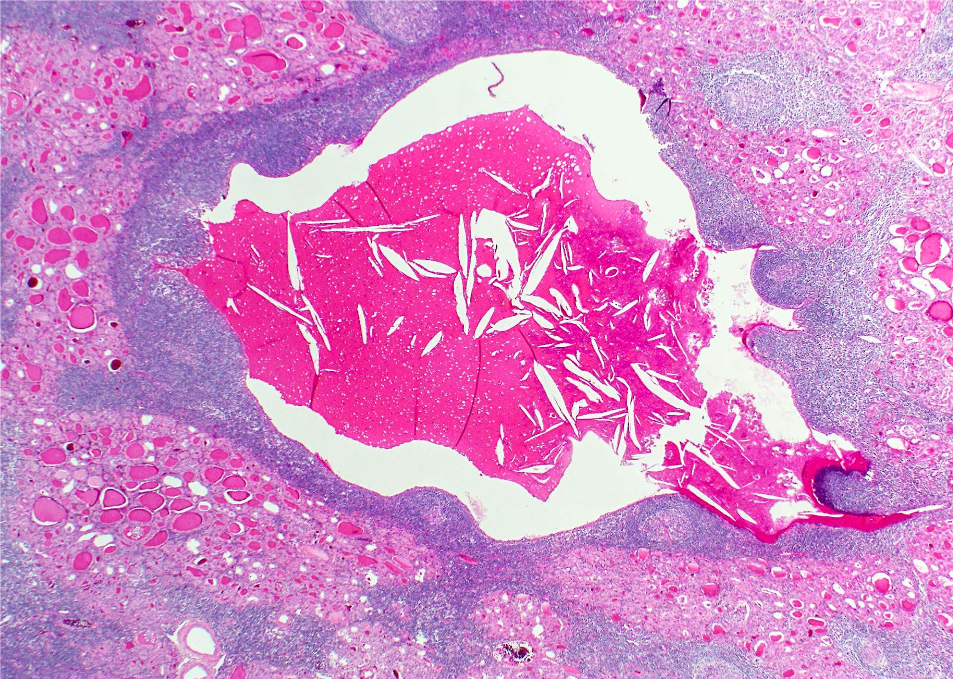
- The cyst structure is identical to branchial cleft cyst (Branchial pouch / cleft anomalies) composed of lymphoid tissue with germinal centers and squamous lining
- Epithelial lining is attenuated stratified squamous (one or two cells to approximately seven cells in thickness) or focal respiratory-type epithelium with ciliated or goblet cells
- Focally can be denuded
- Cyst lumen contains keratin / mucin and debris with cholesterol clefts
- Abundant adjacent lymphoid tissue, lymphoid follicles with prominent germinal centers common in cyst wall (Hum Pathol 1994;25:1238)
- Old cysts have a rim of fibrous tissue
- No thyroid parenchyma within the cyst, may have entrapped follicles in the outer side of cystic wall
- Signs of thyroiditis (often florid Hashimoto thyroiditis) in surrounding thyroid (Am J Surg Pathol 1989;13:45)
- Solid cell nests, basaloid squamous nests and C cell hyperplasia often found in surrounding parenchyma (Am J Surg Pathol 1989;13:1072, Am J Surg Pathol 1990;14:1165)
Microscopic (histologic) images
Cytology description
- Mature superficial squamous cells with intact nuclei, anucleate squames, clusters of neutrophils and lymphocytes, macrophages, amorphous debris in the background (AJNR Am J Neuroradiol 2000;21:1340)
- Some cases yield solely lymphocytes (Endocr J 2000;47:303), squamous cells (Am J Otolaryngol 2006;27:146) or respiratory epithelium (J Clin Ultrasound 2006;34:298)
Positive stains
Negative stains
Differential diagnosis
- Intrathyroidal thyroglossal duct cyst: midline location, thyroid remnants in cyst wall, no germinal centers (Am J Surg Pathol 1989;13:45)
- Cystic tumors:
- Papillary carcinoma
- Follicular carcinoma
- Follicular adenoma (Pathol Int 2000;50:897)
- Cystic goiter
- Cystic thymic remnants: basaloid cells with Hassall corpuscles (Arch Pathol Lab Med 2003;127:251)
- The distinction from branchial cleft cyst rests on the finding of respiratory-type epithelium (Arch Pathol Lab Med 2003;127:251)
- Cytology:
- Hashimoto thyroiditis with squamous metaplasia
- Mucoepidermoid carcinoma
- Squamous cell carcinoma
- Warthin-like variant of papillary thyroid carcinoma (Soonchunhyang Med Sci 2014;20:131)


