Table of Contents
Definition / general | Essential features | Epidemiology | Pathophysiology | Diagnosis | Laboratory | Prognostic factors | Case reports | Microscopic (histologic) description | Microscopic (histologic) images | Cytology description | Immunohistochemistry & special stains | Electron microscopy images | Differential diagnosis | Additional references | Board review style question #1 | Board review style answer #1Cite this page: Bychkov A. Crystals. PathologyOutlines.com website. https://www.pathologyoutlines.com/topic/thyroidcrystals.html. Accessed April 2nd, 2025.
Definition / general
- Calcium oxalate crystals are frequently found in colloid, especially in inactive follicles of aged individuals (Am J Surg Pathol 1993;17:698)
- Useful in differentiating thyroid tissue from parathyroid tissue (no crystals) on frozen section (Am J Surg Pathol 2002;26:813, Am J Surg Pathol 2014;38:1212)
- Patients with chronic renal failure on long term dialysis have increased frequency of intracolloidal crystals due to systemic oxalate deposition; also noted in kidney, myocardium and other sites (Arch Pathol Lab Med 1979;103:58)
- First described by Zeiss in 1877, who mentioned octahedral crystals of calcium oxalate in the thyroid gland (Am J Pathol 1954;30:545)
- Accumulation of cystine crystals is found in cystinosis and leads to destruction and atrophy of follicular cells (J Pediatr 1977;91:204)
Essential features
- Colloid often contains calcium oxalate crystals, particularly in nodular goiter but also in aged or hypoactive benign thyroid and rarely in tumors
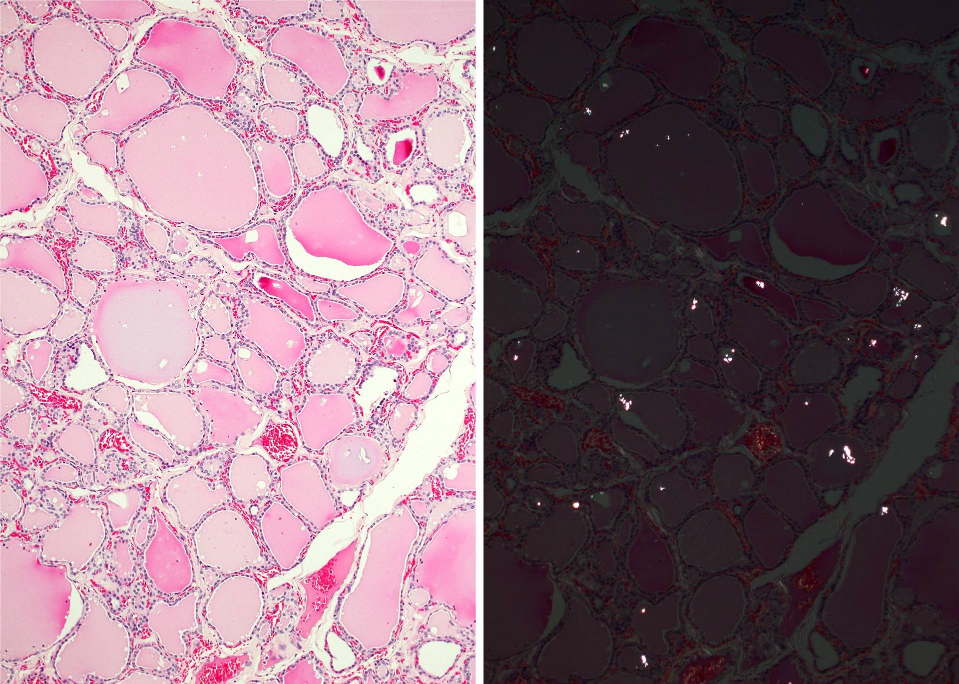
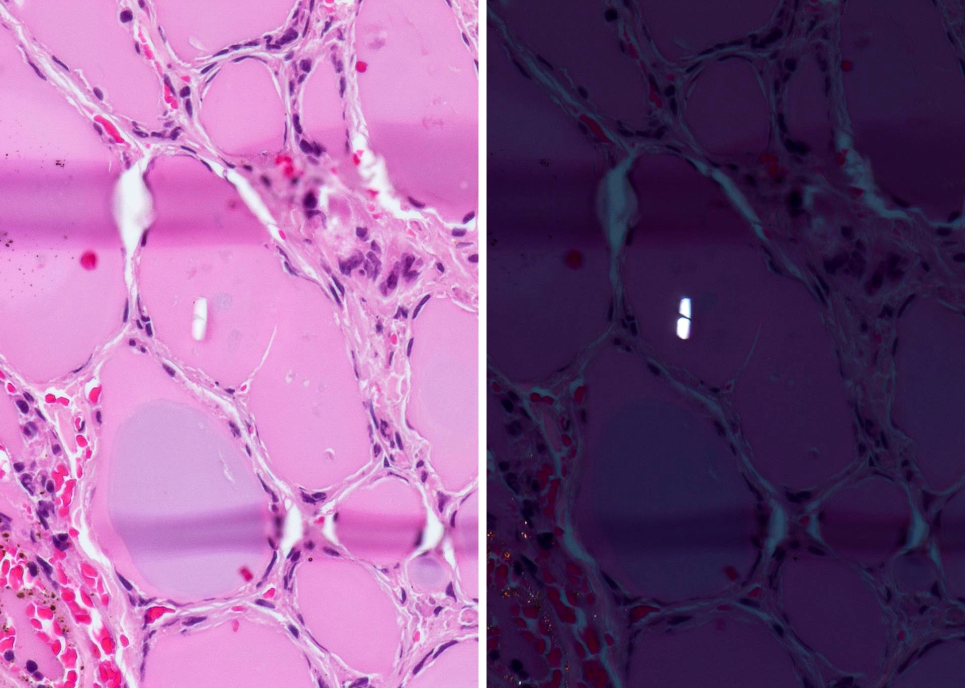
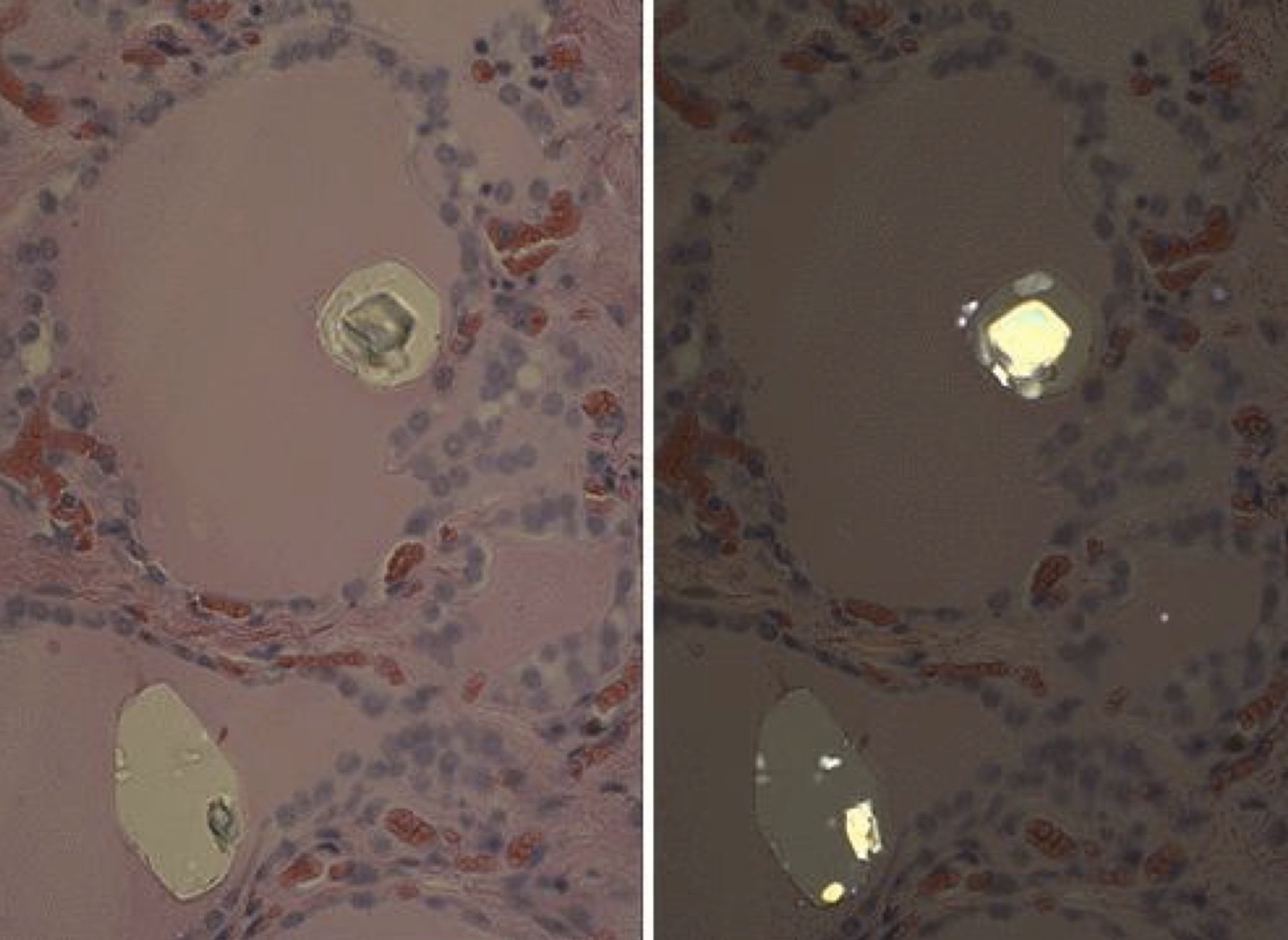
- Appear as intracolloidal birefringent crystals of various shape incidentally found in histologic sections or fine needle aspirates
- Calcium oxalate crystals are useful in differentiating thyroid tissue from parathyroid tissue (no crystals) at frozen section
Epidemiology
- Present in 79% of normal appearing thyroids at autopsy (Am J Clin Pathol 1987;87:443)
- More prevalent in older individuals, with the highest prevalence (85%) in over 70 year olds, while no crystals were seen in those under 10 years old (Virchows Arch A Pathol Anat Histopathol 1993;422:301)
- Heavy deposits of calcium crystals seen almost exclusively in benign conditions (Am J Surg Pathol 1993;17:698)
- Found in 70% of benign nodules (goiter, adenoma) vs. 8% of malignant nodules (FTC > PTC) (Am J Surg Pathol 1993;17:698)
Pathophysiology
- Occurrence of calcium crystals in normal human thyroid is associated with a low functional state of the thyroid follicles (Virchows Arch A Pathol Anat Histopathol 1993;422:301)
- Finding intracolloidal crystals may be a function of increasing age or disease state (Wenig: Atlas of Head and Neck Pathology, Third Edition, 2015)
- Progressive accumulation may advance to dystrophic calcification
Diagnosis
- Incidental microscopic finding (Nose: Diagnostic Pathology - Endocrine, First Edition, 2012)
Laboratory
- No association with abnormal laboratory findings of thyroid function
- Patients with renal failure on dialysis may have disseminated oxalate deposition in visceral organs including thyroid
Prognostic factors
- No prognostic implications
Case reports
- 77 year old woman with multinodular goiter (Diagn Cytopathol 2016;44:814)
Microscopic (histologic) description
- Anisotropic crystals appear as small transparent pale or colorless refractile foci variable in size and shape (polygonal, sand-like, needle shaped, rhomboid or irregular)
- Large round or oval vacuoles within the colloid may contain calcium oxalate crystals
- Crystals are strongly birefringent under polarized light microscopy
- Intrathyroidal crystals are exclusively found within colloid and do not appear within cytoplasm of the follicular epithelial cells or in stroma (Wenig: Atlas of Head and Neck Pathology, Third Edition, 2015)
- Background thyroid (Am J Surg Pathol 1993;17:698):
- There is no association with any specific thyroid condition
- Highest prevalence of crystals was found in nodular goiters and follicular adenomas (up to 80%)
- Rarely associated with malignant tumors, 33% prevalence in follicular carcinoma and 5% in papillary carcinoma
- One study found low prevalence in Graves' disease and Hashimoto thyroiditis but a large amount of crystals in subacute thyroiditis, which were also located in granulomas (Mills: Histology for Pathologists, Fourth Edition, 2012)
- Crystals found at autopsy tend to disappear within hours after death (Virchows Arch A Pathol Anat Histopathol 1993;422:301)
Microscopic (histologic) images
Cytology description
- Variably sized and shaped crystals in the background of colloid (Diagn Cytopathol 2016;44:814)
- In one series, total incidence of birefringent crystals was 45%, including 68% in benign lesions and 21% in malignant tumors (Acta Cytol 1999;43:575)
- Benign diseases show a more scattered distribution of birefringent crystals, compared to the typical focal distribution in cancer
- Occurrence of crystals in thyroid FNAC is lower than that in histologic specimens
Immunohistochemistry & special stains
- Crystals are associated with weak or negatively thyroglobulin stained colloid and are not common in strongly Tg positive colloid (Virchows Arch A Pathol Anat Histopathol 1993;422:301)
Electron microscopy images
Differential diagnosis
- Intracolloidal vacuoles (empty spaces) and dense particulated colloid (basophilic clumps) do not show birefringence under polarized light
Additional references
Board review style question #1
How are calcium oxalate crystals useful in frozen sections from thyroid and parathyroid?
- Differentiate cancer from benign tissue
- Distinguishing thyroid from parathyroid tissue
- Helps to locate psammoma bodies
- Identify age of patient
- No practical utility
Board review style answer #1
B. Distinguishing thyroid from parathyroid tissue. Calcium oxalate crystals are useful in differentiating thyroid tissue (scattered to abundant crystals) from parathyroid tissue (no crystals) at frozen section.
Comment Here
Reference: Crystals
Comment Here
Reference: Crystals


