Table of Contents
Definition / general | Essential features | Terminology | Epidemiology | Pathophysiology | Etiology | Clinical features | Diagnosis | Radiology description | Radiology images | Prognostic factors | Treatment | Gross description | Gross images | Differential diagnosis | Additional referencesCite this page: Özer E. Dandy-Walker Malformation. PathologyOutlines.com website. https://www.pathologyoutlines.com/topic/syndromesdandywalker.html. Accessed April 2nd, 2025.
Definition / general
- Dandy-Walker syndrome (DWS) is a congenital brain malformation involving the cerebellum
- Note: Dandy-Walker does not represent a single entity, as there are three identified types of Dandy-Walker complexes: DWS malformation, DWS mega cisterna magna and DWS variant
Essential features
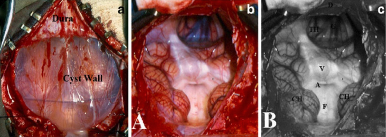
- Dandy-Walker malformation (DWM) has six features (Childs Nerv Syst 2011;27:1665)
- Large, median posterior fossa cyst communicating to the fourth ventricle
- Absence of the lower portion of the vermis at different degrees
- Hypoplasia, anterior rotation and upward displacement of the remnant of the vermis
- Antero-lateral displacement of normal or hypoplastic cerebellar hemispheres
- Large bossing posterior fossa with elevation of the torcular
- Absence or flattening of the angle of the fastigium (highest point of fourth ventricle)
- Large, median posterior fossa cyst communicating to the fourth ventricle
- All six features are not found in each case
- The essential features are: enlargement of the posterior fossa, cystic dilatation of the fourth ventricle and agenesis of the vermis
- Hydrocephalus is present in 80% of the cases and should not be considered a specific component of the malformation
- Other CNS malformations can be observed like occipital encephaloceles, corpus callosum agenesis, schizencephaly and glial heterotopias
- Dandy–Walker variant (DWV): cystic posterior mass with variable hypoplasia of the cerebella vermis and no enlargement of the posterior fossa (
- Dandy Walter mega-cisterna magna: enlarged cisterna magna with normal cerebellar vermis and fourth ventricle
Terminology
- Dandy-Walker malformation, Dandy-Walker syndrome
Epidemiology
- The incidence is 1:25,000 - 1:30,000 births with a slight female predominance
- Familial cases are very uncommon (Childs Nerv Syst 2011;27:1665)
Pathophysiology
- DWM may be caused by many conditions affecting the brain development in an early stage
- The type of insult is less important than the timing and duration of exposure to the noxious agent (J Child Neurol 2011;26:1483)
Etiology
Dandy-Walker malformation can occur due to:
- Mendelian conditions: Walker-Warburg syndrome, Mohr syndrome, Meckel-Gruber syndrome
- Chromosomal aberrations: several duplications involving 5p, 8p, 8q; trisomy 9, duplication on 17q, Turner syndrome
- Environmentally induced malformation syndrome: prenatal exposure to rubella, cytomegalovirus, toxoplasmosis, coumadin, alcohol and maternal diabetes
- Also multifactorial and sporadic disorders: see Congenit Anom (Kyoto) 2007;47:113, Childs Nerv Syst 2011;27:1665
Clinical features
- The symptoms are related to hydrocephalus, cerebellar and cranial nerves dysfunction and to the presence of associated anomalies
- 80 - 90% present in the first year
- DWM is often associated with other brain or systemic anomalies (Cerebellum Ataxias 2016;3:1)
Diagnosis
- May be diagnosed in utero by using 3D ultrasound as early as 14 weeks of gestation
- Fetal MRI is indicated in case of posterior fossa malformation suspected by ultrasonography
Radiology description
- MRI with sagittal views and T2 weighted images are mandatory for studying precisely the content of the posterior fossa
Prognostic factors
- The clinical course is very variable and depends on the severity of the associated central nervous system malformations, with neurologic development ranging from normal to severely intellectually disabled
Treatment
- The goal of treatment is the control of the hydrocephalus and of the posterior fossa cyst
Gross description
- In classic DWM, a huge csyt occupies almost the entire posterior fossa, displacing the brainstem forward and flattening the pons against the clivus
- The neuropathological picture is completed by aplasia of the vermis, heterotopia of the cerebellar cortex and enlargement of the posterior fossa, with a high position of the tentorium and transverse sinuses
Differential diagnosis
- Different cystic lesions may originate in the posterior fossa
- Lesions that do not meet the diagnostic criteria should NOT be considered as DWM, i.e. cysts that do not communicate directly with the fourth ventricle, cystic fourth ventricle associated with a normal posterior fossa and normally inserted tentorium
Additional references


