
Stomach
Other nonneoplastic
Xanthoma
Authors: Giby V. George, M.B.B.S., Aaron R. Huber, D.O.
Editorial Board Members: Claudio Luchini, M.D., Ph.D., Naziheh Assarzadegan, M.D.


Last author update: 19 July 2023
Last staff update: 18 March 2024
Copyright: 2003-2025, PathologyOutlines.com, Inc.
PubMed Search: Xanthoma


Table of Contents
Definition / general | Essential features | Terminology | ICD coding | Epidemiology | Sites | Pathophysiology | Etiology | Clinical features | Diagnosis | Prognostic factors | Case reports | Treatment | Clinical images | Gross description | Microscopic (histologic) description | Microscopic (histologic) images | Positive stains | Negative stains | Sample pathology report | Differential diagnosis | Additional references | Board review style question #1 | Board review style answer #1 | Board review style question #2 | Board review style answer #2Cite this page: George GV, Huber AR. Xanthoma. PathologyOutlines.com website. https://www.pathologyoutlines.com/topic/stomachxanthoma.html. Accessed April 2nd, 2025.
Definition / general
- Gastric xanthoma (GX) is a benign mucosal tumor-like lesion composed of foamy macrophages within the lamina propria
Essential features
- Benign tumor-like lesion most commonly occurring in the antrum of the stomach
- Endoscopically seen as small, yellow-white nodules or plaque-like lesions
- Characterized histologically by accumulation of foamy macrophages (xanthoma cells) within the lamina propria
- Almost always associated with background gastric pathology
Terminology
- Xanthelasma, lipid islands
ICD coding
- ICD-10: K31.9 - disease of stomach and duodenum, unspecified
Epidemiology
- First described as lipid laden macrophages in the gastric mucosa by Orth in 1887
- Mean age of 60 years (53% between 40 and 60 years old)
- Rarely seen in the pediatric population
- Male predominance (3:1)
- Incidence is quite variable depending on the study but thought to be a relatively uncommon incidental finding at endoscopy with an estimated incidence of 0.02 - 0.8%; however, autopsy studies have reported rates of 1.9% and 58%
- Background mucosal pathology can be seen, such as Helicobacter pylori gastritis, intestinal metaplasia, atrophic gastritis or gastric ulcers
- May also be associated with metachronous or synchronous early gastric cancer and rapid growth of gastric cancer (J Clin Med 2021;10:5704, Can J Gastroenterol Hepatol 2020;2020:3578927)
- May be seen after partial gastrectomy
- Pathologists should carefully examine the background mucosa for additional pathologic findings when diagnosing gastric xanthoma since this entity does not occur in normal gastric mucosa
- References: Can J Gastroenterol Hepatol 2020;2020:3578927, Cureus 2022;14:e25026, Oxf Med Case Reports 2018;2018:omy051, J Pediatr Gastroenterol Nutr 2021;73:e26
Sites
- Gastrointestinal tract xanthomas are most commonly found in the stomach (70%) (particularly the antrum [76%] and pylorus) but have also been reported in the cardia, fundus and corpus
- Other less common sites within the gastrointestinal tract include the esophagus, small bowel or colon and rectum
- References: APMIS 2004;112:3, World J Gastroenterol 2015;21:1091, Bosn J Basic Med Sci 2012;12:127, Cureus 2022;14:e25026, Can J Gastroenterol Hepatol 2020;2020:3578927
Pathophysiology
- Pathogenesis and clinical significance remain unclear
- Given the association with background mucosal pathology, one hypothesis suggests that gastric xanthoma is a mucosal reaction to prior injury or inflammation
- When the mucosa is damaged, release of lipid rich material may be phagocytosed and hence, create foamy macrophages
- Another hypothesis suggests that phagocytosis of Helicobacter pylori organisms that have penetrated the lamina propria induces the macrophages to become foamy
- Cutaneous xanthoma and xanthelasma are both strongly associated with disorders of lipid metabolism; however, data regarding such an association in gastric xanthoma is less clear
- References: Cureus 2022;14:e25026, Oxf Med Case Reports 2018;2018:omy051, Bosn J Basic Med Sci 2012;12:127, Can J Gastroenterol Hepatol 2020;2020:3578927, Am J Gastroenterol 2001;96:3216
Etiology
- The exact etiology, pathogenesis and clinical significance of gastric xanthoma is unclear but usually coexists with other gastric pathology such as Helicobacter pylori gastritis, atrophic gastritis, gastric ulcers, intestinal metaplasia and early gastric cancer (see Epidemiology and Pathophysiology)
Clinical features
- Most commonly an incidental finding at endoscopy for another reason
Diagnosis
- Ultimately, the diagnosis is based on the characteristic histopathologic findings (see Microscopic description)
Prognostic factors
- Gastric xanthoma is completely benign and nonneoplastic; however, background gastric pathology may be present
Case reports
- 12 year old girl with abdominal pain (J Pediatr Gastroenterol Nutr 2021;73:e26)
- 34 year old woman with dyspepsia (Am J Gastroenterol 2001;96:3216)
- 44 year old man with intermittent epigastric pain (BMJ Case Rep 2013;2013:bcr2013201017)
- 57 year old man with history of Barrett esophagus (Cureus 2022;14:e25026)
- 78 year old man with chronic anemia (Oxf Med Case Reports 2018;2018:omy051)
Treatment
- No treatment is required for gastric xanthoma as it is a benign condition; treatment is aimed at coexistent gastric pathology
Clinical images
Images hosted on other servers:

Endoscopic yellow plaque-like lesion

Endoscopic yellow nodule and plaque-like lesion
Gross description
- Single or multiple, well demarcated, yellow-white, round or oval nodules or plaques that are typically between 1 and 10 mm on endoscopy (rarely larger than 5 mm) (Oxf Med Case Reports 2018;2018:omy051, Bosn J Basic Med Sci 2012;12:127)
Microscopic (histologic) description
- Histologic hallmark is the presence of lipid laden foamy macrophages (xanthoma cells) within the lamina propria (Ann Diagn Pathol 2013;17:72, Arch Pathol Lab Med 2004;128:937)
Microscopic (histologic) images
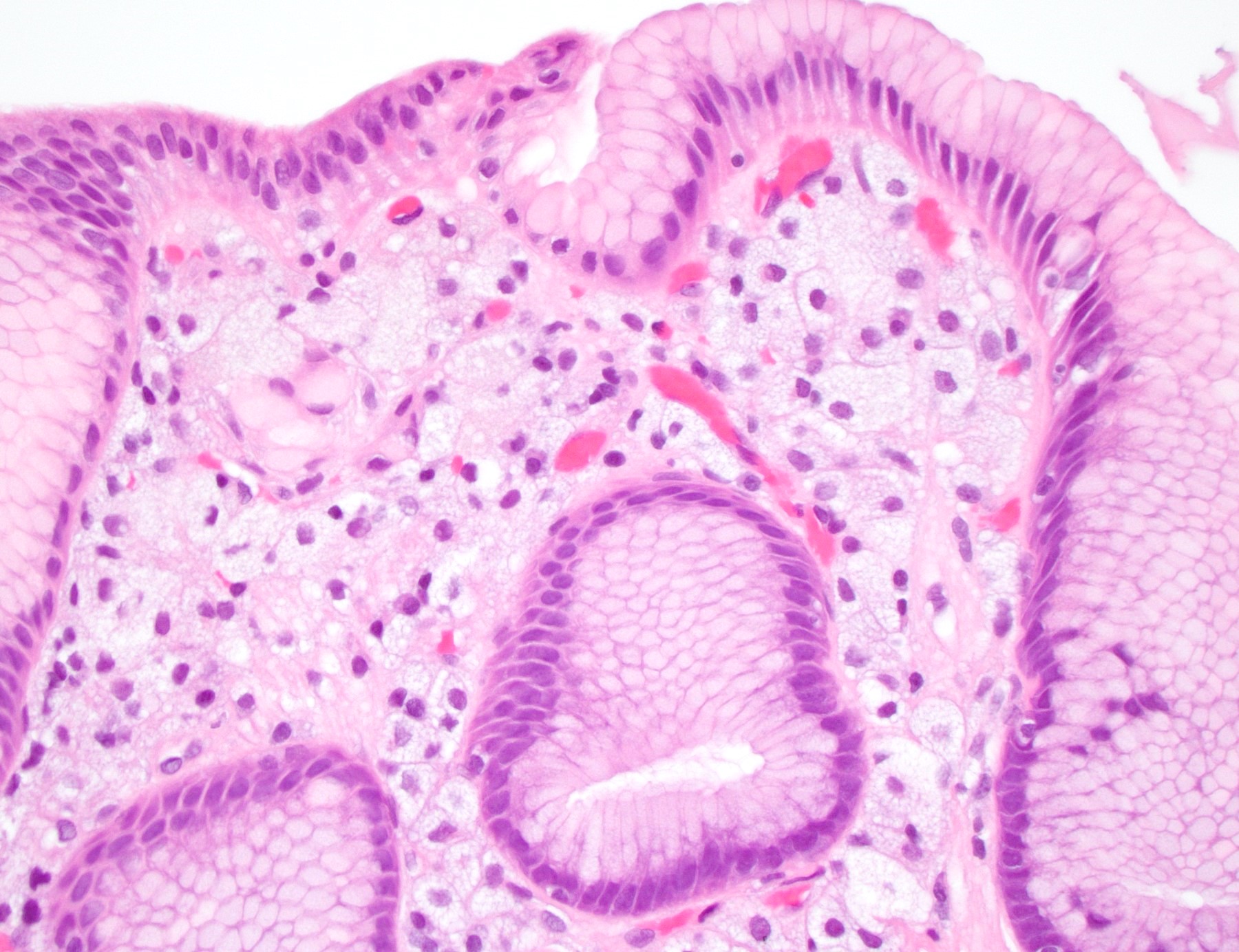
Contributed by Aaron R. Huber, D.O.
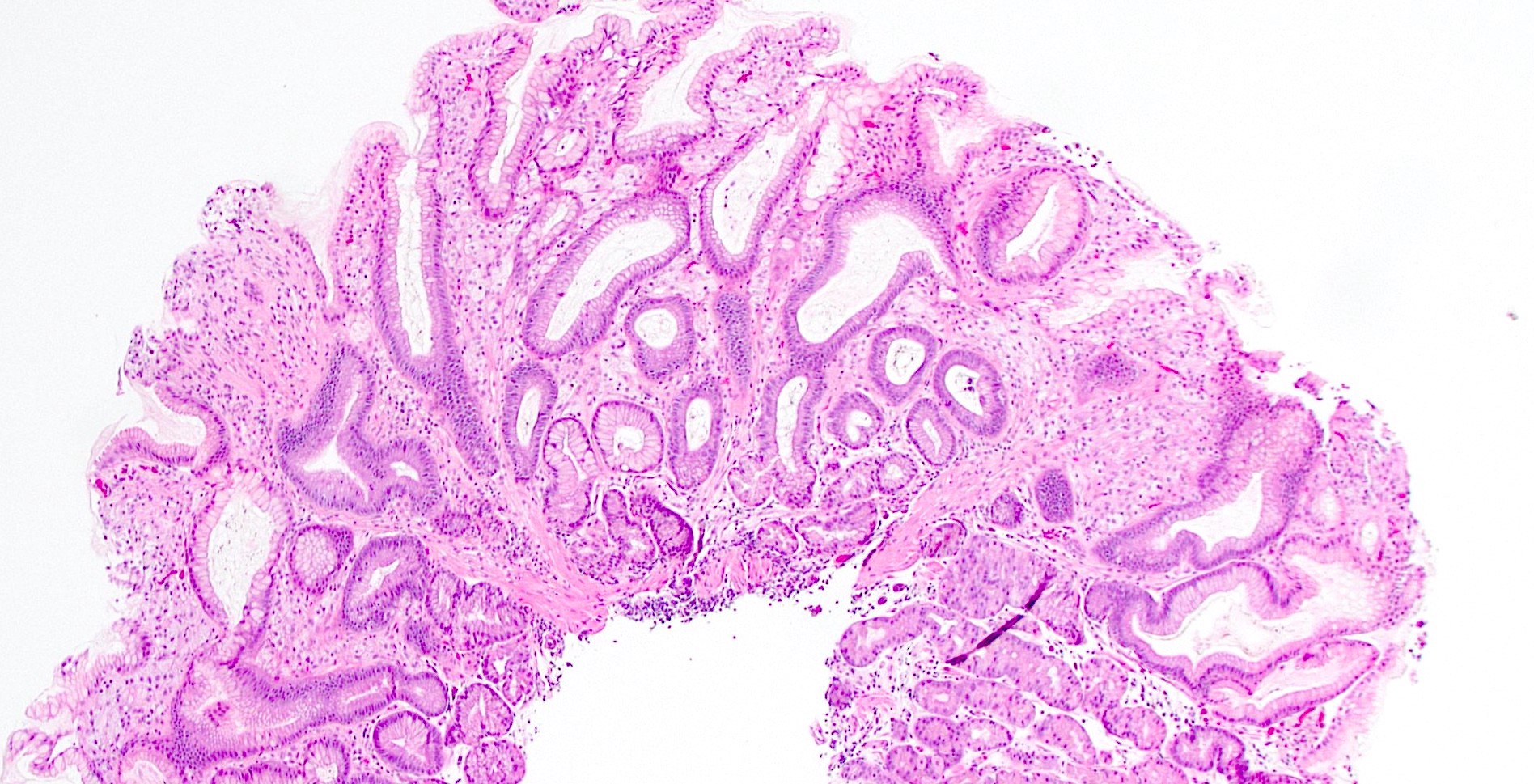
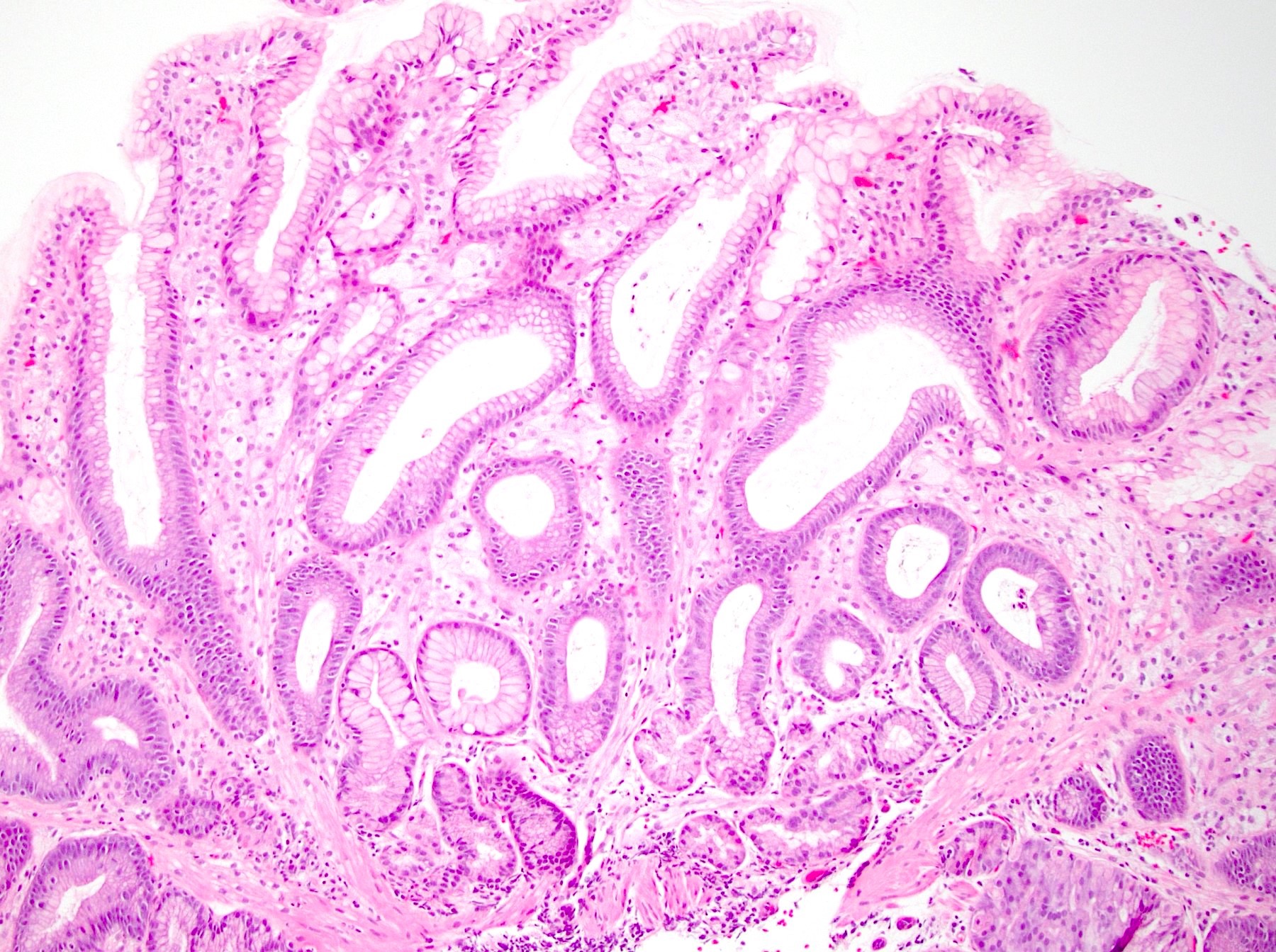
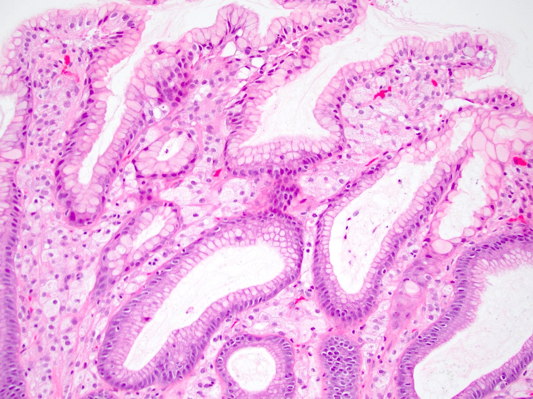
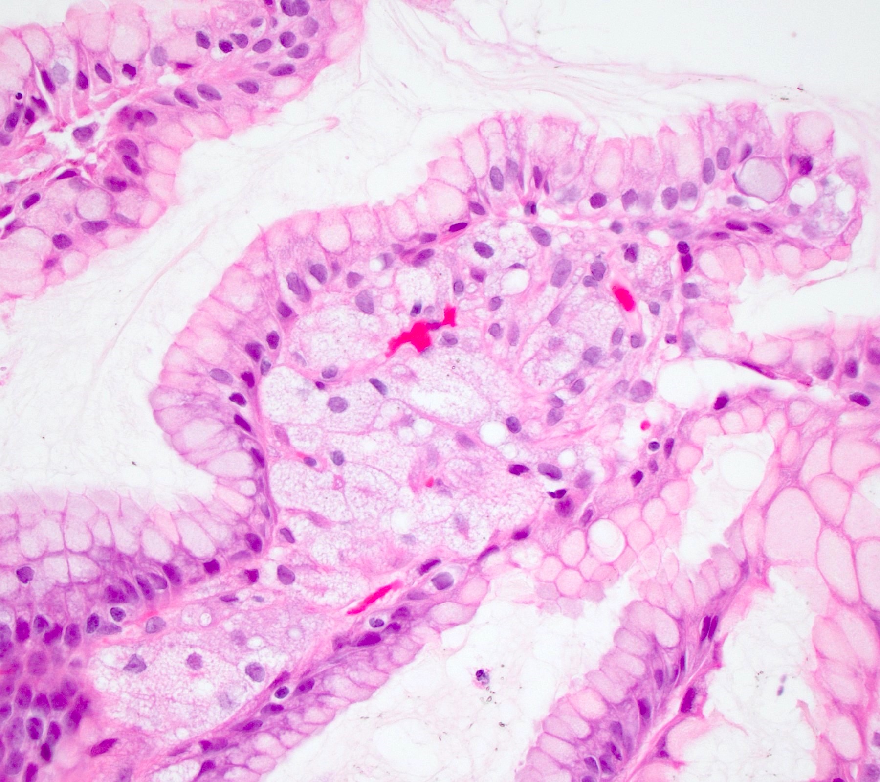
Foamy macrophages within the lamina propria
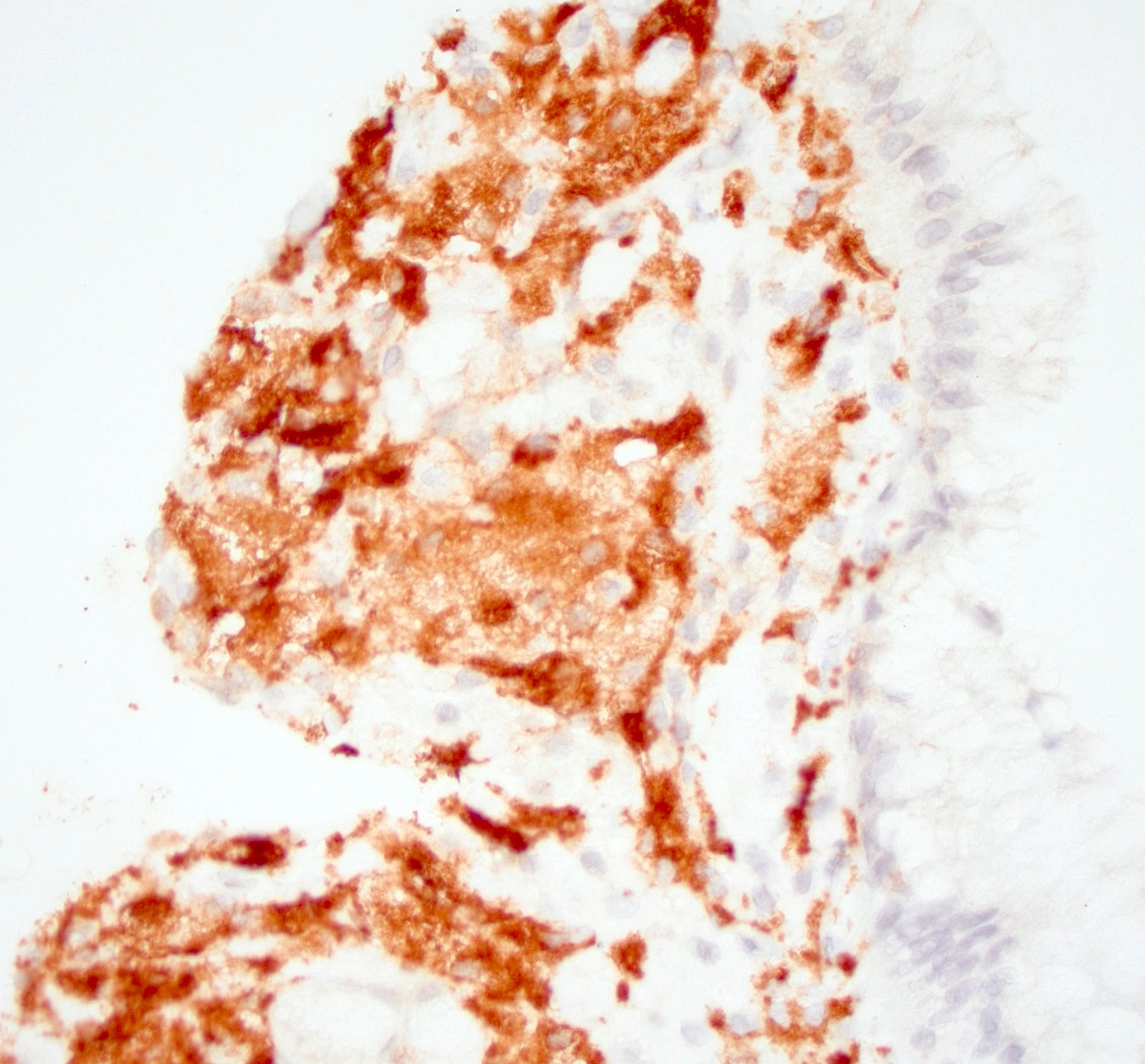
CD68
Sample pathology report
- Stomach, nodule, biopsy:
- Gastric xanthoma (see comment)
- Comment: H&E stained sections demonstrate clusters of foamy macrophages within the lamina propria. CD68 immunohistochemical stain highlights the macrophages, while keratin AE1 / AE3 is negative within these cells. The morphologic and immunophenotypic findings are compatible with gastric xanthoma.
Differential diagnosis
- Signet ring cell carcinoma:
- Metastatic carcinoma:
- Granular cell tumor:
- Gastric lipoma:
- Rare, typically involves the submucosa of the antrum (unlikely to be sampled in endoscopic mucosal biopsies) (BJR Case Rep 2019;5:20180109)
- Adipocytes will be S100 positive
Additional references
Board review style question #1
A 56 year old man underwent an esophagogastroduodenoscopy for reflux symptoms. A biopsy was taken of a small, sessile yellow-white nodule in the stomach. The histology is depicted in the H&E image. The unusual stroma cells were keratin negative and CD68 and CD163 positive. What is the best diagnosis?
- Granular cell tumor
- Metastatic breast carcinoma
- Normal gastric mucosa
- Signet ring cell carcinoma
- Xanthoma
Board review style answer #1
E. Xanthoma. The H&E image demonstrates an accumulation of lipid laden macrophages within the lamina propria indicative of xanthoma. Answer B is incorrect because although metastatic breast carcinoma, especially lobular carcinoma, is common in the stomach, keratin would be positive and CD68 or CD163 would be negative. In addition, more specific markers such as GATA3 would be positive. Answer D is incorrect because although signet ring cell carcinoma is one of the top differential diagnostic considerations, the cells in signet ring cell carcinoma are cytologically malignant, whereas those in gastric xanthoma are bland with abundant foamy cytoplasm. In addition, the signet ring carcinoma cells would be keratin positive and CD68 and CD163 negative. Answer A is incorrect because granular cell tumor is more commonly seen in the esophagus and has granular cytoplasm with pyknotic nuclei. These cells are CD68 positive but also S100 positive, unlike xanthoma cells (S100 negative). Answer C is incorrect because normal gastric mucosa would not demonstrate these findings.
Comment Here
Reference: Xanthoma
Comment Here
Reference: Xanthoma
Board review style question #2
A 45 year old woman underwent an esophagogastroduodenoscopy for reflux symptoms. A biopsy was taken of a yellow-white plaque-like lesion in the gastric antrum. The histology demonstrated antral mucosa with clusters of foamy histiocytes within the lamina propria. Which of the following is true about the pathologic entity described?
- May be associated with Helicobacter pylori gastritis
- Most commonly occurs in teenagers and is rare in adults
- Occurs commonly in patients with disorders of lipid metabolism, similar to cutaneous examples
- Occurs in normal gastric mucosa
Board review style answer #2
A. May be associated with Helicobacter pylori gastritis. Answer D is incorrect because gastric xanthoma (GX) essentially never occurs in normal gastric mucosa and may be associated with Helicobacter gastritis, atrophic gastritis, gastric ulcers, intestinal metaplasia and gastric cancer. Answer B is incorrect because gastric xanthoma has been reported in children; however, it is much more common in adults. Answer C is incorrect because unlike cutaneous examples of xanthelasma or xanthoma, evidence linking gastric xanthoma with disorders of lipid metabolism is inconclusive.
Comment Here
Reference: Xanthoma
Comment Here
Reference: Xanthoma
