Table of Contents
Definition / general | Essential features | ICD coding | Epidemiology | Sites | Pathophysiology | Etiology | Diagrams / tables | Diagnosis | Prognostic factors | Case reports | Treatment | Clinical images | Microscopic (histologic) description | Microscopic (histologic) images | Positive stains | Sample pathology report | Differential diagnosis | Board review style question #1 | Board review style answer #1 | Board review style question #2 | Board review style answer #2Cite this page: Nazli S, Hagen CE. Proton pump inhibitors. PathologyOutlines.com website. https://www.pathologyoutlines.com/topic/stomachPPI.html. Accessed April 1st, 2025.
Definition / general
- Chronic proton pump inhibitor (PPI) therapy results in characteristic histopathologic changes in the gastric mucosa
Essential features
- Decrease in hydrochloric acid (HCl) due to PPI intake causes an increase in gastrin production by the antral G cells promoting an achlorhydric and hypergastrinemic state
- Resulting endoscopic findings include gastric polyps, cobblestone-like mucosa and black spots
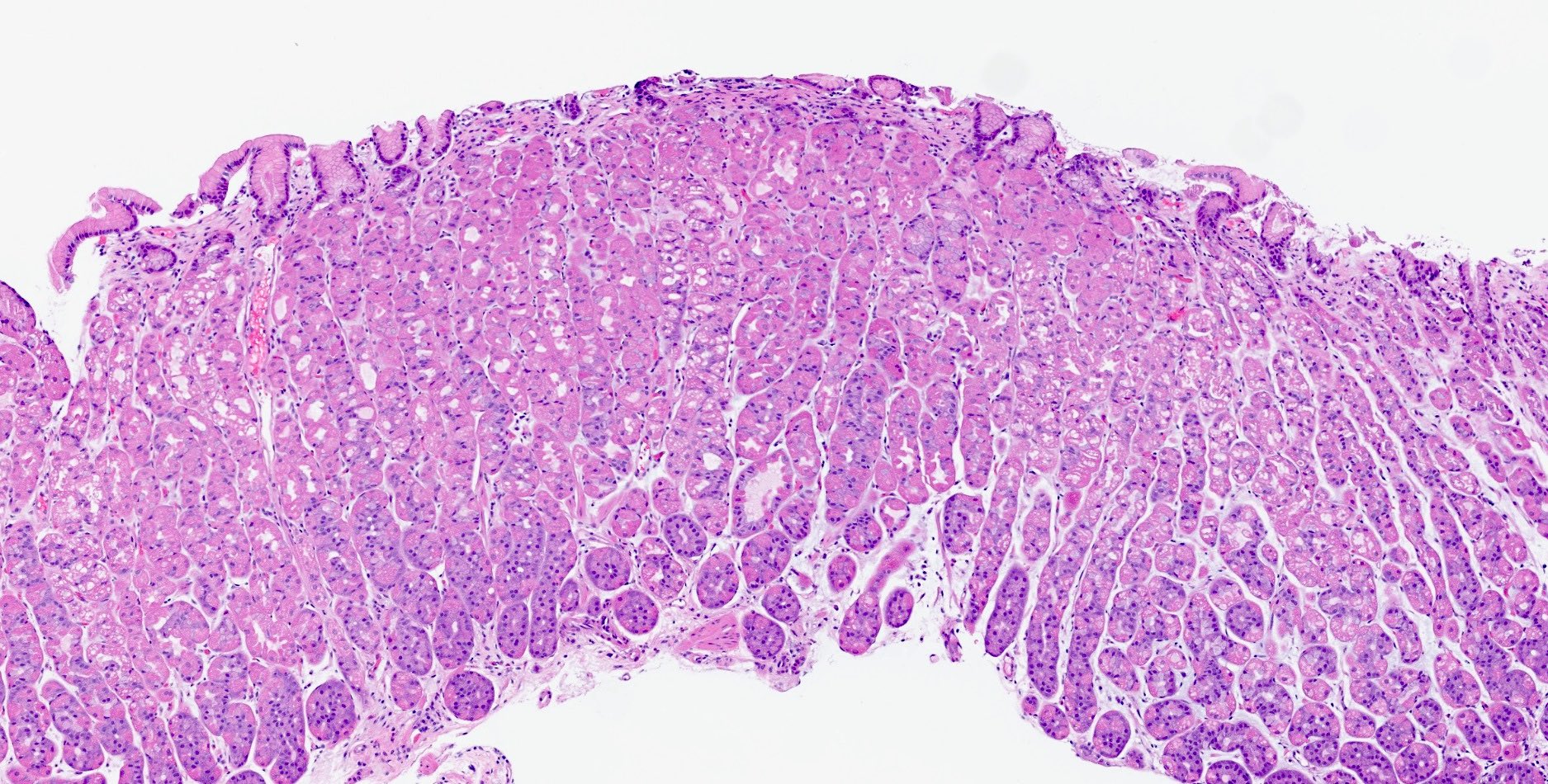
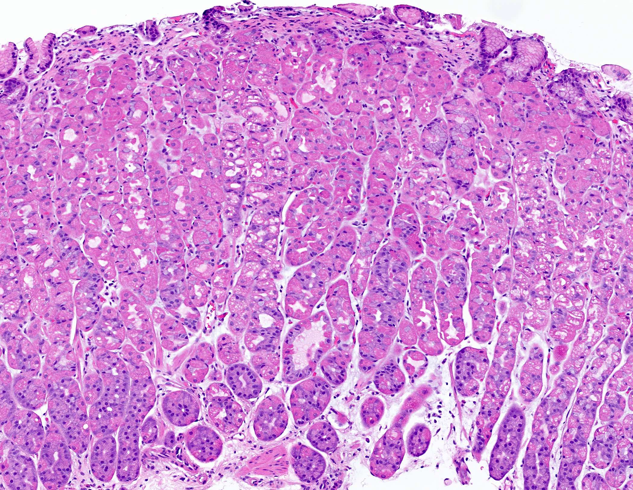
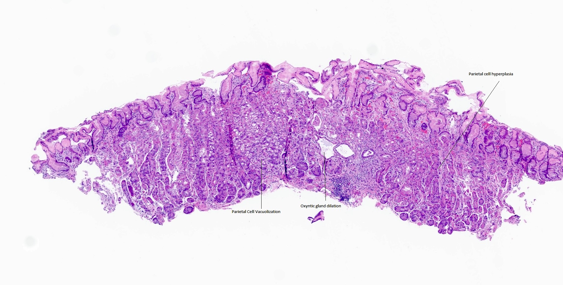
- Characteristic histologic changes include parietal cell hyperplasia / hypertrophy, cystic dilation of oxyntic glands with fundic gland polyp formation, foveolar hyperplasia and hyperplastic polyp formation and cytoplasmic vacuolization of parietal cells
ICD coding
- ICD-10: K31.89 - other diseases of stomach and duodenum
Epidemiology
- PPIs are used worldwide for acid related disease and changes can be seen with no age or gender predilection
Sites
- Gastric mucosa predominantly in the fundus and body of the stomach
Pathophysiology
- Proton pump inhibitors are used in a myriad of medical conditions including reflux esophagitis, peptic ulcer disease, stress ulcer prophylaxis, H. pylori infection and hiatal hernia, among others
- Mechanistically, proton pump inhibitors inhibit the parietal cell acid secretion leading to less stomach acid generation
- With the loss of the negative feedback on the antral G cells, gastrin secretion is increased
- Gastrin exerts trophic changes in both parietal and enterochromaffin-like cells within the oxyntic mucosa
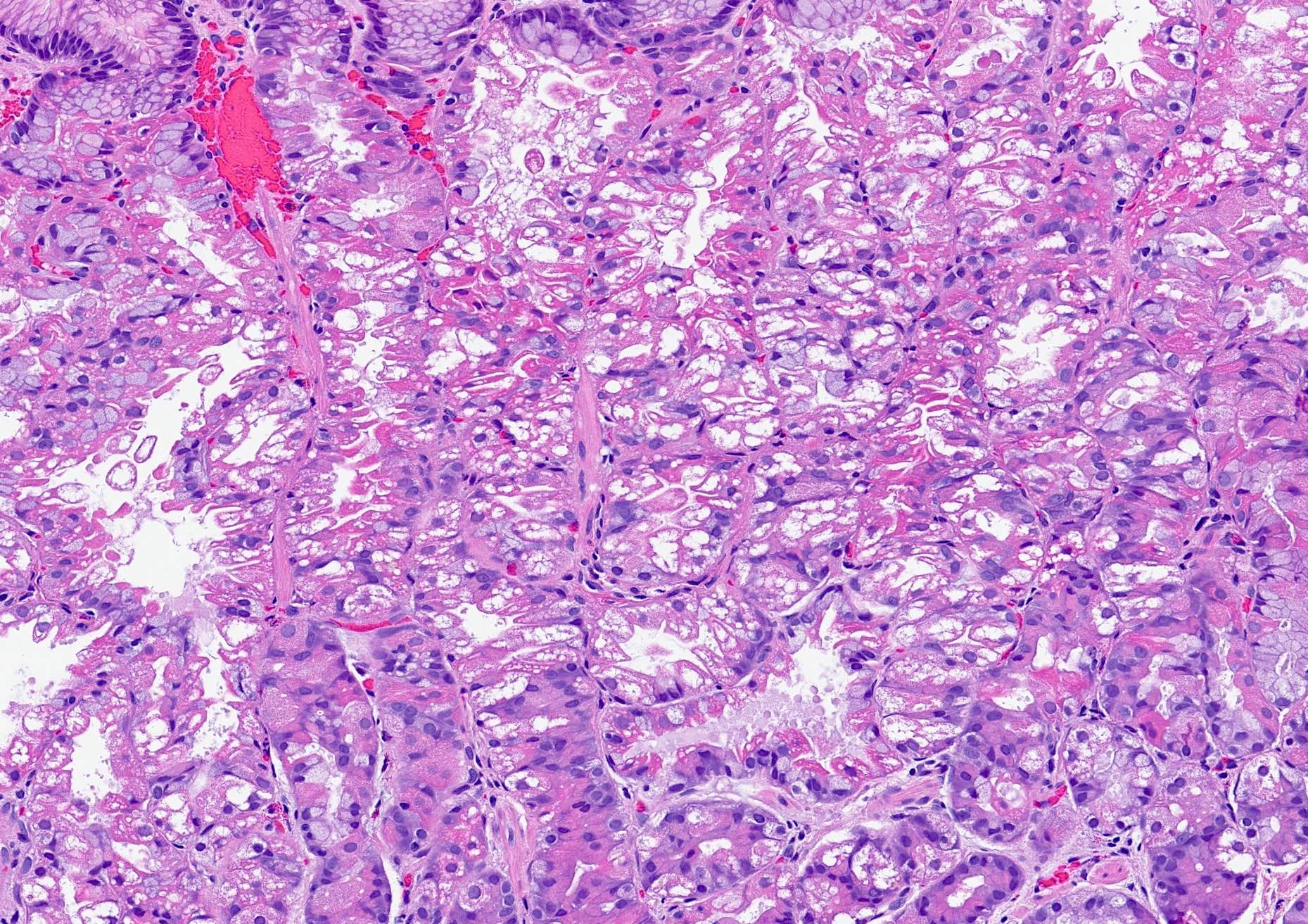
- Ultrastructural changes described as degeneration within the parietal cells may be seen and are due to irreversible binding with proton pump inhibitors; this leads to dilated secretory canaliculi within the parietal cells, which histologically can appear as vacuolization
- Reference: Histopathology 2013;63:735
Etiology
- PPIs are a potent suppressor of acid secretion with minimal toxicity making them a commonly prescribed agent
- Class of drug includes omeprazole, rabeprazole, pantoprazole, esomeprazole and lansoprazole
- Reference: Gastrointest Endosc Clin N Am 2020;30:239
Diagrams / tables
Diagnosis
- Endoscopic biopsy with histologic examination
- Correlation with clinical use of PPI
Prognostic factors
- No association between gastric fundic gland polyps and gastrointestinal neoplasia in > 100,000 patients (Clin Gastroenterol Hepatol 2009;7:849)
Case reports
- 32 year old woman with anemia (Gastrointest Endosc 2007;66:394)
- 37 year old man with PPI induced changes in foveolar type gastric adenocarcinoma (DEN Open 2023;4:e293)
- 42 year old man with PPI associated hyperplastic polyp (Case Rep Gastroenterol 2021;15:539)
- 52 year old man with multiple gastric neuroendocrine tumors associated with long term use of PPI and potassium competitive acid blocker (Intern Med 2024;63:2001)
Treatment
- Discontinuation of the offending drug; polyps regress when treatment stops (Hum Pathol 2000;31:684)
Clinical images
Microscopic (histologic) description
- Parietal cell hypertrophy and hyperplasia
- Protrusion of the parietal cytoplasm into the gland lumina creating an irregular hobnail appearance (parietal cell protrusions)
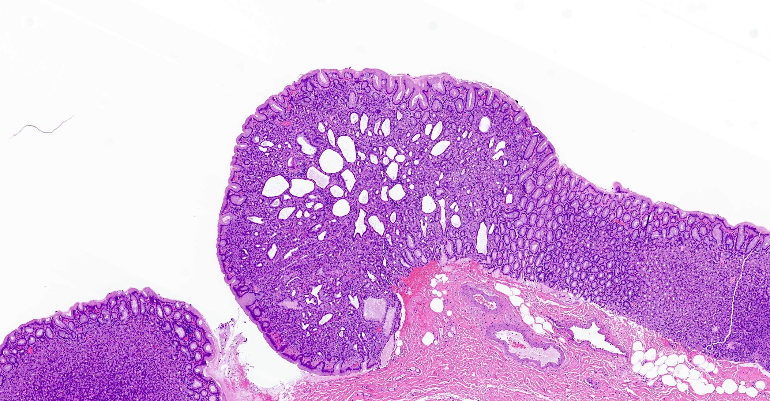
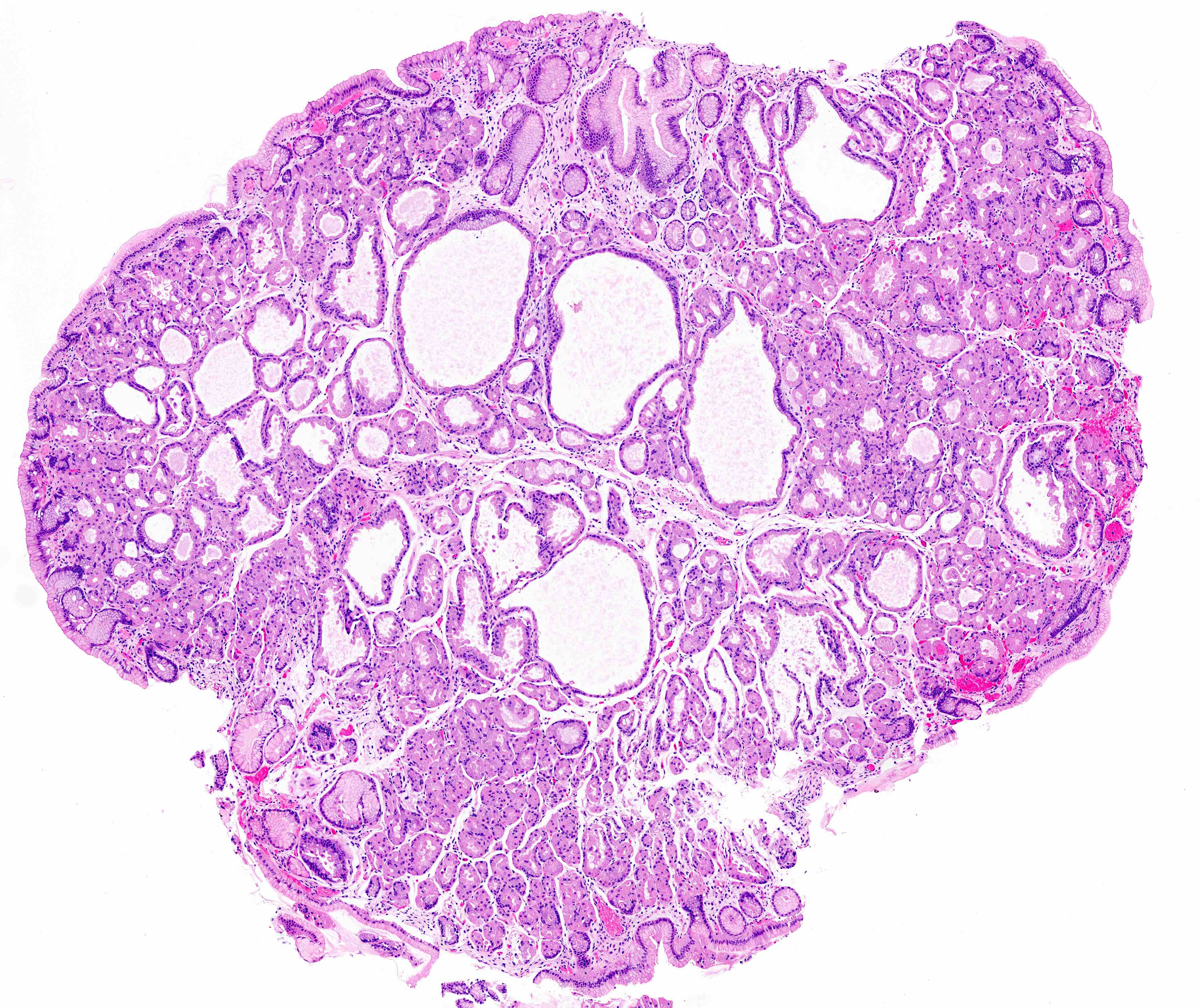
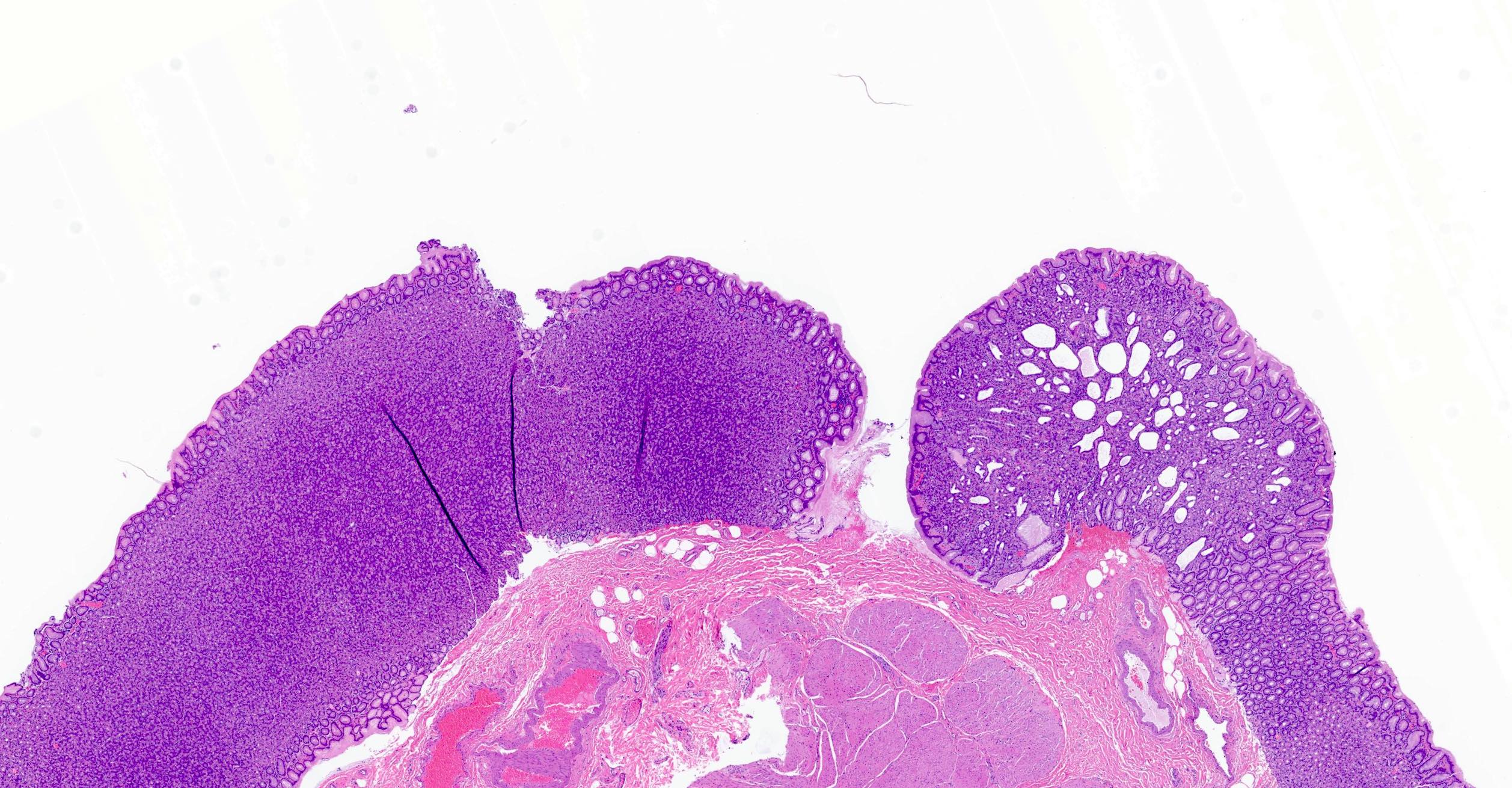
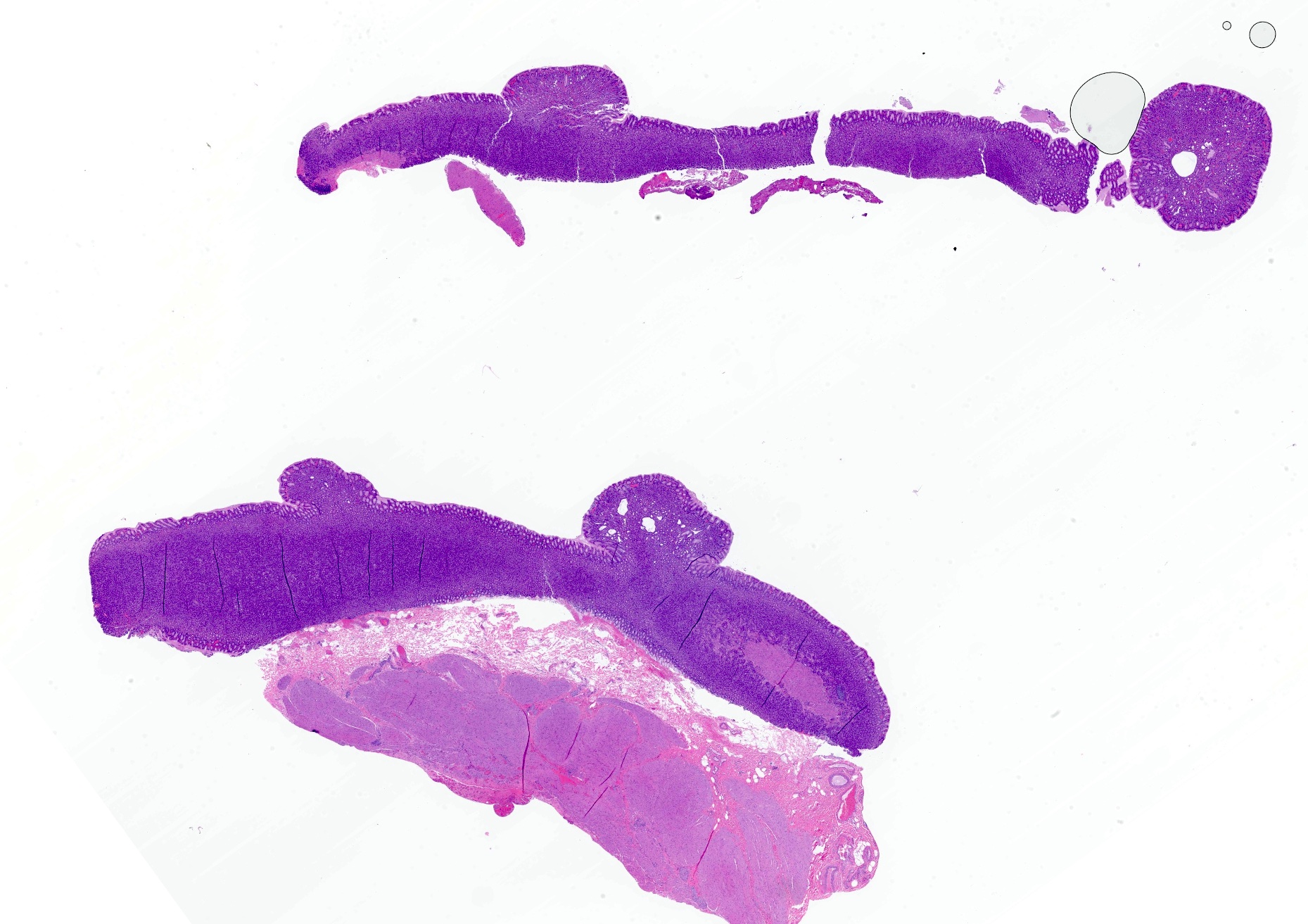
- Cystic dilation of fundic glands and fundic gland polyp formation
- Cytoplasmic vacuolization of parietal cells
- Foveolar epithelial hyperplasia or hyperplastic polyps
- Neuroendocrine cell (ECL) hyperplasia
- References: Hum Pathol 2000;31:684, Gut Liver 2021;15:646, Histopathology 2013;63:735
Microscopic (histologic) images
Positive stains
- Anti-H+ / K+ ATPase (proton pump) antibodies, although not routinely required as features can be readily identified on H&E and history of PPI use
Sample pathology report
- Stomach, polyp, endoscopic biopsy:
- Fundic gland polyp
- Stomach, endoscopic biopsy:
- Fundic mucosa with proton pump inhibitor effect (see comment)
- Comment: Fragments of fundic type mucosa show parietal cell hyperplasia and focal oxyntic gland dilation consistent with proton pump inhibitor effect. No intestinal metaplasia, dysplasia or malignancy is identified. Some of the parietal cells have a foamy to vacuolated appearance. Parietal cell vacuolization is a known consequence of proton pump inhibitor therapy and is a result of dilated secretory canaliculi within the parietal cells.
Differential diagnosis
- Zollinger-Ellison syndrome:
- Massive oxyntic gland hyperplasia
- Neuroendocrine tumors (gastrinomas) of small bowel or pancreas
- Gastric adenocarcinoma and proximal polyposis of the stomach (GAPPS):
- Autosomal dominant mode of inheritance
- Fundic gland polyps restricted to the oxyntic mucosa of gastric body and fundus
- Significant risk of progression to gastric adenocarcinoma
- Familial adenomatous polyposis (FAP):
- Autosomal dominant with germline APC mutation
- Endoscopically, hundreds to thousands of polyps throughout the gastrointestinal (GI) tract
- Gastric polyps may consist of fundic gland polyps and adenomas; fundic polyps may show low grade epithelial dysplasia
- Reference: Histopathology 2022;80:827
Board review style question #1
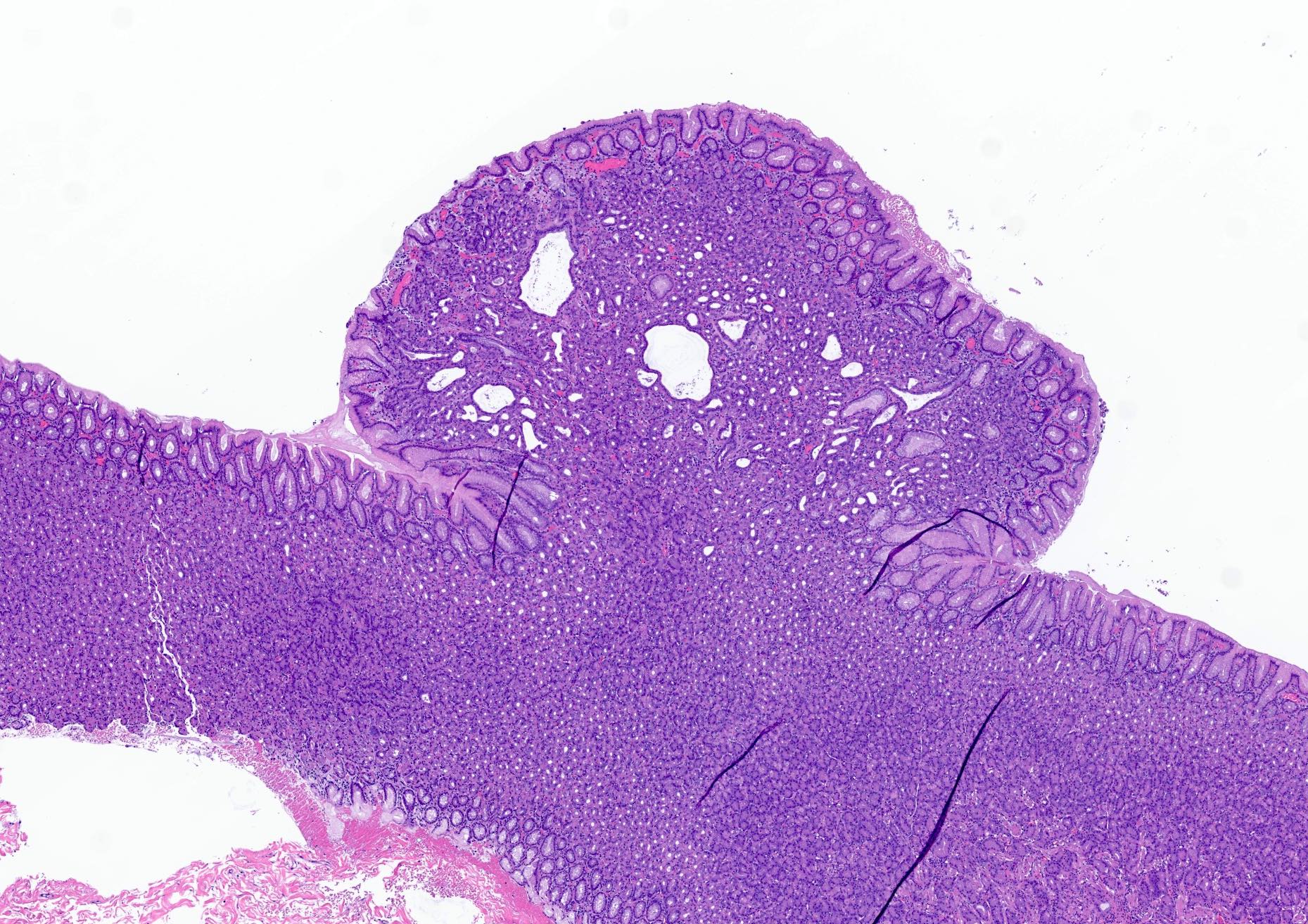
A 70 year old man with gastroesophageal reflux disease (GERD) taking omeprazole was noted to have multiple gastric polyps. A biopsy was performed and is shown above. He has no known family history of gastrointestinal tract malignancy. What is the most likely diagnosis?
- Familial adenomatous polyposis (FAP) syndrome
- Gastric adenocarcinoma and proximal polyposis of the stomach (GAPPS) syndrome
- Hyperplastic polyp
- Sporadic fundic gland polyp
Board review style answer #1
D. Sporadic fundic gland polyp. The image shows a gastric polyp characterized by cystically dilated oxyntic glands consistent with fundic gland polyp. Given the patient's negative family history and proton pump inhibitor (PPI) use, this is likely a sporadic fundic gland polyp related to said PPI use. Answers A and B are incorrect because fundic gland polyps can be seen in FAP and GAPPS syndrome but the patient's older age, negative family history and reported PPI use make this unlikely. Answer C is incorrect because morphologically, the histologic features are not compatible with a hyperplastic polyp, which typically has prominent foveolar hyperplasia and an edematous inflammatory stroma.
Comment Here
Reference: Proton pump inhibitors
Comment Here
Reference: Proton pump inhibitors
Board review style question #2
A 65 year old woman taking pantoprazole undergoes gastric biopsy. Parietal cells are noted to have cytoplasmic vacuolization. What is the underlying etiology of this vacuolization?
- Dilated secretory canaliculi
- Hyperchlorhydria
- Hypogastrinemia
- Parietal cell dysplasia
Board review style answer #2
A. Dilated secretory canaliculi. Degeneration within the parietal cells may be seen as a result of irreversible binding with proton pump inhibitors; this leads to dilated secretory canaliculi within the parietal cells, which histologically can appear as vacuolization. Answers B and C are incorrect because proton pump inhibitor (PPI) use induces a hypergastrinemic and hypochlorhydric state. Answer D is incorrect because the cytoplasmic vacuolization of parietal cells may raise concern for neoplasia but is benign in nature.
Comment Here
Reference: Proton pump inhibitors
Comment Here
Reference: Proton pump inhibitors


