Table of Contents
Definition / general | Essential features | Terminology | ICD coding | Epidemiology | Sites | Pathophysiology | Etiology | Diagnosis | Radiology description | Prognostic factors | Case reports | Treatment | Gross description | Microscopic (histologic) description | Microscopic (histologic) images | Positive stains | Negative stains | Molecular / cytogenetics description | Sample pathology report | Differential diagnosis | Board review style question #1 | Board review style answer #1 | Board review style question #2 | Board review style answer #2Cite this page: Martinez-Ciarpaglini C. Gastric carcinoma with lymphoid stroma. PathologyOutlines.com website. https://www.pathologyoutlines.com/topic/stomachLEL.html. Accessed April 3rd, 2025.
Definition / general
- Gastric carcinoma composed of small nests of cancer cells broadly distributed in a background of dense and prominent lymphoid stroma, histologicically similar to nasopharyngeal carcinoma (Arch Pathol Lab Med 2008;132:706)
Essential features
- Infrequent subtype of gastric cancer composed of small trabeculae and nests of epithelial cells embedded in a dense lymphoid infiltrate reminiscent of lymphoid tissue
- Strongly associated with Epstein-Barr virus (EBV) infection and microsatellite instability (mutually exclusive)
- PDL1 is usually overexpressed, making these tumors prone to immune checkpoint blockade therapy
Terminology
- Lymphoepithelioma-like carcinoma
- Medullary carcinoma
- Undifferentiated carcinoma with lymphoid stroma
ICD coding
- ICD-O: 8140/3 - adenocarcinoma, NOS
Epidemiology
- Constitutes about 4% of all gastric carcinomas (Arch Pathol Lab Med 2008;132:706)
- Mean age 70 years (Am J Surg Pathol 2018;42:453, J Surg Res 2017;210:159)
- Strongly associated with Epstein-Barr virus (EBV) infection and microsatellite instability (MSI) (Am J Surg Pathol 2018;42:453)
- EBV positive cases are more frequent in men (Gastric Cancer 2015;18:246)
- MSI associated cases are more frequent in women and older patients (ESMO Open 2019;4:e000470)
Sites
- EBV associated cases are frequently located in the cardia and middle portion of the stomach (Arch Pathol Lab Med 2008;132:706)
- Microsatellite instability associated cases are more common in the antrum (Arch Pathol Lab Med 2008;132:706, ESMO Open 2019;4:e000470)
Pathophysiology
- EBV seems to be involved in the early stage of carcinogenesis and remains expressed in all primary tumor cells and their metastases (Arch Pathol Lab Med 2008;132:706)
- EBV infection of gastric cells by lymphocytes with reactivated EBV is suspected to be the first step of EBV associated gastric carcinoma development (Cancer Sci 2008;99:195)
- EBV expression consistently absent in normal epithelium or dysplastic lesions (J Exp Clin Cancer Res 2009;28:14, Arch Pathol Lab Med 2008;132:706)
Etiology
- EBV and microsatellite instability in gastric carcinoma are mutually exclusive (Am J Surg Pathol 2018;42:453)
- In Asia, most cases (around 80%) are related to EBV infection, a significantly higher rate compared with Western countries (7 - 39%) (Gastric Cancer 2015;18:246, Am J Surg Pathol 2018;42:453, ESMO Open 2019;4:e000470)
- EBV can be demonstrated in 1.8 - 18% of all sporadic gastric carcinomas (Mod Pathol 2003;16:641, ESMO Open 2019;4:e000470)
- Around 38% of cases are associated with microsatellite instability (Am J Surg Pathol 2018;42:453)
- Microsatellite instability is detected in 15 - 30% of all gastric carcinomas (J Surg Oncol 2018;117:829, ESMO Open 2019;4:e000470)
Diagnosis
- Although most cases seems to be localized at the time of diagnosis, distinctive clinical signs and symptoms associated with this type of tumor have not been reported (Am J Surg Pathol 2018;42:453, J Surg Res 2017;210:159)
- Endoscopy: frequently described as a hypoechoic lesion in the submucosa and the muscularis mucosa, mimicking features of subepithelial tumors (World J Gastroenterol 2014;20:1365)
Radiology description
- CT scan: well circumscribed mass with a large thickness to length ratio with the low density stripe of the normal gastric wall abruptly terminated at the edge of the lesion (Medicine (Baltimore) 2019;98:e14839)
Prognostic factors
- Microsatellite instability associated cases have a better outcome (reduced risk of mortality) independently of other factors such as clinical stage or nodal metastasis (ESMO Open 2019;4:e000470, Appl Immunohistochem Mol Morphol 2017;25:12)
- EBV positive cases have a lower incidence of lymph node metastasis and seem to show longer overall survival, although other results are contradictory (J Surg Res 2017;210:159, Gastric Cancer 2015;18:246, J Surg Oncol 2018;117:829, ESMO Open 2019;4:e000470)
Case reports
- 41 year old man with a submucosal mass in the gastric antrum (Clin Gastroenterol Hepatol 2018;16:e87)
- 53 year old man with epithelioid granulomas in a gastric carcinoma with lymphoid stroma (Oncol Lett 2013;5:549)
- 56 year old HIV positive woman with a gastric mass (Pathology 2009;41:593)
- 64 year old man with a gastric mass and previous history of EBV associated nasopharyngeal undifferentiated carcinoma (Pathology 2010;42:684)
Treatment
- Localized cases are prone to curative submucosal dissection (World J Gastroenterol 2014;20:1365)
- High sensitivity to immune checkpoint blockade therapy with pembrolizumab (anti-PD1 antibody) has been observed in advanced EBV and microsatellite instability gastric cancer (overall response rate of 85.7% in microsatellite instability and overall response rate of 100% in EBV positive cases) (Nat Med 2018;24:1449)
Gross description
- Large ulcerated tumors with well delineated margins and pushing borders (Arch Pathol Lab Med 2008;132:706, J Surg Res 2017;210:159)
Microscopic (histologic) description
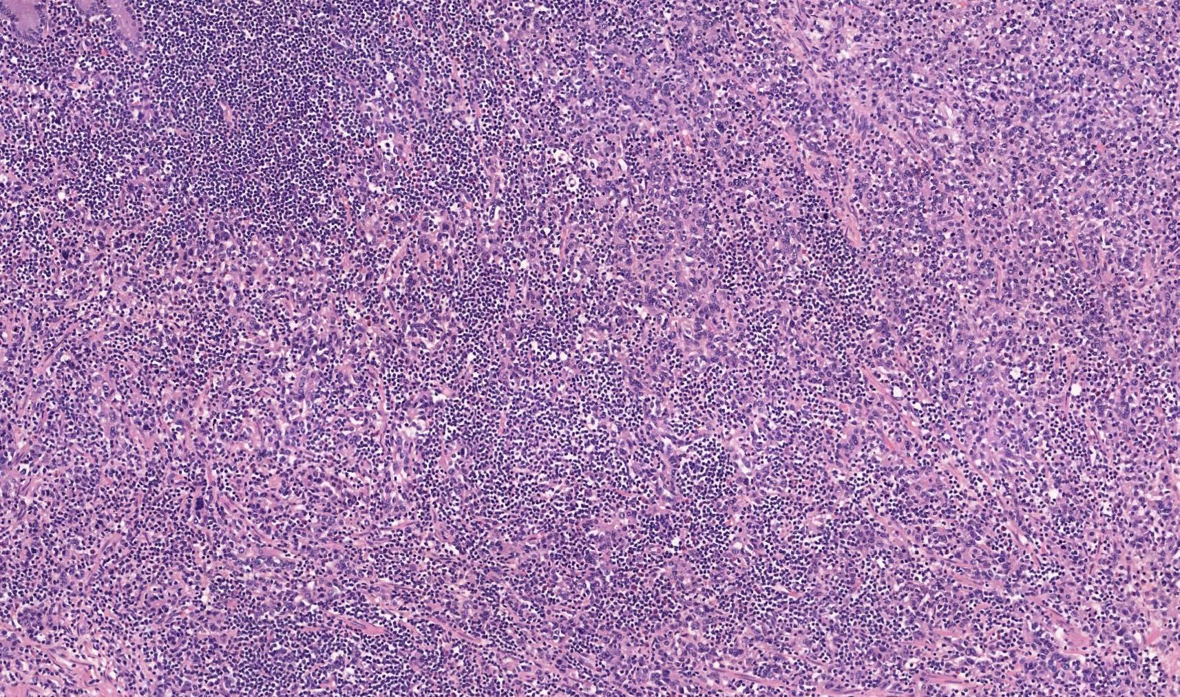
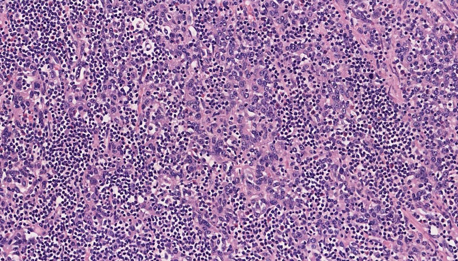
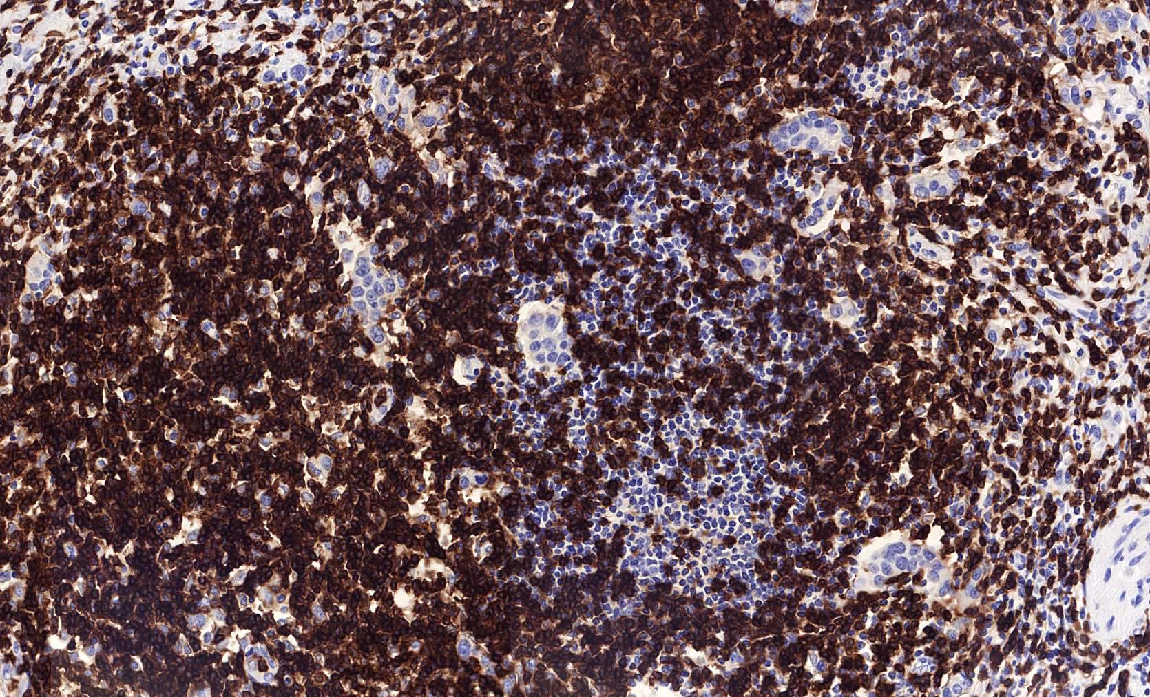
- Dense lymphoid infiltrate in a nondesmoplastic stroma reminiscent of lymphoid tissue (mean 500 tumor infiltrating lymphocytes per high power field) (Mod Pathol 2003;16:641)
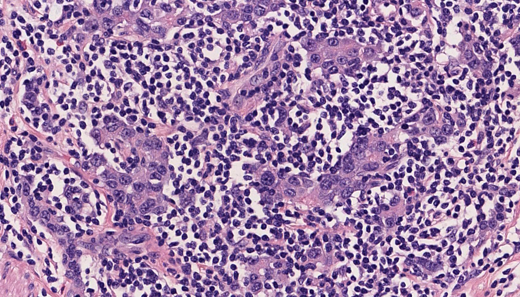
- Tumor cells are large and oval, contain vesicular to clear nuclei, have prominent nucleoli and abundant eosinophilic cytoplasm with poorly defined cell borders (Arch Pathol Lab Med 2008;132:706)
- Neoplastic cells are arranged primarily in microalveolar, thin trabecular and primitive tubular patterns uniformly distributed throughout the lymphoid stroma (Arch Pathol Lab Med 2008;132:706)
- Discrete areas of glandular differentiation may be seen (Am J Surg Pathol 2018;42:453)
- Small lymphocytes can also infiltrate into cancer cell nests (Arch Pathol Lab Med 2008;132:706)
- Epithelioid granulomas are sometimes observed within the lymphoid stroma (Arch Pathol Lab Med 2008;132:706)
- May rarely show osteoclast-like giant cells (Pathol Int 2010;60:551)
Microscopic (histologic) images
Positive stains
- CD3 and CD8: the lymphocytes infiltrating into tumor cell nests are predominantly cytotoxic (Cancer Lett 2003;200:33)
- Microsatellite instability can be evaluated immunohistochemically with the use of commercially available antibodies for the mismatch repair proteins (MSH2, MSH6, MLH1 and PMS2) (ESMO Open 2019;4:e000470)
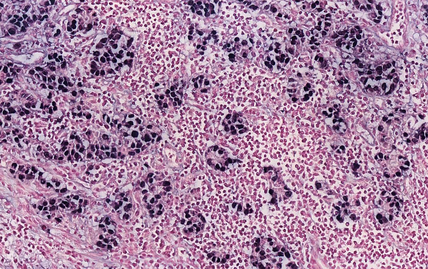
- EBV encoded RNA (EBER) in situ hybridization is the most sensitive method for detecting EBV in paraffin embedded tissues and stains all tumor cells (Arch Pathol Lab Med 2008;132:706, Cancer Lett 2003;200:33)
- PDL1 / L2 and their receptors (PD1 / 2) are frequently overexpressed in EBV gastric cancer (Oncotarget 2016;7:32925)
Negative stains
- HER2 is usually negative (J Surg Res 2017;210:159, Am J Surg Pathol 2018;42:453)
- LMP1 is negative in most cases (J Exp Clin Cancer Res 2009;28:14)
Molecular / cytogenetics description
- For microsatellite instability evaluation in gastric cancer, the immunohistochemical study of MMR proteins and PCR show an excellent concordance (ESMO Open 2019;4:e000470)
- EBV positive tumors show the higher prevalence of DNA hypermethylation among all gastric tumors (Nature 2014;513:202)
- The main molecular alterations in EBV associated cases include hypermethylation of the promoter region of the gene CDKN2A (p16INK4A) and mutation in phosphatidylinositol-3-kinase catalytic subunit alpha (PIK3CA) in about 5 - 10% (Oncotarget 2016;7:32925, Nature 2014;513:202, ESMO Open 2019;4:e000470)
- MLH1 hypermethylation is characteristic of microsatellite instability associated cases (Nature 2014;513:202)
- Microsatellite instability cases show high tumor mutation burden and KRAS alterations (56%) (Am J Surg Pathol 2018;42:453)
Sample pathology report
- Gastric mass, upper body, endoscopic biopsy:
- Gastric carcinoma with lymphoid stroma
- In situ hybridization study for EBER is diffusely positive in all tumor cells
Differential diagnosis
- Reactive lymphoid hyperplasia:
- No epithelial component (AE1 / AE3 immunostaining can confirm)
- Lymphoid infiltrate in gastric carcinoma with lymphoid stroma may obscure the neoplastic proliferation (Arch Pathol Lab Med 2008;132:706)
- Mucosal associated marginal zone lymphoma (MALT lymphoma):
- Intraepithelial lymphocytes may mimic the appearance of lymphoepithelial lesions, especially in superficial and small endoscopic biopsies
- Distinction is based on the recognition of atypia in epithelial cells (Arch Pathol Lab Med 2008;132:706)
- AE1 / AE3 is useful to evaluate the broad distribution of malignant epithelial cells
- Predominant CD3+ T cell lymphoid population helps to exclude a MALT lymphoma, which is a B cell proliferation
Board review style question #1
- The image shows a gastric endoscopic biopsy of a well circumscribed tumor located in the antrum. Epstein-Barr in situ hybridization study was completely negative. Which molecular alteration is probably present in this lesion?
- BRAF mutation
- HER2 overexpression
- Microsatellite instability
- MYC translocation
Board review style answer #1
C. Microsatellite instability
Reference: Gastric carcinoma with lymphoid stroma (lymphoepithelioma-like carcinoma, medullary carcinoma)
Comment Here
Reference: Gastric carcinoma with lymphoid stroma (lymphoepithelioma-like carcinoma, medullary carcinoma)
Comment Here
Board review style question #2
- Which of the following proteins is frequently overexpressed in gastric carcinoma with lymphoid stroma?
- HER2
- p16
- p53
- PDL1
Board review style answer #2


