Table of Contents
Definition / general | Essential features | Terminology | Clinical features | Interpretation | Uses by pathologists | Prognostic factors | Microscopic (histologic) images | Positive staining - normal | Positive staining - disease | Negative staining | Molecular / cytogenetics description | Sample pathology report | Board review style question #1 | Board review style answer #1 | Board review style question #2 | Board review style answer #2 | Board review style question #3 | Board review style answer #3 | Board review style question #4 | Board review style answer #4Cite this page: Batra H, Parwani A. Uroplakin II. PathologyOutlines.com website. https://www.pathologyoutlines.com/topic/stainsuroplakinII.html. Accessed April 1st, 2025.
Definition / general
- Uroplakin II, a 15kDa protein that is part of the uroplakin family (UP1a, UP1b, UPII and UPIII), which forms the urothelial plaque covering the apical surface of the urothelium
Essential features
- Uroplakins are terminal differentiation products that are exclusively expressed in urothelial cells (Kidney Int 2009;75:1153)
- 15 kDa transmembrane protein involved in terminal differentiation of urothelium
- Exists as a heterodimer along with UP1a (Kidney Int 2009;75:1153)
- UPII IHC has significantly higher sensitivity (57% versus 50%) than UPIII and the same specificity (~99%) for urothelial carcinomas (Histopathology 2014;65:132)
- Helpful in pinpointing the origin where secondary involvement by prostate adenocarcinoma or metastatic carcinoma may be positive for GATA3
Terminology
- UPII, UPK2, UROII
Clinical features
- Highly specific and moderately sensitive for urothelial carcinomas
- Helpful in pinpointing the primary origin as urothelium in cases where GATA3 is inconclusive
Interpretation
- Moderate to strong, predominantly membranous and cytoplasmic staining reaction in virtually all umbrella cells in the urethra (Appl Immunohistochem Mol Morphol 2022;30:326)
- At least weak to moderate cytoplasmic and membranous staining reaction of the majority of intermediate urothelial cells (Hum Pathol 2007;38:1703)
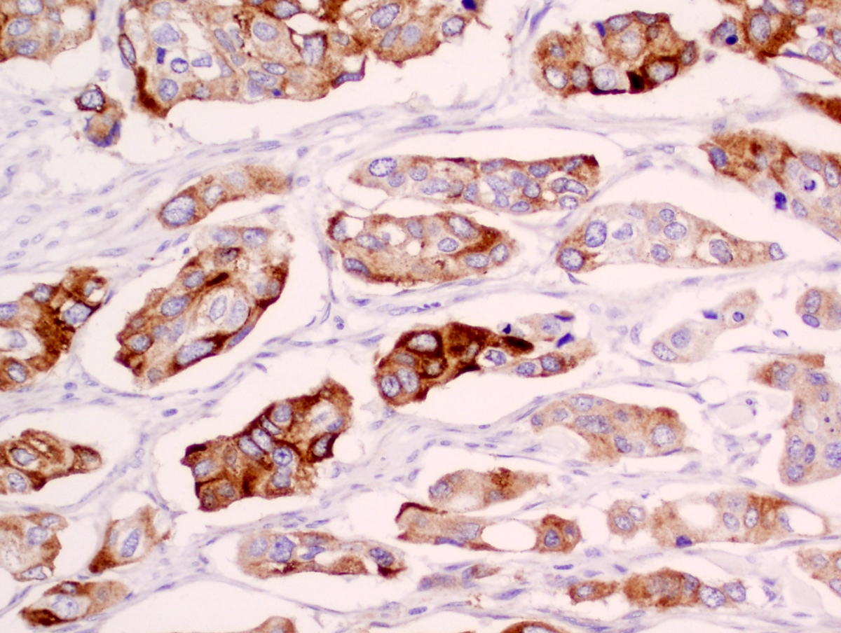
- Diffuse membranous staining in urothelial carcinoma cells (Appl Immunohistochem Mol Morphol 2022;30:326)
Uses by pathologists
- Highly specific and moderately sensitive for urothelial carcinomas
- Helpful in ascertaining the primary origin as urothelium, especially in cases where GATA3 is inconclusive
- Currently recommended by ISUP as a second line additional marker to confirm urothelial primary (Am J Surg Pathol 2014;38:e20)
Prognostic factors
- Loss of UPII seen more frequently in higher grade lesions (Arch Pathol Lab Med 2014;138:943)
Microscopic (histologic) images
Positive staining - normal
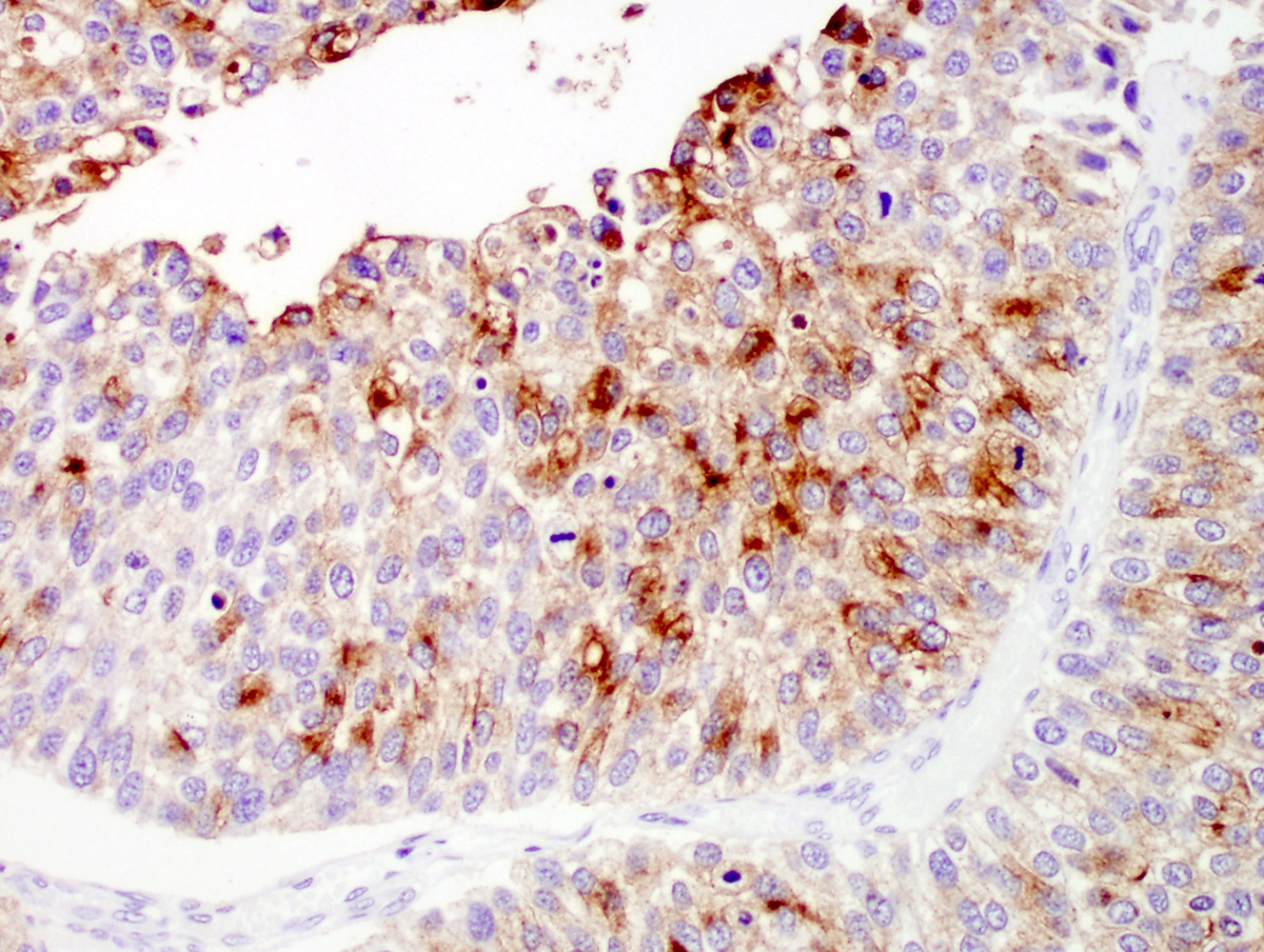
- Apical expression in normal bladder epithelium (umbrella cells)
- Intermediate cells in normal bladder epithelium
Positive staining - disease
- Urothelial carcinoma
- Ovarian Brenner tumor (Am J Surg Pathol 2003;27:1434)
- Pleomorphic invasive lobular carcinoma (single study) (Dis Markers 2016;2016:2940496)
- Apocrine metaplasia of breast (single study) (Dis Markers 2016;2016:2940496)
- Apocrine carcinoma of breast (single study) (Dis Markers 2016;2016:2940496)
- Adenocarcinoma of the colon (< 5% of cases) (Anticancer Res 2018;38:4759)
- Squamous cell carcinoma of the lung (< 5% of cases) (Anticancer Res 2018;38:4759)
- Endometrioid carcinoma of the uterine corpus (< 5% of cases) (Anticancer Res 2018;38:4759)
- Classical hepatocellular carcinoma of the liver (10%) (seen only in intracytoplasmic hyaline bodies) (Anticancer Res 2018;38:4759)
Negative staining
- Prostatic carcinoma (Anticancer Res 2018;38:4759)
- Squamous cell carcinoma of bladder (Hum Pathol 2013;44:164)
- Renal cell carcinoma (Histopathology 2014;65:132)
- Classic invasive lobular carcinoma (Dis Markers 2016;2016:2940496)
Molecular / cytogenetics description
- UPII mRNA expression in tissue and peripheral blood is associated with lymph node metastasis (J Pathol 2003;199:41)
Sample pathology report
- Lymph node, pelvic node dissection:
- Metastatic urothelial carcinoma (see comment)
- Comment: Histologic sections show a lymph node with deposits of tumor cells in diffuse sheets and few with a trabecular pattern. The cells are round to polygonal, demonstrating moderate pleomorphism, inconspicuous nucleoli and high N:C ratios. Immunohistochemistry is positive for CK20, p63, GATA3, S100, uroplakin III and uroplakin II and negative for PSA, PSAP, NKX3.1 and prostein, suggesting a urothelial origin. Please correlate clinicoradiologically. Follow up is advised.
Board review style question #1
Uroplakin II forms a heterodimer with which other uroplakin?
- UPIa
- UPIb
- UPIII
- UPV
Board review style answer #1
Board review style question #2
Uroplakin II is expressed normally in which of the following?
- Apical region (umbrella cells) of urothelium
- Muscularis propria of bladder
- Normal breast tissue
- Prostate
Board review style answer #2
Board review style question #3
Board review style answer #3
Board review style question #4
Which of the following is true regarding uroplakin II, as compared to uroplakin III?
- Equally sensitive IHC marker for urothelial carcinoma
- Less sensitive IHC marker for urothelial carcinoma
- Less specific IHC marker for urothelial carcinoma
- More sensitive IHC marker for urothelial carcinoma
Board review style answer #4


