Table of Contents
Definition / general | Essential features | Terminology | Clinical features | Interpretation | Uses by pathologists | Microscopic (histologic) images | Positive staining - normal | Positive staining - disease | Negative staining | Board review style question #1 | Board review style answer #1Cite this page: Lobo J, Henrique R. PLAP. PathologyOutlines.com website. https://www.pathologyoutlines.com/topic/stainsplap.html. Accessed April 1st, 2025.
Definition / general
- Placental-like alkaline phosphatase, a marker of germ cell tumors, especially seminoma
- Sensitive but not a specific marker
Essential features
- Placental-like alkaline phosphatase, part of a family of alkaline phosphatases with 4 members
- High levels in serum of seminoma patients (sensitive) but also detected in several normal and disease conditions (not specific)
- Sensitive immunohistochemical marker for germ cell neoplasms (testicular, ovarian and extra gonadal) and germ cell neoplasia in situ (GCNIS) but not specific
- Virtually always positive (strong and diffuse) in seminomas; useful for discriminating from spermatocytic tumor
Terminology
- Placental-like alkaline phosphatase (PLAP)
- Also known as Regan isoenzyme (Ann Clin Biochem 2014;51:611)
Clinical features
- High levels found in seminomas but not a reliable serum biomarker (sensitive but not specific):
- Elevated in physiological conditions (high levels in smokers)
- Elevated in several diseases, including various malignancies (Eur J Cancer 1990;26:1049, Clin Biochem 1987;20:387, Br J Urol 1996;77:138)
Interpretation
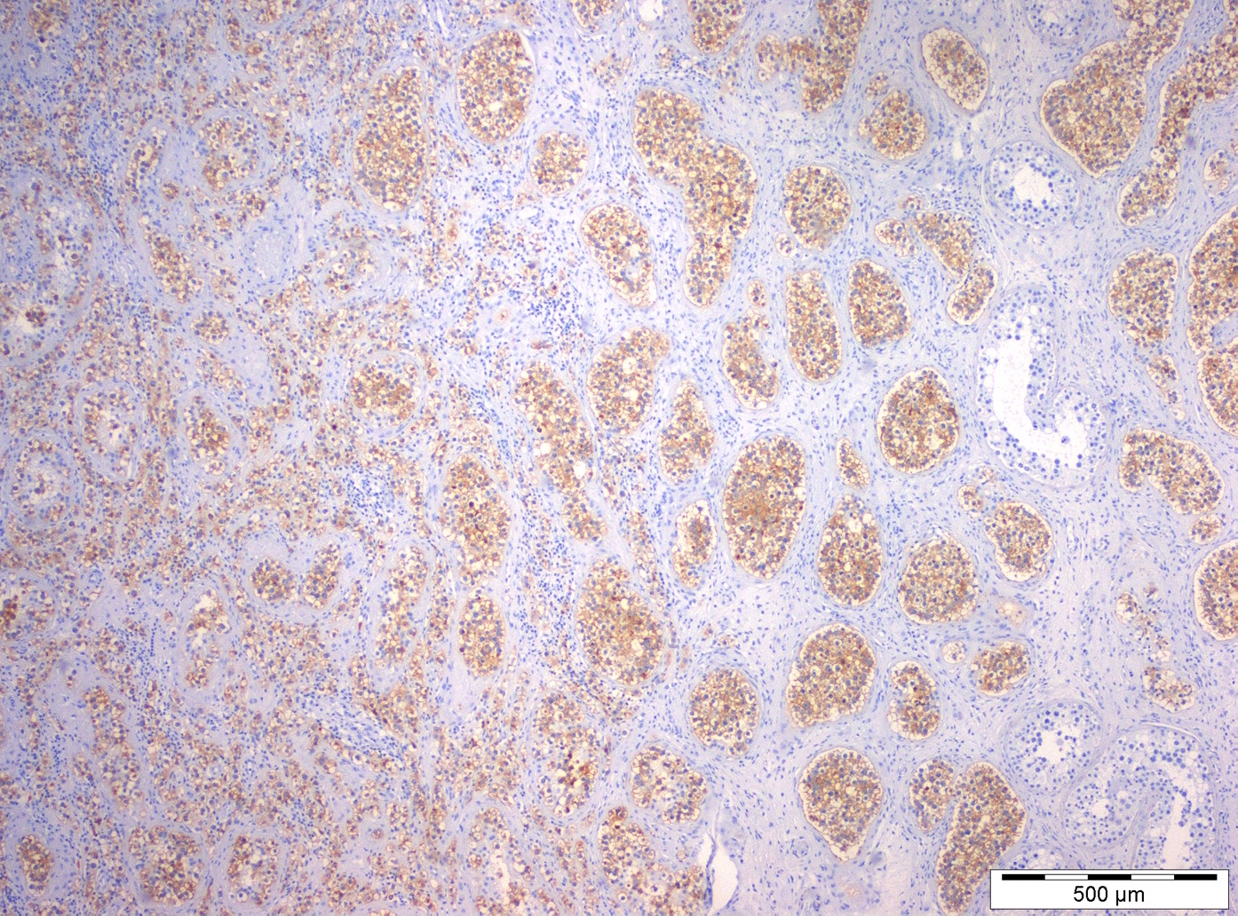
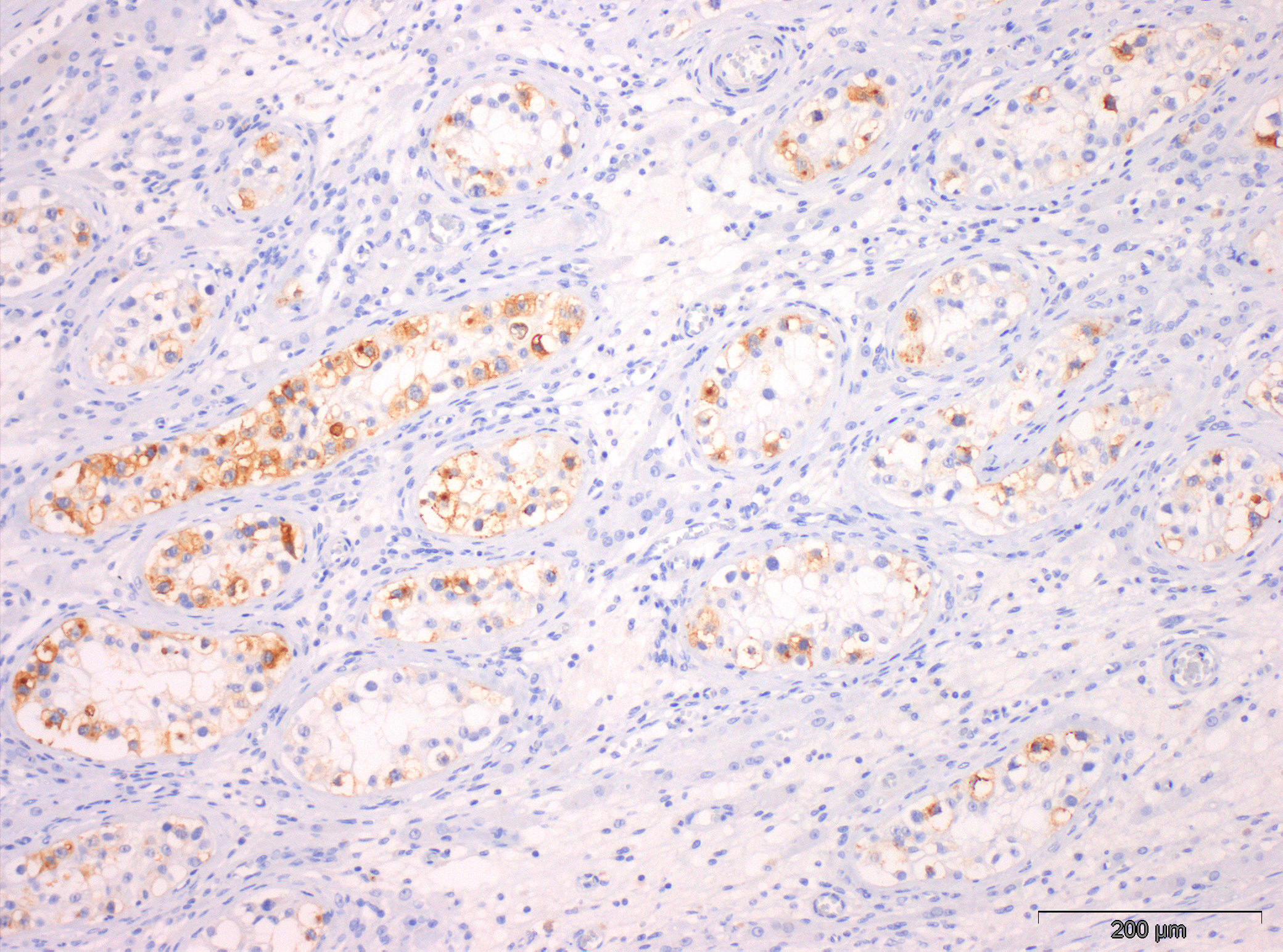
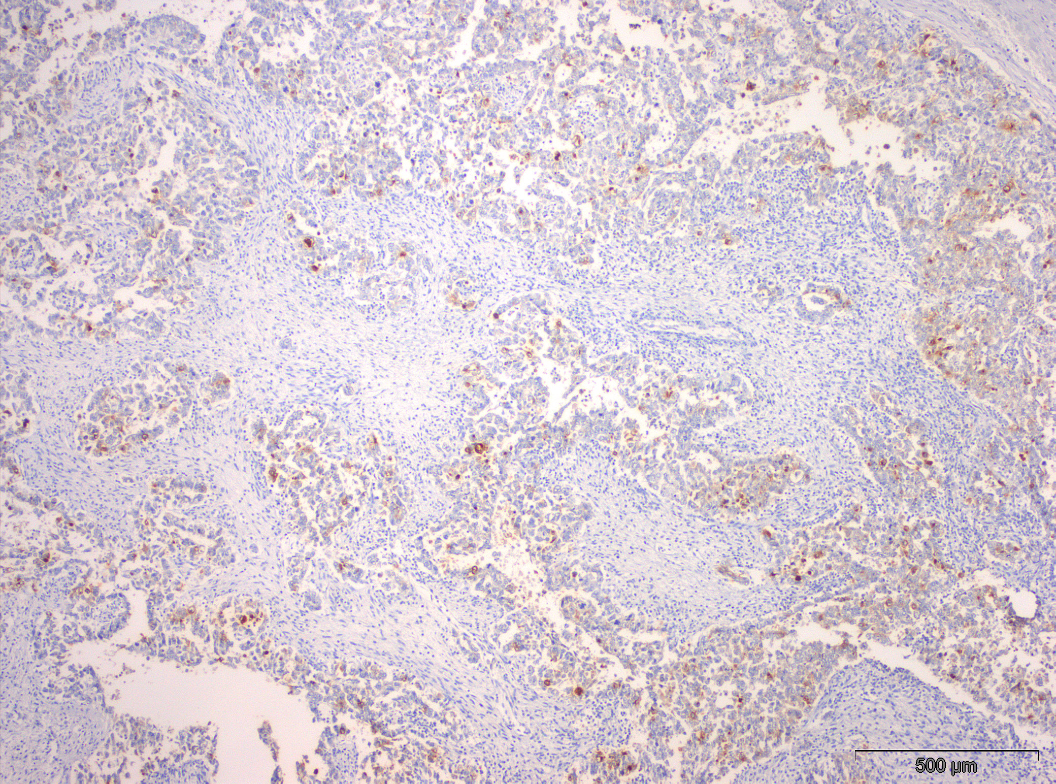
- Membranous and cytoplasmic staining are expected
Uses by pathologists
- Supporting diagnosis of germ cell neoplasms, including the precursor lesion germ cell neoplasia in situ and overt germ cell tumors (testicular, ovarian and extra gonadal)
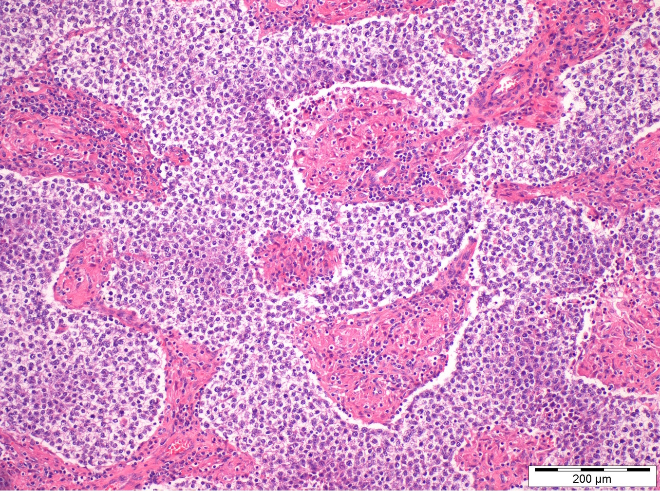
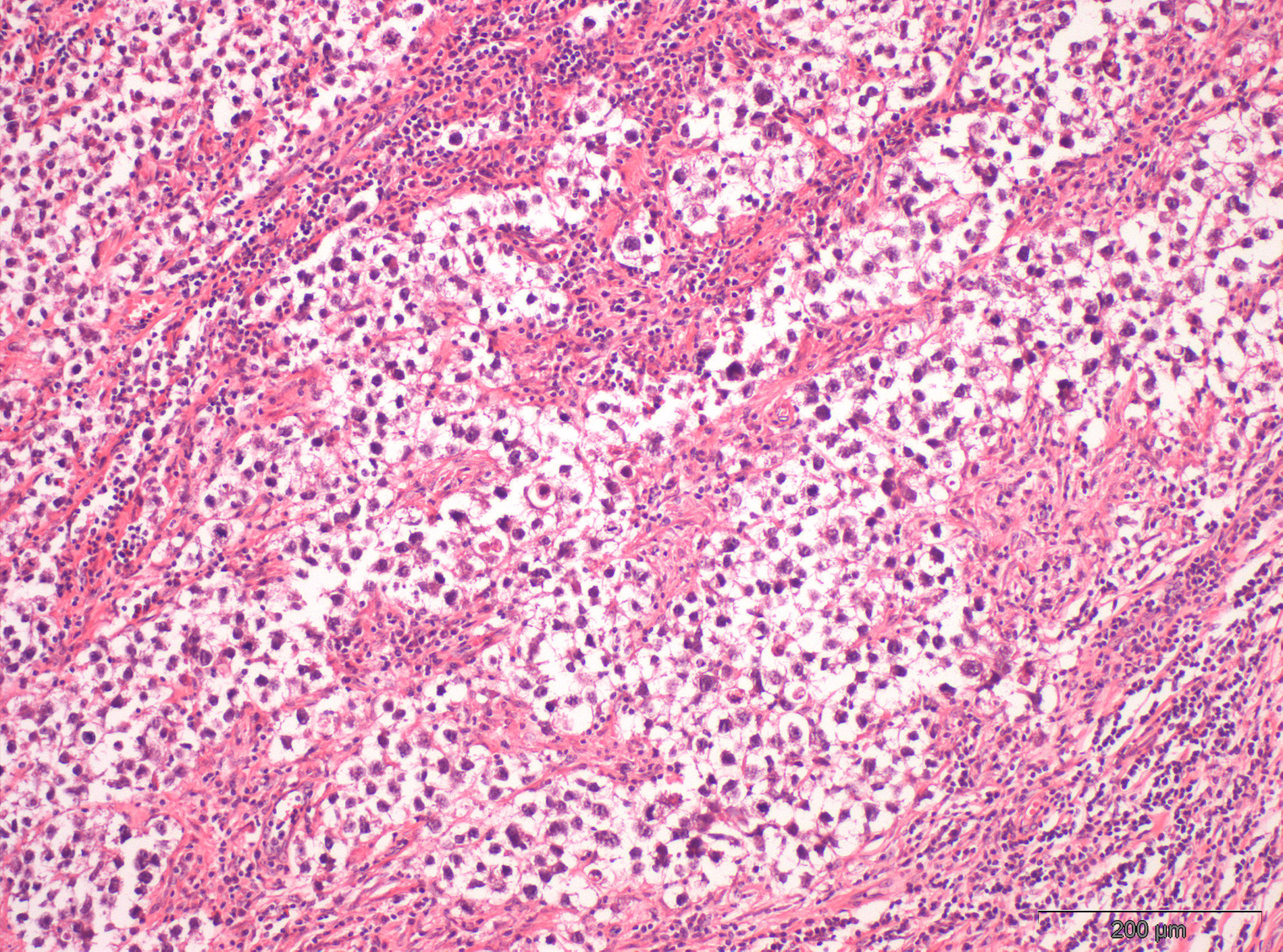
- Mainly a marker of seminoma (also dysgerminoma and germinoma of the brain) - positive in up to 100% of seminomas, strong diffuse positivity; useful for distinguishing seminoma from spermatocytic tumor
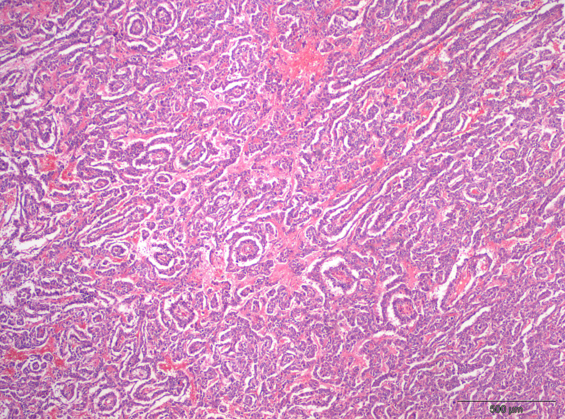
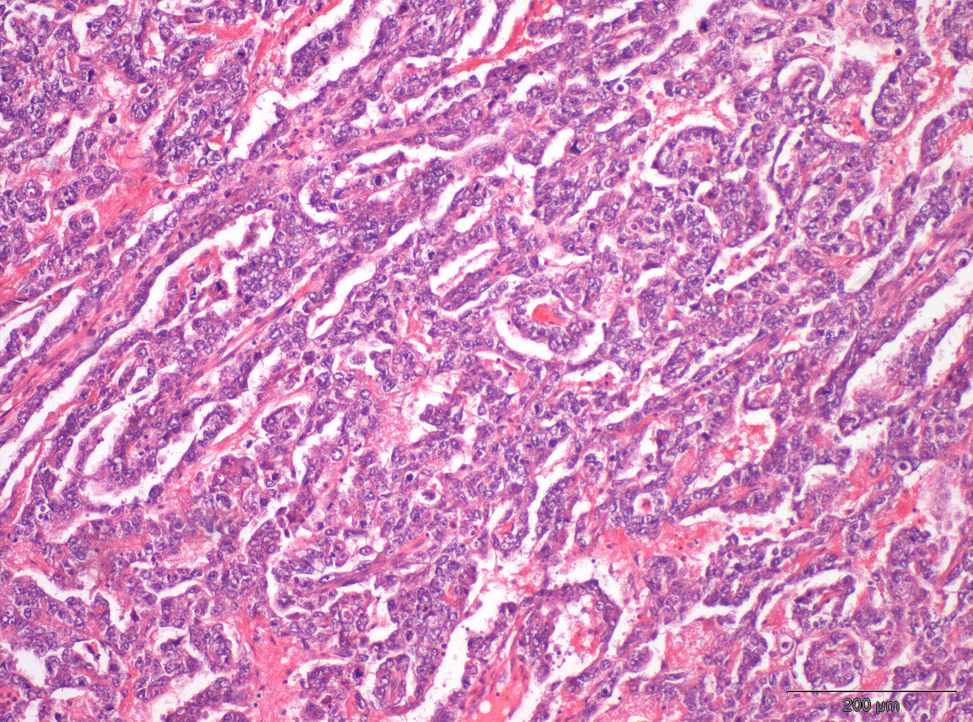
- Also frequent low intensity and focal expression in embryonal carcinoma and yolk sac tumor (in 85 - 97% of the cases)
- Focal positivity in cytotrophoblast cells of choriocarcinomas and in immature teratoma elements may be seen (Urol Res 1990;18:87, Am J Surg Pathol 1987;11:21, Eur J Obstet Gynecol Reprod Biol 1998;81:123, J Neurosurg 1985;63:733)
Microscopic (histologic) images
Positive staining - normal
- Term placenta is the best positive control (MAbs 2014;6:86)
- Cross reactivity with smooth or skeletal muscle fibers is acceptable (Am J Surg Pathol 2002;26:1627)
- Several normal tissues show expression (APMIS 1990;98:797, Hum Pathol 1987;18:946)
- Positivity in normal fetal testis is expected (Int J Androl 1987;10:29, APMIS 1990;98:797)
Positive staining - disease
- Germ cell tumors:
- Testicular:
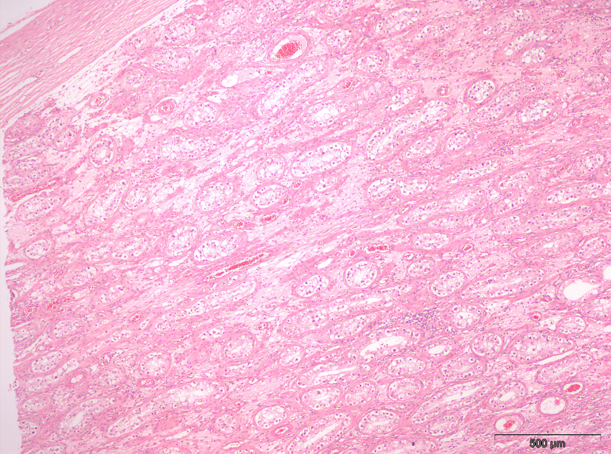
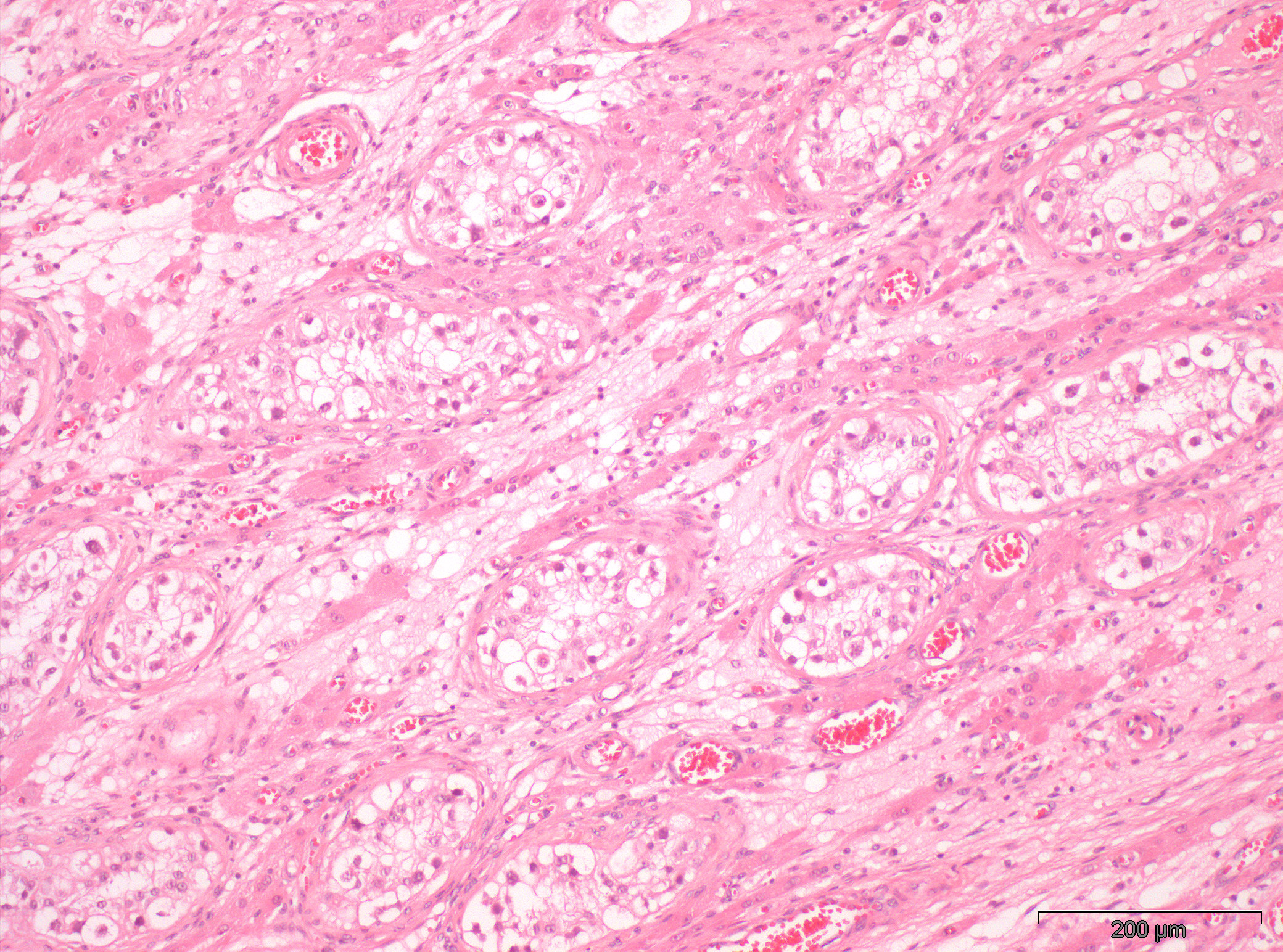
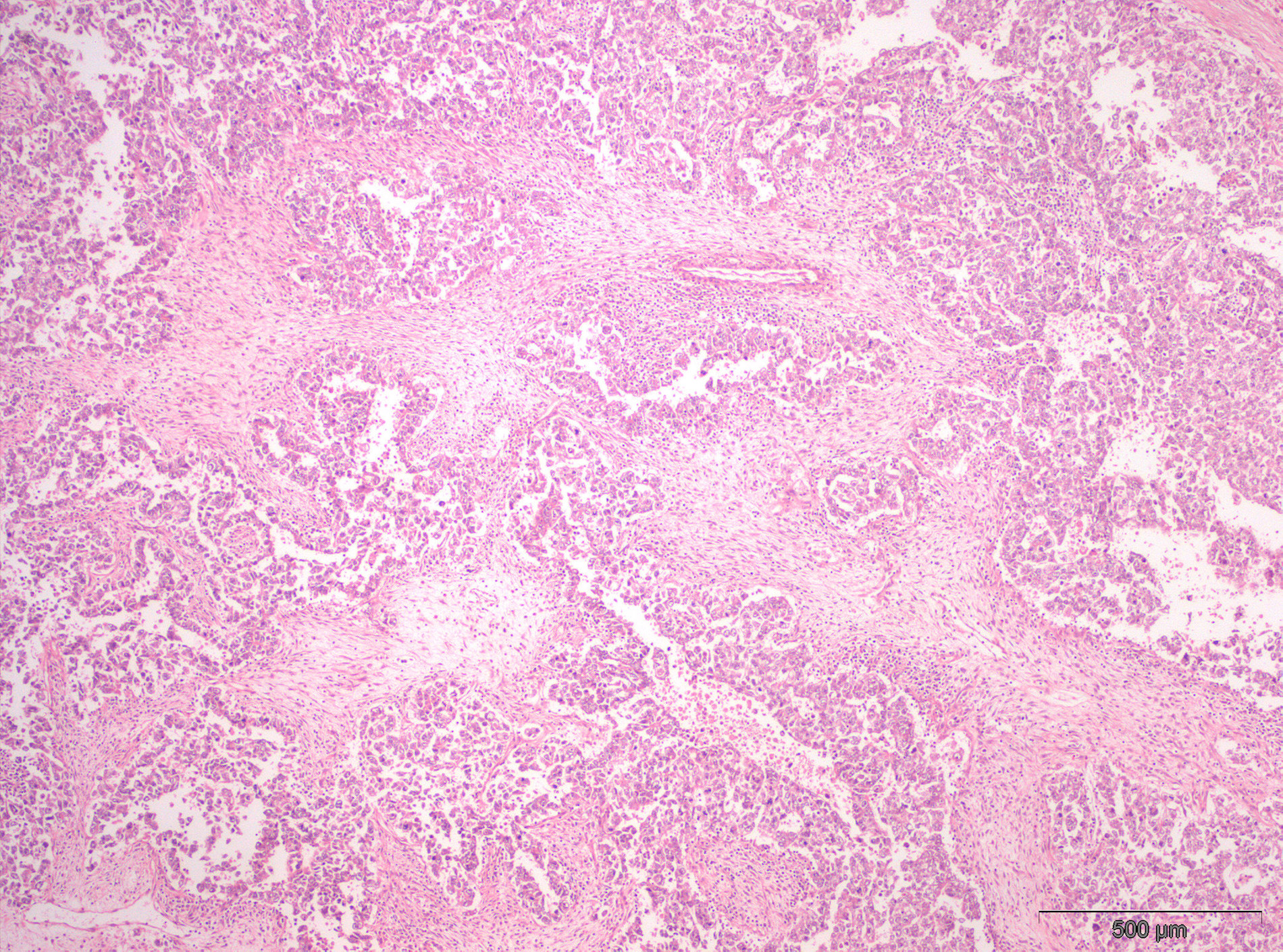
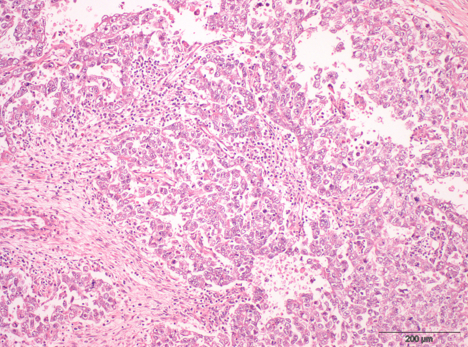
- Germ cell neoplasia in situ 100% (strong, diffuse)
- Seminoma (strong, diffuse)
- Embryonal carcinoma 96 - 97% (focal) (Am J Surg Pathol 1987;11:21, Hum Pathol 1988;19:663)
- Germ cell components of gonadoblastoma (Am J Surg Pathol 1987;11:21, Hum Pathol 1988;19:663)
- Ovarian:
- Dysgerminoma 100%
- Embryonal carcinoma
- Germ cell components of gonadoblastoma (Mol Cancer 2007;6:12)
- Extragonadal:
- Germinoma of brain 83% (Int J Clin Exp Pathol 2014;7:6965)
- Testicular:
- Placental site nodule 100% (Int J Gynecol Pathol 1994;13:191)
- Leiomyoma 100%, rhabdomyosarcoma 100%, desmoplastic small round cell tumor 79% (Am J Surg Pathol 2002;26:1627)
Negative staining
- Adult testis, epididymis and vas deferens and adult ovary, endometrium, gastrointestinal tract, kidney, lung, prostate, thyroid, adrenal gland, spleen, lymph node, brain, salivary glands, breast (APMIS 1990;98:797)
- Yolk sac tumor, testicle and ovary 25 - 85% (low intensity, focal) (Am J Surg Pathol 1987;11:21, Hum Pathol 1988;19:663)
- Choriocarcinoma 45% focal
- Teratoma, mature and immature 4 - 5% (Am J Surg Pathol 1987;11:21, Hum Pathol 1988;19:663)
- Spermatocytic tumor (APMIS 1990;98:797, Urol Res 1990;18:87)
- Epithelioid trophoblastic tumor focal (Am J Surg Pathol 1998;22:1393)
- Leiomyosarcoma 46%, myofibroblastic tumor 29%, gastrointestinal stromal tumor 13%, Wilms tumor 13%, synovial sarcoma 9% (Am J Surg Pathol 2002;26:1627)
Board review style question #1
- Which of the following sentences is true about the immunohistochemistry marker PLAP?
- It is specific for seminoma histology
- Staining is observed in germ cell tumors of the testis but not in ovarian ones
- Nuclear staining is expected
- Staining is evident across histologies, including in spermatocytic tumor
- It is useful for detecting germ cell neoplasia in situ
Board review style answer #1
E. PLAP is positive and useful for spotting germ cell neoplasia in situ. Despite being a sensitive marker, it is not specific for seminoma, being positive in several other tumors (both germ cell and nongerm cell malignancies) and normal tissues. Staining is membrane and cytoplasmic. It is useful for distinguishing seminoma from spermatocytic tumor, an important differential diagnosis, since spermatocytic tumor is negative.
Comment Here
Reference: PLAP
Comment Here
Reference: PLAP


