Table of Contents
Definition / general | Essential features | Terminology | Clinical features | Interpretation | Uses by pathologists | Microscopic (histologic) images | Positive staining - normal | Positive staining - disease | Negative staining | Sample pathology report | Board review style question #1 | Board review style answer #1Cite this page: Asirvatham JR. p40. PathologyOutlines.com website. https://www.pathologyoutlines.com/topic/stainsp40.html. Accessed March 29th, 2025.
Definition / general
- Truncated, nontransactivating p63 isoform (Mod Pathol 2012;25:405)
- May stimulate cell proliferation, block apoptosis and favor unrestrained tumor growth (Int J Surg Pathol 2013;21:229)
Essential features
- Nuclear marker with expression in squamous, urothelial, myoepithelial / basal cell differentiation
- More specific for squamous cell differentiation than p63 in lung carcinoma (Mod Pathol 2012;25:405)
- Recommended for subtyping non small cell carcinoma (J Thorac Oncol 2019;14:377)
Terminology
- Also called ΔNp63
- There are other molecules that are also called p40:
- Eukaryotic initiation factor 3 (eIF3-p40) (J Virol 2007;81:11569)
- Interleukin 12 / cytokine p40 (or its antibodies used to treat psoriasis) (J Biol Chem 2011;286:29806, Semin Cutan Med Surg 2010;29:48)
- Lysosomal membrane p40 (Biochem J 2008;414:431)
- Phagocyte NADPH oxidase subunit p40 phox (Blood 2009;114:3309)
- Klebsiella pneumoniae toll-like receptor2 ligand p40 (J Immunother 2009;32:875)
- Lactobacillus casei BL23 (J Mol Microbiol Biotechnol 2010;19:231)
- Mycoplasma p40 (Microbiology (Reading) 2011;157:473)
- Saccharomyces cerevisiae p40 / ARPC1 (J Biol Chem 2010;285:8481)
Clinical features
- Mutations cause ectrodactyly ectodermal dysplasia cleft syndrome, ankyloblepharon ectodermal dysplasia clefting syndrome, nonsyndromic split hand / split foot malformation (J Cell Sci 2011;124:2200)
Interpretation
- Nuclear stain
Uses by pathologists
- Usually positive in squamous, urothelial and myoepithelial / basal cell differentiation
- Part of recommended panel for subtyping non small cell lung carcinoma (J Thorac Oncol 2019;14:377)
Microscopic (histologic) images
Contributed by Jaya Ruth Asirvatham, M.B.B.S. and Andrey Bychkov, M.D., Ph.D.
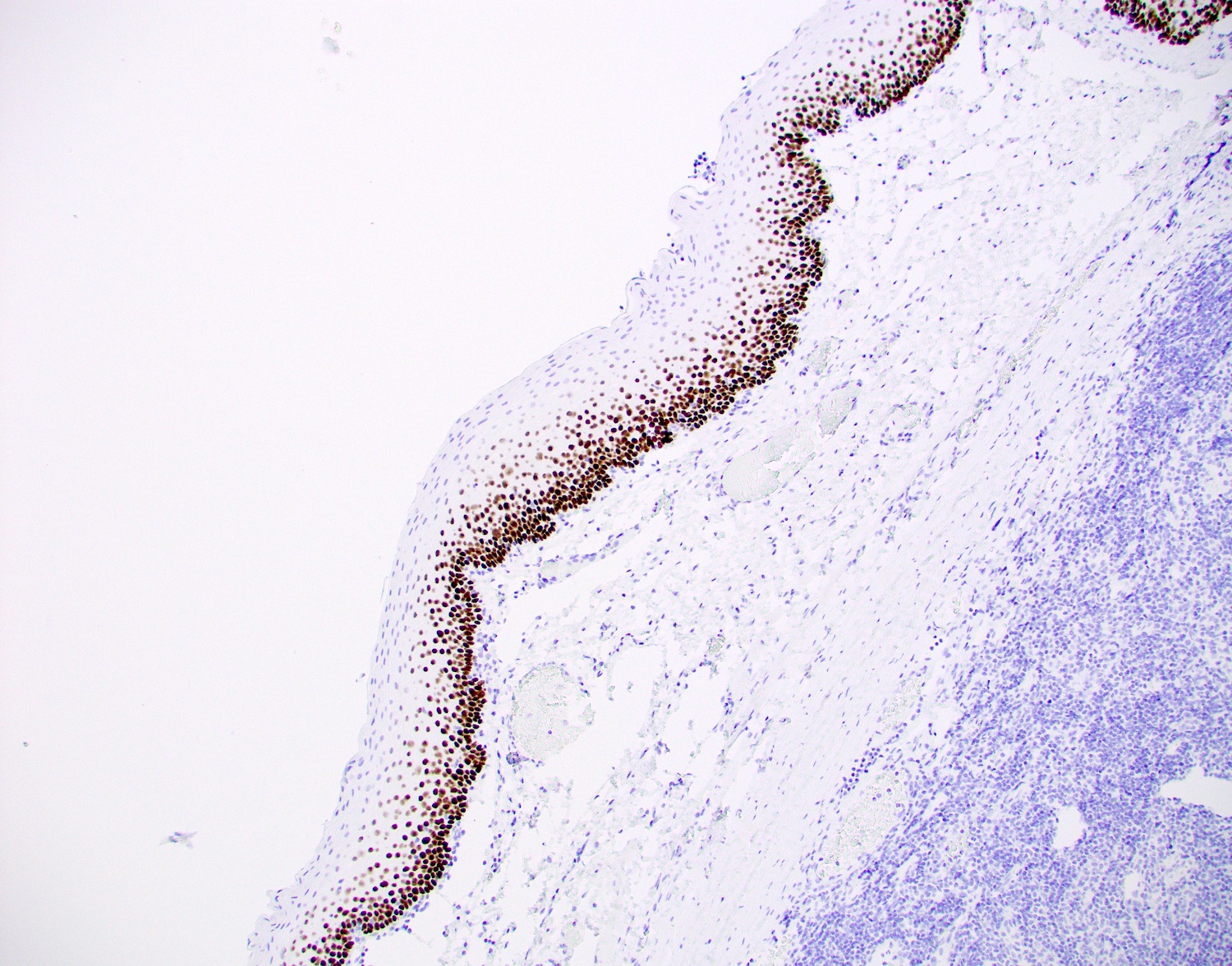
Squamous mucosa

Squamous cell carcinoma, tonsil
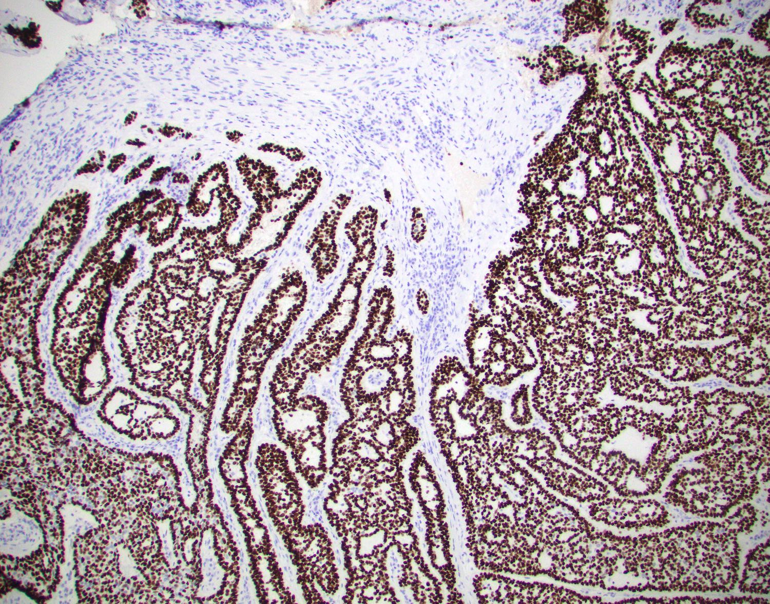
Basaloid neoplasm
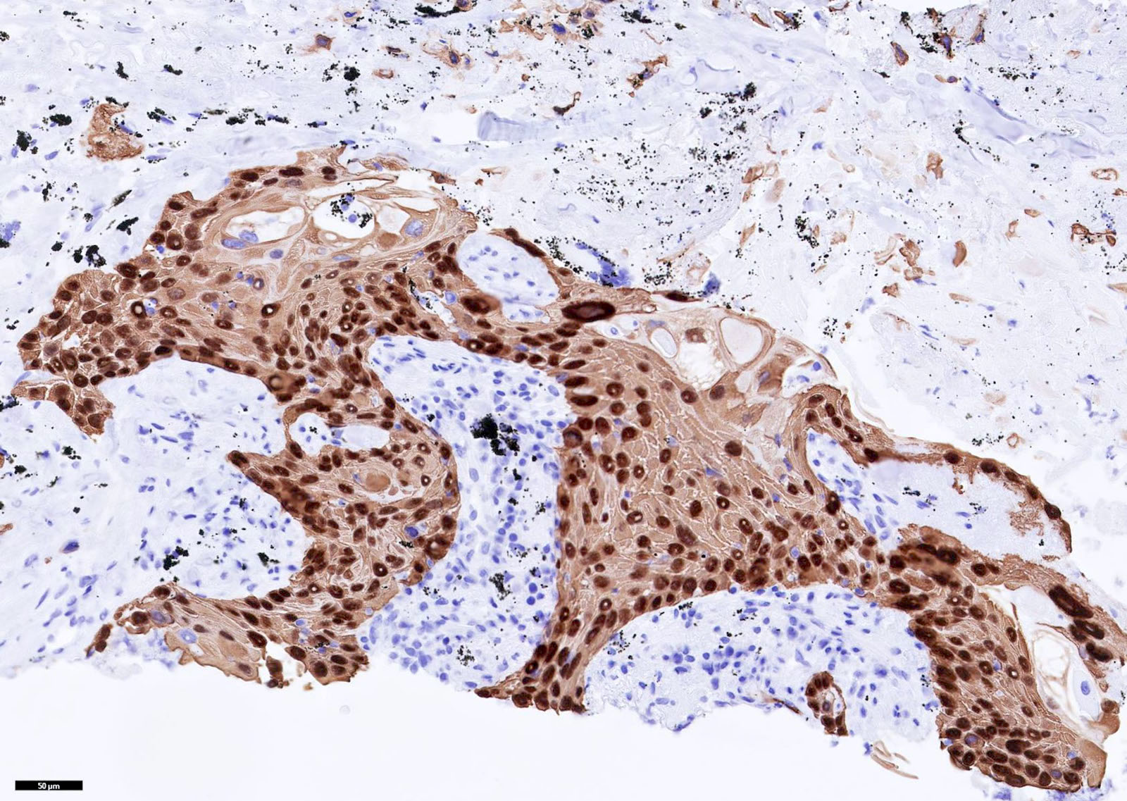
Lung squamous cell carcinoma
Positive staining - normal
- Squamous cells (Mod Pathol 2012;25:405)
- Myoepithelial / basal cells in breast, salivary gland and prostate (Am J Surg Pathol 2001;25:1054)
- Cytotrophoblasts (Am J Surg Pathol 2004;28:1177)
Positive staining - disease
- Generally positive in squamous, basal / myoepithelial and urothelial cell differentiation
- Squamous cell carcinoma of any site
- Bone / head and neck / soft tissue: adamantinoma-like Ewing family tumor (Am J Surg Pathol 2013;37:772, Am J Surg Pathol 2019;43:187, Head Neck Pathol 2022 Jan 13 [Epub ahead of print])
- Breast: myoepithelial carcinoma, metaplastic carcinoma
- Gynecological:
- Choriocarcinoma (Malays J Pathol 2017;39:175)
- Stratified mucin producing intraepithelial lesion (SMILE) (Cells 2021;10:2039)
- Lung:
- Squamous cell carcinoma (p40 [strong] and > 50% of cells, TTF1-)
- Adenosquamous (p40 strong and < 50%) (J Thorac Oncol 2012;7:281, Am J Surg Pathol 2012;36:895)
- Adenocarcinoma (5% are positive but in < 5% of cells) (Mod Pathol 2012;25:405)
- Metastatic urothelial or squamous cell carcinoma, undifferentiated thymic carcinomas and choriocarcinomas may also be positive for p40 (Malays J Pathol 2017;39:175)
- Bronchiolar adenoma (Am J Surg Pathol 2018;42:1010)
- Head and neck: squamous cell / sarcomatoid carcinomas (Pathol Res Pract 2022;229:153733)
- Nasal cavity, paranasal sinuses, nasopharynx (Head Neck Pathol 2014;8:141):
- Ewing / PNET (25%, 1/4 cases) (Head Neck Pathol 2014;8:141)
- HPV related multiphenotypic sinonasal carcinoma (Am J Surg Pathol 2017;41:1690)
- Nasopharyngeal carcinoma (close to 100%) (Head Neck Pathol 2014;8:141)
- NUT carcinoma (75%) (Head Neck Pathol 2014;8:141)
- Olfactory neuroblastoma (28%, 13/46 cases; if positive, often focal) (Head Neck Pathol 2014;8:141)
- Sinonasal undifferentiated carcinoma (55%) (Head Neck Pathol 2014;8:141)
- Small cell carcinoma (50%, 1/2 cases) (Head Neck Pathol 2014;8:141)
- SMARCB1 deficient sinonasal carcinoma (44%) (Am J Surg Pathol 2014;38:1282)
- Salivary gland (Oral Surg Oral Med Oral Pathol Oral Radiol 2022;133:189):
- Adenoid cystic carcinoma (83%)
- Myoepithelial carcinoma (~100%)
- Basal cell adenoma and basal cell carcinoma (100%)
- Oncocytoma (100%, 7/7 cases)
- Myoepithelioma (100%)
- Epithelial myoepithelial carcinoma (~100%)
- Oncocytoma (100%)
- Pleomorphic adenoma
- Mucoepidermoid adenoma
- Nasal cavity, paranasal sinuses, nasopharynx (Head Neck Pathol 2014;8:141):
- Thymic neoplasms (Ann Diagn Pathol 2015;19:216):
- Thymoma (97%)
- Undifferentiated thymic carcinomas that lack squamous features (50%)
- Skin:
- Primary cutaneous mucinous carcinoma (25%) (Am J Dermatopathol 2021;43:e175)
- Basal cell carcinoma
- Skin adnexal tumors such as hidradenoma (Am J Surg Pathol 2020;44:711)
- Urothelial carcinoma (up to 87%, with or without squamous differentiation) (Pathology 2016;48:543, Adv Anat Pathol 2020;27:114)
Negative staining
- Lung: adenocarcinoma (5% are positive but in < 5% of cells) (Mod Pathol 2012;25:405)
- Sarcomatoid mesothelioma (6% are positive) (Int J Surg Pathol 2021;29:820)
Sample pathology report
- Cervix, biopsy:
- Invasive squamous cell carcinoma, poorly differentiated
- Ancillary studies: The malignant cells are diffusely positive for cytokeratin 5/6, p40 and p16 and are negative for cytokeratin 7, supporting the diagnosis.
Board review style question #1
A 55 year old male construction worker with a 20 pack year smoking history and no prior diagnosis of malignancy, presents with a lung mass, which is biopsied. The biopsy demonstrates a poorly differentiated malignant neoplasm. An immunohistochemical panel comprising CK7, CK20, CK5/6, TTF1, p40, napsin, calrentin and BerEP4 is performed.
Which profile supports the diagnosis of squamous cell carcinoma?
- CK7+, TTF1+, napsin+, other stains negative
- CK5/6+, p40+ in more than 50% of cells, strong, other stains negative
- CK7+, CK5/6+, TTF1+, p40+ in less than 50% of cells strong, other stains negative
- CK5/6+, calretinin+, others negative
- CK20+, others negative
Board review style answer #1
B. CK5/6+, p40+ in more than 50% of cells, strong, other stains negative. Strong p40 staining in greater than 50% of cells is expected in squamous cell carcinoma. Profile A is seen in lung adenocarcinoma. Profile C is supportive of lung adenosquamous carcinoma. Profile D is consistent with mesothelioma. Metastatic colorectal carcinoma is a possibility with profile E. A diagnosis of primary squamous cell carcinoma of the lung needs to be made in the appropriate clinical context, as metastatic squamous cell carcinoma or direct extension from a thymic carcinoma may have the same profile. PAX8 and CD117 are often positive in thymic carcinomas and will be negative in primary lung squamous cell carcinoma.
Comment Here
Reference: p40
Comment Here
Reference: p40
