Table of Contents
Definition / general | Uses by pathologists | Positive staining - normal | Positive staining - disease | Negative stains | Microscopic (histologic) imagesCite this page: Pernick N. Cytokeratin MNF 116. PathologyOutlines.com website. https://www.pathologyoutlines.com/topic/stainsmnf116.html. Accessed March 30th, 2025.
Definition / general
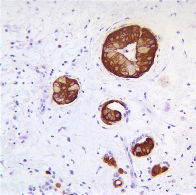
- Broad spectrum cytokeratin marker which stains high and low molecular weight cytokeratins (CK 5, 6, 8, 17 and probably 19)
Uses by pathologists
- Detect micrometastases in lymph nodes (BJU Int 2006;98:70)
- Detect positive margins in Mohs’ surgery (Dermatol Surg. 2003;29:375)
- Double immunostaining with laminin or collagen type IV is useful to detect microinvasion in VIN or CIN (Arch Pathol Lab Med 2005;129:747)
Positive staining - normal
- Most epithelial cells, including lung type II epithelial cells (Am J Respir Cell Mol Biol 1998;18:786)
- Trophoblast (Acta Obstet Gynecol Scand 2003;82:722)
- Uterine smooth muscle (Histopathology 1995;27:407)
Positive staining - disease
- Most carcinomas (J Histochem Cytochem 2001;49:1369)
- Mesothelioma (Am J Dermatopathol 1997;19:261)
- Pituitary adenoma (Eur J Endocrinol 2003;148:357)
Negative stains
- Myofibroblastic tumors (or weak, J Cutan Pathol 2003;30:393)


