Table of Contents
Definition / general | Interpretation | Uses by pathologists | Microscopic (histologic) images | Positive staining - normal | Positive staining - disease | Negative stainingCite this page: Pernick N. HepPar1. PathologyOutlines.com website. https://www.pathologyoutlines.com/topic/stainsheppar1.html. Accessed March 30th, 2025.
Definition / general
- Hepatocyte Paraffin 1
- Also called Hep Par1, Hep
- Recognizes mitochondrial antigen of hepatocytes
- Highly sensitive (92%); negative in higher nuclear grade tumors (Am J Surg Pathol 2002;26:978)
- Moderately specific; false positive cases were CK7+ or CK20+ (adenocarcinoma), chromogranin+ or synaptophysin+ (neuroendocrine)
- May be biomarker for early Barrett esophagus (Am J Clin Pathol 2012;137:111)
Interpretation
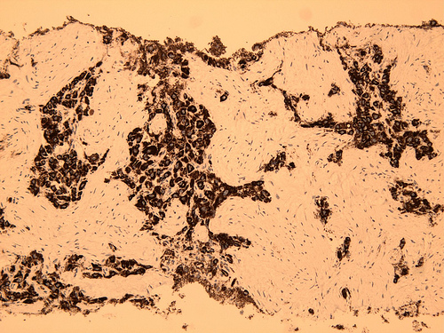
- Granular cytoplasmic staining due to mitochondrial binding (Am J Clin Pathol 2006;125:722)
Uses by pathologists
- Determine hepatocellular origin, particularly in panel with alpha-fetoprotein and CEA or CD10 (canalicular pattern, more specific than Hep Par1)
- Differentiate hepatocellular carcinoma from cholangiocarcinoma or metastases to liver, as part of panel (Malays J Pathol 2006;28:87)
Microscopic (histologic) images
Positive staining - normal
- Hepatocytes, small intestinal epithelium
Positive staining - disease
- Most hepatocellular carcinomas and its metastases (Int J Surg Pathol 2010;18:433), some colon adenocarcinomas and some nonhepatocellular carcinomas metastatic to liver
Negative staining
- Bile duct adenoma, hepatoid adenocarcinoma (often)


