Table of Contents
Definition / general | Essential features | Pathophysiology | Clinical features | Interpretation | Uses by pathologists | Microscopic (histologic) images | Virtual slides | Positive staining - not malignant | Positive staining - disease | Negative staining | Sample pathology report | Board review style question #1 | Board review style answer #1Cite this page: Baniak N. Glypican 3. PathologyOutlines.com website. https://www.pathologyoutlines.com/topic/stainsglypican3.html. Accessed March 30th, 2025.
Definition / general
- Heparan sulfate proteoglycan on the cell surface; highly expressed in embryonic tissues but minimally expressed in normal adult tissues (Int J Mol Sci 2021;22:5780, Arch Pathol Lab Med 2015;139:537)
Essential features
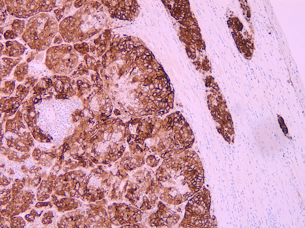
- Cytoplasmic or membranous staining
- Used as part of a panel in diagnosing differentiating hepatocellular carcinoma from benign liver lesions and metastatic neoplasms
- Used to identify a yolk sac tumor component in primary or metastatic germ cell tumors
Pathophysiology
- Oncofetal protein encoded by GPC3 gene; plays a role in the regulation of cell division and apoptosis (Diagn Pathol 2015;10:34)
Clinical features
- Mutations of the GPC3 gene are responsible for Simpson-Golabi-Behmel syndrome (Virchows Arch 2015;466:67)
Interpretation
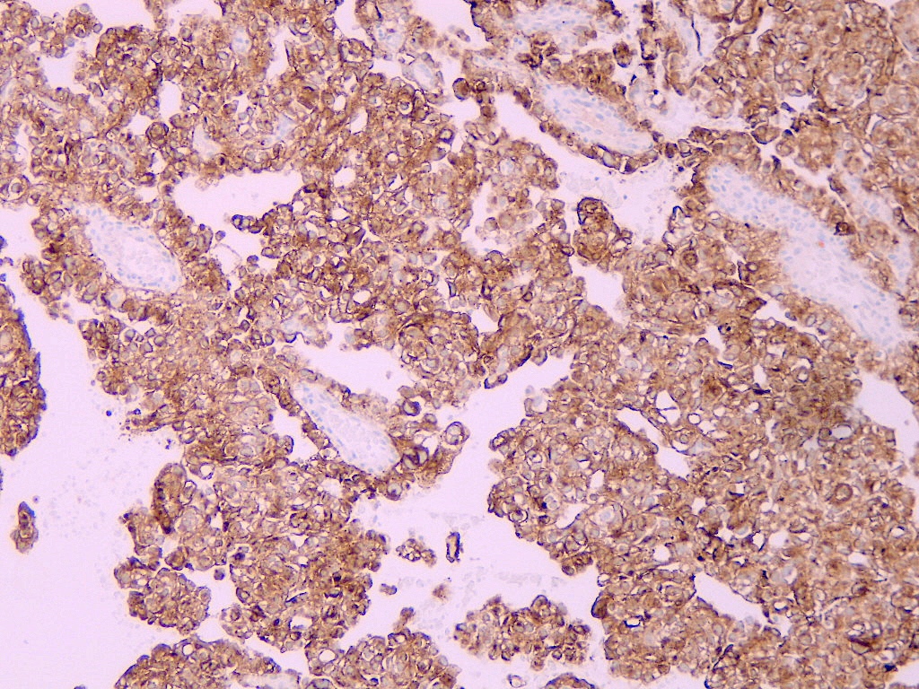
- Cytoplasmic or membranous staining (Mod Pathol 2020;33:2354, Int J Mol Sci 2021;22:5780)
Uses by pathologists
- Distinguish hepatocellular carcinoma from nonmalignant liver lesions (adenoma, focal nodular hyperplasia) as part of a panel (Am J Clin Pathol 2008;130:219, Am J Surg Pathol 2008;32:433, Arch Pathol Lab Med 2015;139:537)
- In 1 study, glypican 3 negativity alone was 100% sensitive and 58% specific; the combination of intact reticulin with either glypican 3 or glutamine synthetase negativity was 92% sensitive and 95% specific in the distinction of hepatic adenoma from hepatocellular carcinoma (Arch Pathol Lab Med 2015;139:537)
- Diagnosis of hepatoid carcinoma of various sites, such as stomach and lungs (Cancer Sci 2009;100:626, Respir Med 2016;119:175)
- Distinguish hepatocellular carcinoma from metastatic carcinoma (Mod Pathol 2008;21:626, J Pak Med Assoc 2018;68:1029)
- In 1 study, sensitivity was 82%, specificity was 94.3%, positive predictive value was 94.8% and negative predictive value was 80.6%; diagnostic accuracy was 87.5%
- Does not distinguish metastatic hepatoid carcinoma of various sites from hepatocellular carcinoma
- SALL4 has been touted as a potential marker to distinguish hepatoid adenocarcinoma from hepatocellular carcinoma (Am J Surg Pathol 2010;34:533)
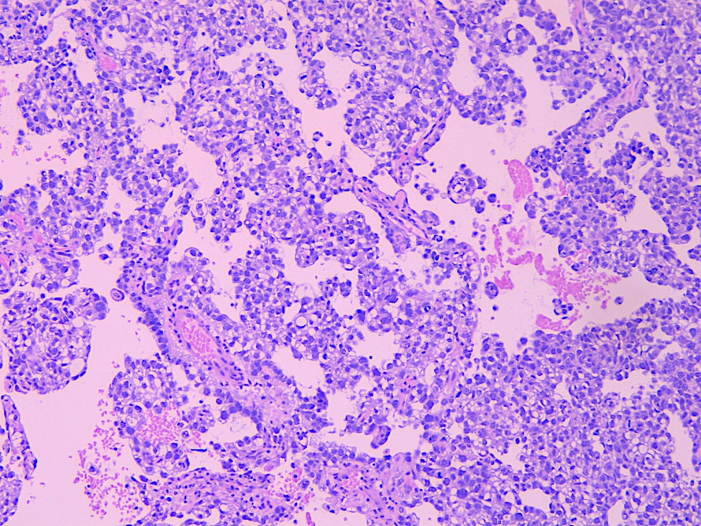
- Subtyping testicular and ovarian germ cell tumors as part of a panel: yolk sac tumor (positive) from embryonal carcinoma, seminoma, teratoma, which are usually negative (Int J Surg Pathol 2013;21:342, Hum Pathol 2010;41:716)
- Differentiating glandular yolk sac tumor (86%) from adenocarcinoma, NOS (7%); as part of a panel along with AFP, EMA, CK7 (Am J Surg Pathol 2014;38:1396)
- Typically in the postchemotherapy setting, distinguishing sarcomatoid yolk sac tumor from sarcoma arising from germ cell tumors (postchemotherapy sarcomatous tumors) (Am J Surg Pathol 2015;39:251)
Microscopic (histologic) images
Positive staining - not malignant
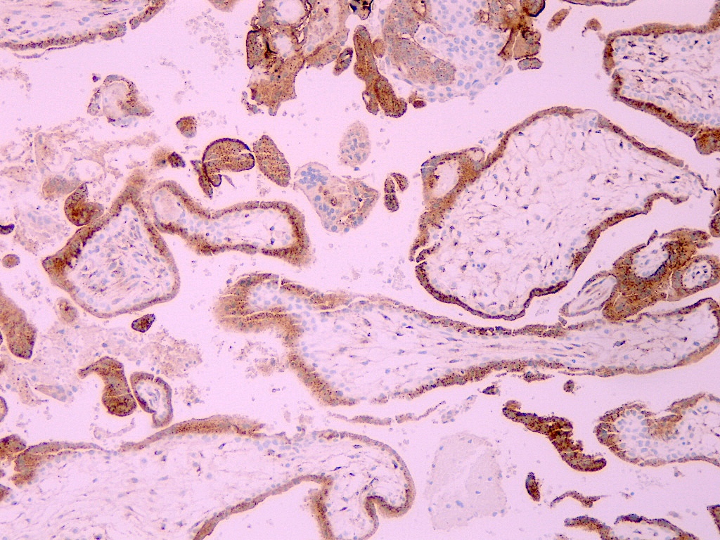
- Placenta; syncytiotrophoblast and cytotrophoblast positive
- Exception: Simpson-Golabi-Behmel syndrome type 1 (Placenta 2022;126:119)
- Nonneoplastic liver with high grade hepatitis C activity (> 80%) (Hum Pathol 2008;39:209)
Positive staining - disease
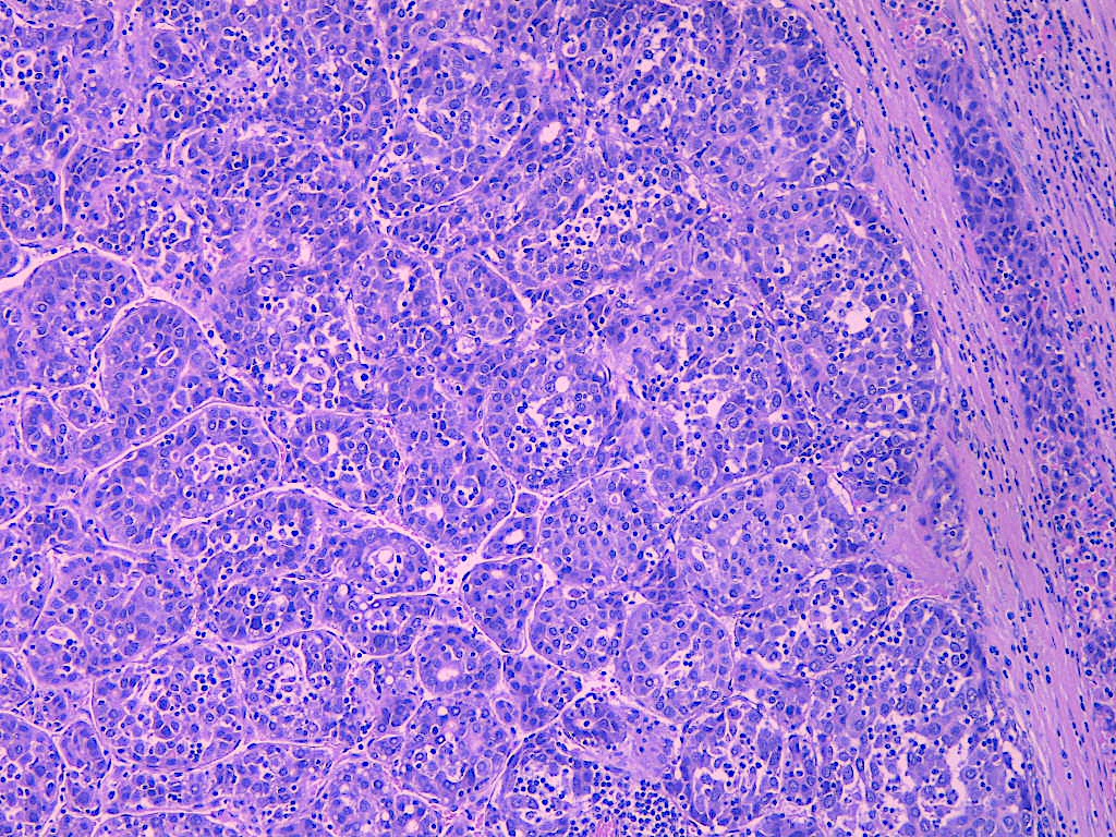
- Hepatocellular carcinoma (HCC): 64 - 97% (Hum Pathol 2013;44:542, Am J Clin Pathol 2008;129:899, Pathol Oncol Res 2015;21:379, Malays J Pathol 2016;38:229)
- Varies with the degree of differentiation; in general, poorly differentiated HCC is more commonly positive than well differentiated HCC (Arch Pathol Lab Med 2015;139:537, Int J Mol Sci 2021;22:5780)
- Sensitivity ranges from 73 - 97% and specificity from 93 - 100% (Pathol Oncol Res 2015;21:379, Malays J Pathol 2016;38:229)
- Yolk sac tumor: ovarian (> 90%), testicular (83 - 100%), uterine (100%), other extragonadal sites (> 90%) (Am J Clin Pathol 2008;129:899, Int J Surg Pathol 2013;21:342, Mod Pathol 2020;33:2354, Mod Pathol 2019;32:1847)
- Choriocarcinoma: 75 - 80% (Mod Pathol 2020;33:2354, Am J Surg Pathol 2014;38:e50)
- Placental site trophoblastic tumor: 80%, cytoplasmic staining in larger multi and mononucleated cells (Diagn Pathol 2010;5:64)
- Placental site nodule (Diagn Pathol 2010;5:64)
- Pancreatic acinar cell carcinoma: 58% (Hum Pathol 2013;44:542)
- Lung squamous cell carcinoma: 54% (Am J Clin Pathol 2008;129:899)
- Liposarcoma: 52% (Am J Clin Pathol 2008;129:899)
- Adrenal cortical adenoma: 27 - 60% (4/15 and 6/10 cases) (Am J Clin Pathol 2008;129:899, Ann Diagn Pathol 2018;35:92)
- Pediatric tumors:
- Atypical teratoid rhabdoid tumors: 100% (Pediatr Dev Pathol 2013;16:272)
- Hepatoblastoma: 100% (Pediatr Dev Pathol 2013;16:272)
- Malignant rhabdoid tumor: 67% (Pediatr Dev Pathol 2013;16:272)
- Wilms tumor (primary): 38 - 77% (Pediatr Dev Pathol 2013;16:272, Virchows Arch 2015;466:67)
- Wilms tumor (metastatic): 93% (Virchows Arch 2015;466:67)
- Metanephric adenoma: 50% (Virchows Arch 2015;466:67)
- Hepatoid adenocarcinoma:
- Gastric (Cancer Sci 2009;100:626)
- Lung (Respir Med 2016;119:175)
- Pancreas (Int J Surg Pathol 2019;27:28)
Negative staining
- Head and neck:
- Laryngeal squamous cell carcinoma: 16% (Am J Clin Pathol 2008;129:899)
- Salivary gland adenoid cystic carcinoma: 2% (Am J Clin Pathol 2008;129:899)
- Thoracic:
- Lung adenocarcinoma: 10% (Am J Clin Pathol 2008;129:899)
- Pleural mesothelioma: 7% (Am J Clin Pathol 2008;129:899)
- Thymoma: 4% (Am J Clin Pathol 2008;129:899)
- Breast:
- Breast carcinoma (lobular, medullary, no special type): 8 - 20% (Am J Clin Pathol 2008;129:899)
- Breast phyllodes tumor: 0% (Am J Clin Pathol 2008;129:899)
- Gastrointestinal:
- Esophageal squamous cell carcinoma: 8% (Am J Clin Pathol 2008;129:899)
- Esophageal adenocarcinoma: 0% (Am J Clin Pathol 2008;129:899)
- Gastric adenocarcinoma, intestinal type: 20% (Am J Clin Pathol 2008;129:899)
- Gastric adenocarcinoma, diffuse type: 4% (Am J Clin Pathol 2008;129:899)
- Colorectal adenocarcinoma: 0 - 2% (Am J Clin Pathol 2008;129:899, Malays J Pathol 2016;38:229)
- Gallbladder adenocarcinoma: 3% (Am J Clin Pathol 2008;129:899)
- Intrahepatic cholangiocarcinoma: 0 - 25% (Am J Clin Pathol 2008;129:899, Hum Pathol 2013;44:542, Malays J Pathol 2016;38:229)
- Extrahepatic cholangiocarcinoma: 0 - 9% (Am J Clin Pathol 2008;129:899, Hum Pathol 2013;44:542)
- Pancreatic ductal adenocarcinoma: 0% (Am J Clin Pathol 2008;129:899, Hum Pathol 2013;44:542)
- Hepatic adenoma: 0% (Mod Pathol 2019;32:1847)
- Focal nodular hyperplasia: 0% (Mod Pathol 2019;32:1847)
- Genitourinary / adrenal:
- Kidney:
- Chromophobe renal cell carcinoma: 20% (Am J Clin Pathol 2008;129:899)
- Clear cell renal cell carcinoma: 2% (Am J Clin Pathol 2008;129:899)
- Oncocytoma: 11% (Am J Clin Pathol 2008;129:899)
- Papillary renal cell carcinoma: 4% (Am J Clin Pathol 2008;129:899)
- Pheochromocytoma: 7% (Am J Clin Pathol 2008;129:899)
- Adrenal cortical carcinoma: 0 - 12% (Am J Clin Pathol 2008;129:899, Ann Diagn Pathol 2018;35:92)
- Urothelial carcinoma: 13 - 44% (Am J Clin Pathol 2008;129:899, Diagn Pathol 2015;10:34)
- Seminoma: 8 - 9% (Int J Surg Pathol 2013;21:342, Am J Clin Pathol 2008;129:899)
- Embryonal carcinoma: 5 - 46% (Int J Surg Pathol 2013;21:342, Mod Pathol 2020;33:2354, Am J Surg Pathol 2014;38:e50)
- Prostatic adenocarcinoma: 3% (Am J Clin Pathol 2008;129:899)
- Kidney:
- Gynecological:
- Ovary:
- Brenner tumor: 33% (Am J Clin Pathol 2008;129:899)
- Endometrioid carcinoma: 9% (Am J Clin Pathol 2008;129:899)
- Mucinous carcinoma: 6% (Am J Clin Pathol 2008;129:899)
- Serous carcinoma: 12% (Am J Clin Pathol 2008;129:899)
- Endometrium:
- Endometrioid carcinoma: 0 - 6% (Am J Clin Pathol 2008;129:899, Int J Gynecol Cancer 2018;28:1318)
- Serous carcinoma: 0 - 13% (Am J Clin Pathol 2008;129:899, Int J Gynecol Cancer 2018;28:1318)
- Clear cell carcinoma: 25% (Int J Gynecol Cancer 2018;28:1318)
- Vulvar squamous cell carcinoma: 12% (Am J Clin Pathol 2008;129:899)
- Cervical squamous cell carcinoma: 15% (Am J Clin Pathol 2008;129:899)
- Cervical adenocarcinoma: 0% (Am J Clin Pathol 2008;129:899)
- Ovary:
- Soft tissue and skin:
- Skin basal cell carcinoma: 14% (Am J Clin Pathol 2008;129:899)
- Merkel cell carcinoma: 33% (Am J Clin Pathol 2008;129:899)
- Melanoma: 0 - 29% (Am J Clin Pathol 2008;129:899, Int J Surg Pathol 2013;21:342)
- Schwannoma: 26% (Am J Clin Pathol 2008;129:899)
- Gastrointestinal stromal tumor: 11% (Am J Clin Pathol 2008;129:899)
- Glomus tumor: 11% (Am J Clin Pathol 2008;129:899)
- Kaposi sarcoma: 13% (Am J Clin Pathol 2008;129:899)
- Leiomyosarcoma: 10% (Am J Clin Pathol 2008;129:899)
- Neurofibroma: 14% (Am J Clin Pathol 2008;129:899)
- Rhabdomyosarcoma: 27% (Am J Clin Pathol 2008;129:899)
- Central nervous system:
- Astrocytoma: 11% (Am J Clin Pathol 2008;129:899)
- Ependymoma: 8% (Am J Clin Pathol 2008;129:899)
- Glioblastoma: 2% (Am J Clin Pathol 2008;129:899)
- Meningioma: 6% (Am J Clin Pathol 2008;129:899)
- Oligodendroglioma: 3% (Am J Clin Pathol 2008;129:899)
- Ganglioneuroma: 0% (Am J Clin Pathol 2008;129:899)
- Medulloblastoma: 0% (Am J Clin Pathol 2008;129:899)
- Optic glioma: 0% (Am J Clin Pathol 2008;129:899)
- Pediatric:
- Alveolar rhabdomyosarcoma: 30% (Pediatr Dev Pathol 2013;16:272)
- Embryonal rhabdomyosarcoma: 18% (Pediatr Dev Pathol 2013;16:272)
- Congenital mesoblastic nephroma: 33% (Virchows Arch 2015;466:67)
- Following all 0%:
- Choroid plexus neoplasm, diffuse astrocytoma (grade II / III), dysembryoplastic neuroepithelial tumor, ependymoma, ganglioglioma, germinoma, glioblastoma, medulloblastoma, meningioma, neurocytoma, oligodendroglioma, pilocytic astrocytoma, Ewing sarcoma, neuroblastoma, synovial sarcoma (Pediatr Dev Pathol 2013;16:272)
- Nonneoplastic tissue:
- Cirrhotic liver: 12% (Am J Clin Pathol 2008;129:899)
- Normal liver: 0% (Am J Clin Pathol 2008;129:899)
- Biliary tract mucosa: 17% (Am J Clin Pathol 2008;129:899)
- Nonneoplastic urothelium: 0% (Diagn Pathol 2015;10:34)
Sample pathology report
- Retroperitoneal lymph node, excision:
- Metastatic germ cell tumor comprised of teratoma (90%) and yolk sac tumor (10%)
- Immunohistochemistry performed shows the following staining profile in the yolk sac component:
- Positive: glypican 3
- Negative: OCT 3/4
Board review style question #1
Board review style answer #1


