Stains & CD markers
BerEP4 / EpCAM

Copyright: 2003-2025, PathologyOutlines.com, Inc.
PubMed Search: BerEP4 / EpCAM
BerEP4 / EpCAM
Author: Nat Pernick, M.D.
Last author update: 1 August 2015
Last staff update: 4 March 2025 (update in progress)
Copyright: 2003-2025, PathologyOutlines.com, Inc.
PubMed Search: BerEP4 / EpCAM
Table of Contents
Definition / general | Uses by pathologists | Microscopic (histologic) images | Cytology images | Positive staining - normal | Positive staining - disease | Negative stainingCite this page: Pernick N. BerEP4 / EpCAM. PathologyOutlines.com website. https://www.pathologyoutlines.com/topic/stainsepcam.html. Accessed March 30th, 2025.
Definition / general
- Antibody to cell membrane glycoproteins expressed on healthy epithelia and in various carcinomas
- Also known as epithelial cell adhesion molecule, BerEp4 (Ber-EP4, J Clin Pathol 1990;43:213), MOC31 (MOC-31, Acta Neuropathol 1991;83:46), CD326, TACSTD1 protein
- MOC31 and BerEP4 are both anti-EpCAM antibodies, MOC31 appears to be superior (Appl Immunohistochem Mol Morphol 2009;17:202)
- Anti-EpCam antibodies are in clinical trials for patients with cancer (Br J Cancer 2007;96:417)
Uses by pathologists
- Membranous staining
- Sensitive and specific for lung adenocarcinoma (positive) vs. mesothelioma (negative, Am J Surg Pathol 2001;25:43)
- May help distinguish, as part of a panel, hepatocellular carcinoma (usually negative) from metastatic adenocarcinoma to liver or cholangiocarcinoma (usually positive, Mod Pathol 2002;15:1279)
- Immunoexpression may predict poor survival in carcinomas of breast, gallbladder (Am J Clin Pathol 2008;129:424), ovary, pancreas
Microscopic (histologic) images
Contributed by Jijgee Munkhdelger, M.D., Ph.D. and Andrey Bychkov, M.D., Ph.D.
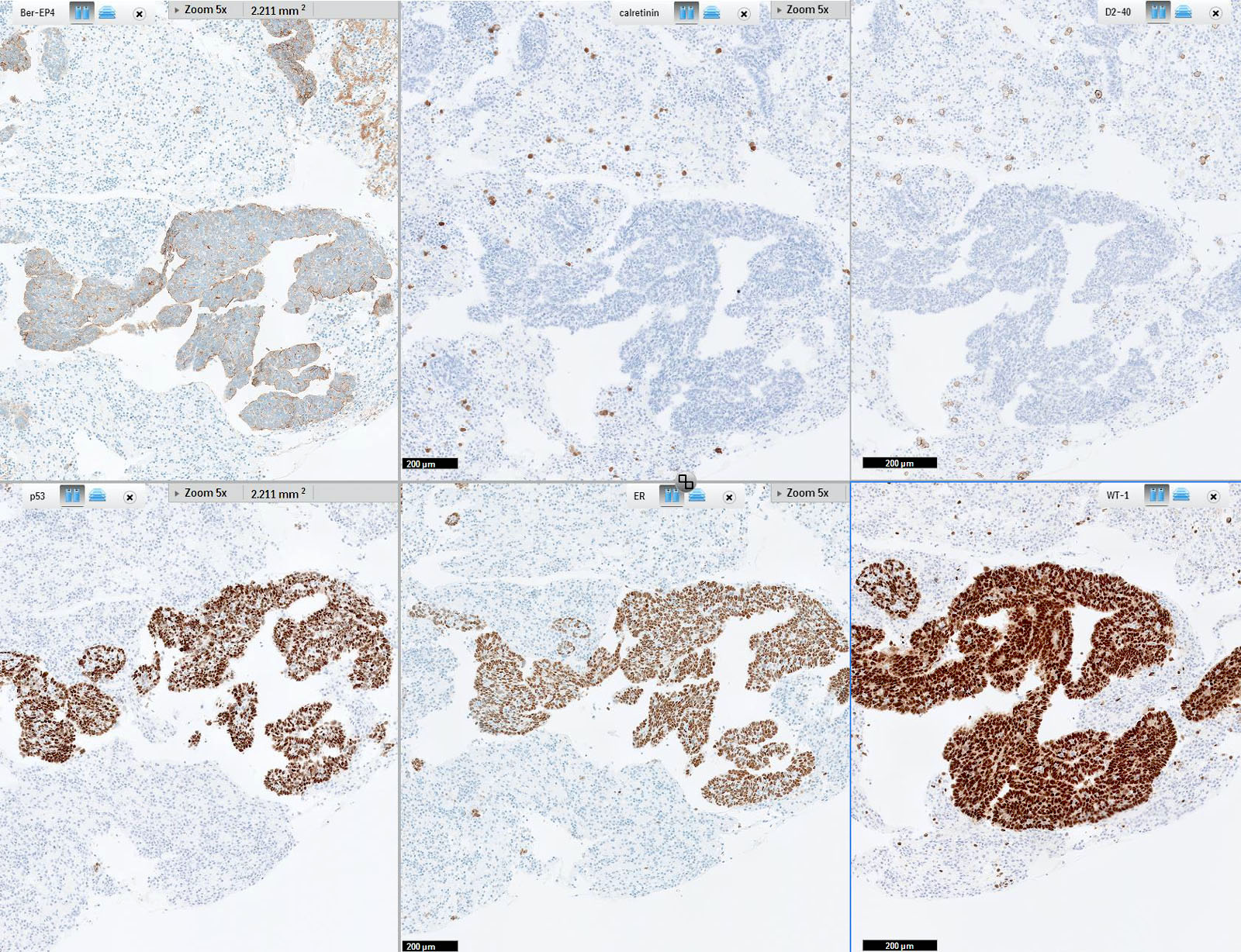
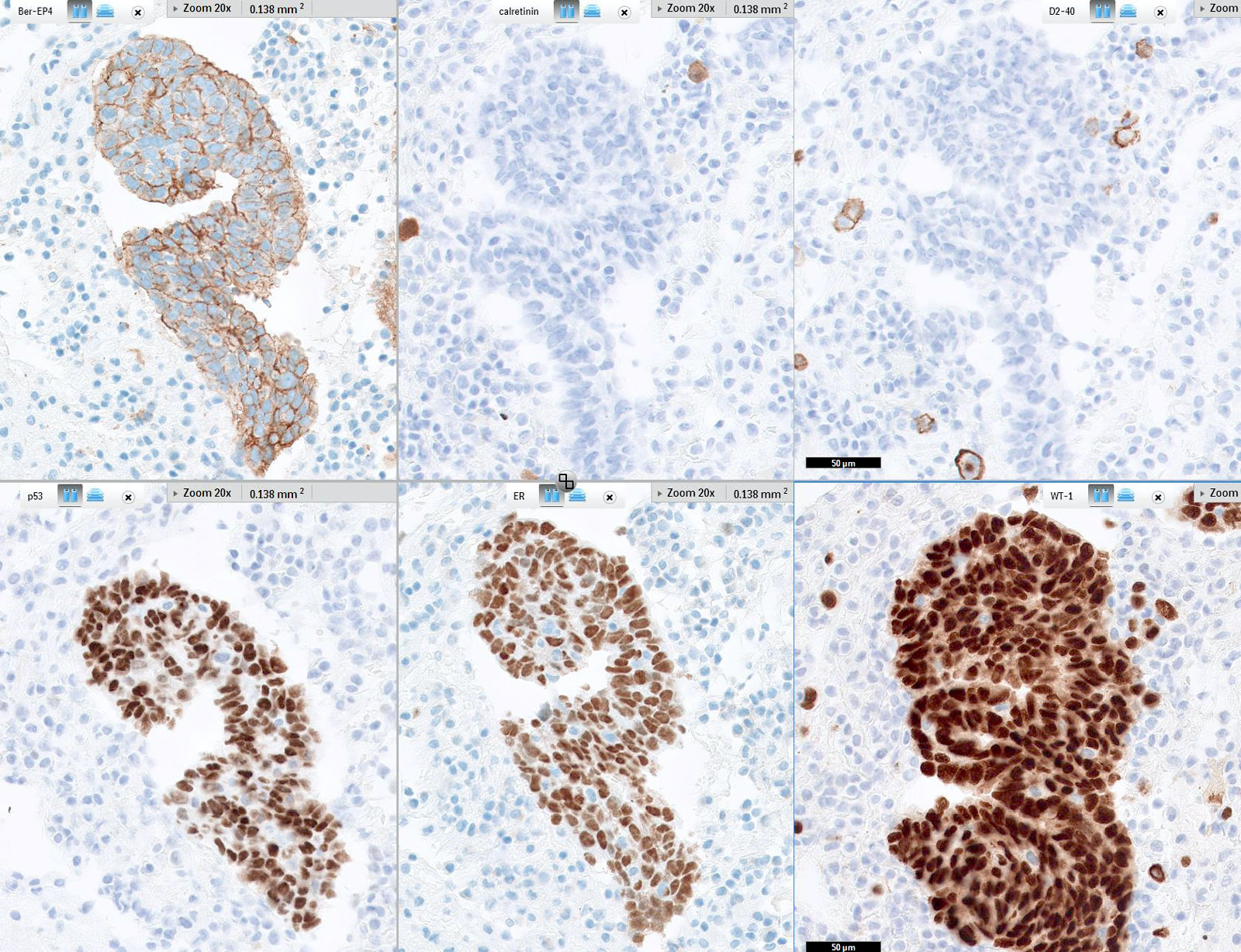
Ovarian serous carcinoma immunoprofile
Images hosted on other servers:

Basaloid squamous cell carcinoma is BerEP4+

Breast: ductal and
lobular carcinoma -
primary and
metastases
(EpCAM)


Breast cancer: increased EpCAM expression in metastases


Colon: loss of EPCAM expression in cancers associated with Lynch syndrome caused by heterozygous EPCAM germline deletions

Esophagus: normal
and Barrett metaplasia;
gastric mucosa
Unknown site: adenocarcinoma (MOC31)
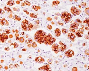
Cytology images
Images hosted on other servers:


Reactive mesothelial cells (BerEP4 / MOC31 negative) versus metastatic adenocarcinoma (positive)
Positive staining - normal
- Basolateral surface of epithelial cells
Positive staining - disease
- Most carcinomas (Hum Pathol 2004;35:122, J Clin Pathol 2011;64:415)
- Barrett metaplasia (J Clin Pathol 2006;59:260)
- Chromophobe renal cell carcinoma (75%), papillary renal cell carcinoma (55%), clear cell renal carcinoma (18%), metastatic renal cell carcinoma (14%, Am J Surg Pathol 2005;29:83)
- Synovial sarcoma (Am J Surg Pathol 2001;25:610)
Negative staining
- Epidermal keratinocytes, gastric parietal cells, hepatocytes, myoepithelial cells, squamous epithelia, thymic cortical epithelium (Front Biosci 2008;13:3096)
- Lymphoma, mesothelioma, most soft tissue sarcomas
- Adenomatoid tumor; renal oncocytoma
