Table of Contents
Definition / general | Essential features | Pathophysiology | Interpretation | Uses by pathologists | Microscopic (histologic) images | Virtual slides | Cytology images | Positive staining - normal | Positive staining - disease | Negative staining | Sample pathology report | Board review style question #1 | Board review style answer #1Cite this page: Baniak N. Calretinin. PathologyOutlines.com website. https://www.pathologyoutlines.com/topic/stainscalretinin.html. Accessed April 2nd, 2025.
Definition / general
- Calcium binding protein (Hum Pathol 2003;34:994)
Essential features
- Nuclear and cytoplasmic staining
- Used primarily in the diagnosis of Hirschsprung disease and to confirm sex cord stromal or mesothelial lineage
- Also positive in adrenal cortical lesions, mesonephric carcinoma, female adnexal tumor of probable Wolffian origin (FATWO) and STK11 adnexal tumor
Pathophysiology
- 29 kilodalton calcium binding protein of the calmodulin superfamily (Hum Pathol 2003;34:994)
- Function is not clearly understood but it is thought to act as a buffer or sensor of intracellular calcium ions (Hum Pathol 2006;37:312, Hum Pathol 2003;34:994)
Interpretation
- Nuclear and cytoplasmic (Mod Pathol 2007;20:248)
Uses by pathologists
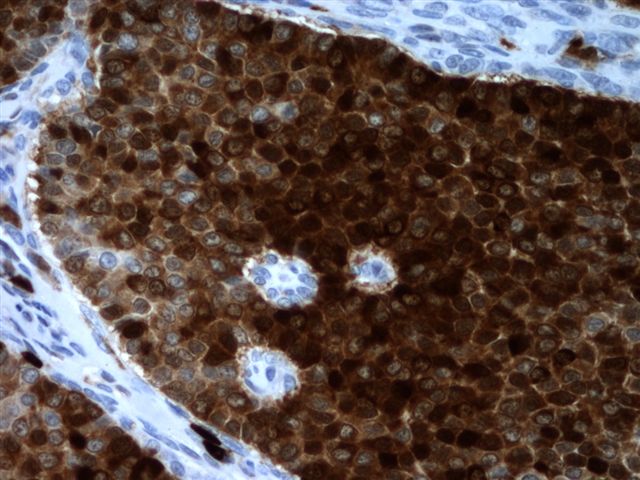
- Differentiate (as part of a panel) pleural mesothelioma (positive) from lung adenocarcinoma (negative) (Mod Pathol 2007;20:248, Indian J Pathol Microbiol 2021;64:655, Am J Surg Pathol 2003;27:150, Arch Pathol Lab Med 2018;142:89)
- Differentiate (as part of a panel) peritoneal mesothelioma (positive) from ovarian serous carcinoma (negative); however, calretinin can stain a significant minority of serous carcinomas (Am J Surg Pathol 2007;31:1139, Am J Surg Pathol 1998;22:1203)
- Confirm mesothelial origin in pleural or peritoneal specimens (Hum Pathol 2003;34:994)
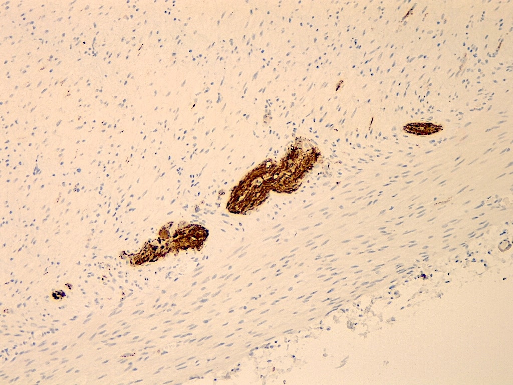
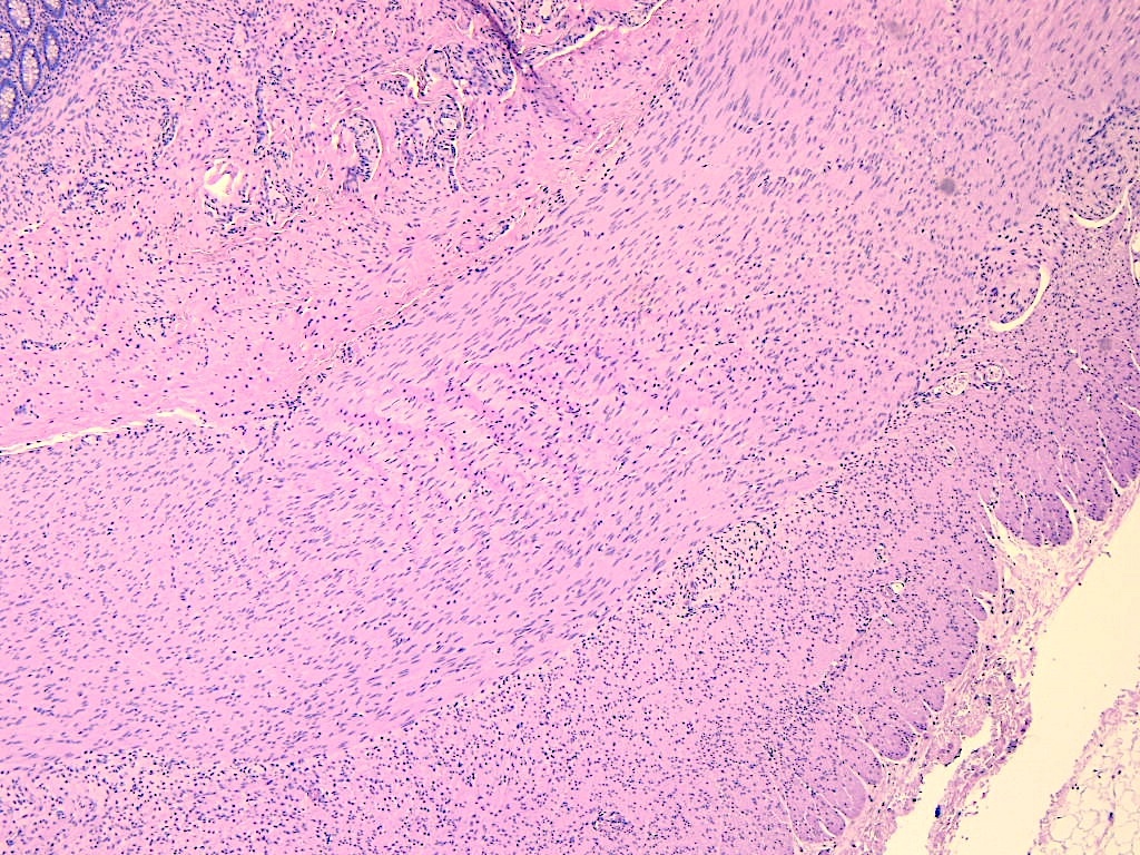
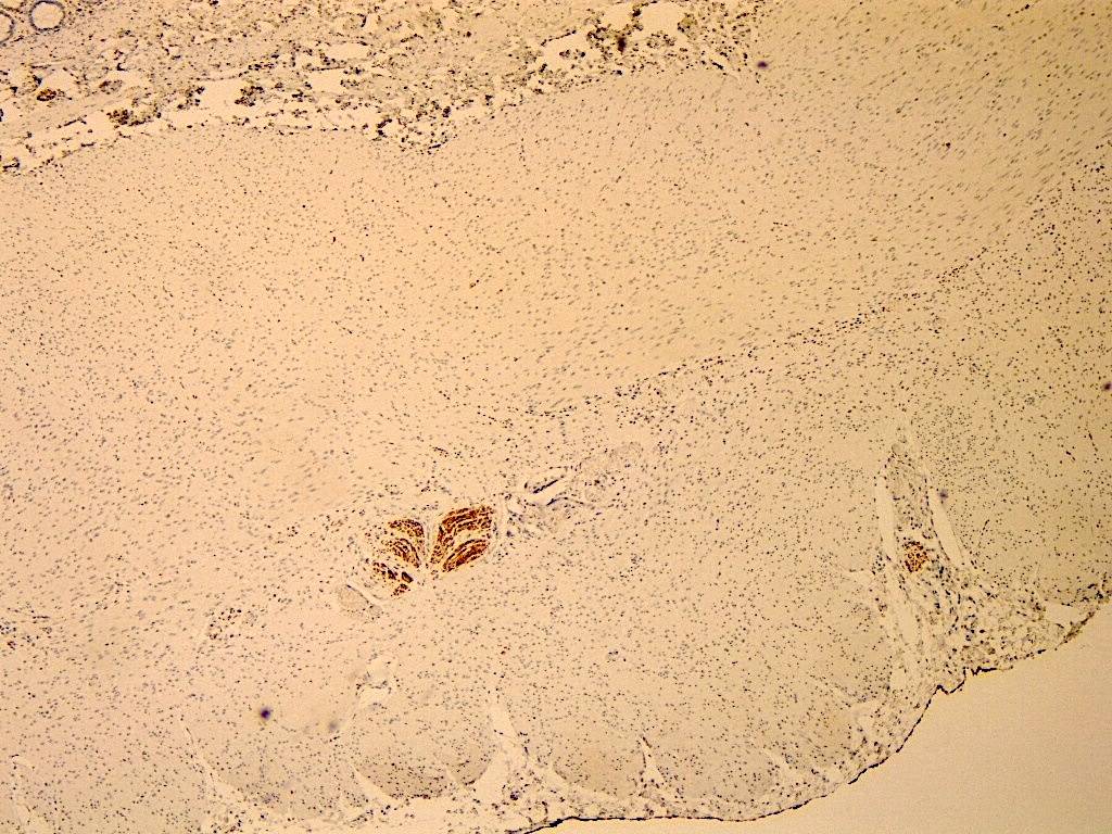
- Diagnosis of Hirschsprung disease (showing an absence of mucosal innervation) (Pediatr Dev Pathol 2021;24:19, Mod Pathol 2009;22:1379)
- Differentiate (as part of a panel) adrenal cortical lesions (positive) from metastatic renal cell carcinoma (negative) (Am J Surg Pathol 2011;35:678)
- Differentiate (as part of a panel) adrenal cortical lesions (positive) from pheochromocytoma (negative) (Am J Surg Pathol 2010;34:423)
- Diagnosing (as part of panel) sex cord stromal tumors (ovarian and testis) (Hum Pathol 2003;34:994, Am J Surg Pathol 2021;45:1303, Hum Pathol 2005;36:195)
- Used as part of a panel in diagnosing cervical mesonephric carcinoma (Am J Surg Pathol 2018;42:1596, Am J Surg Pathol 2001;25:379)
- Used as part of a panel in the diagnosis of STK11 adnexal tumors and FATWO (Am J Surg Pathol 2021;45:1061, Mod Pathol 2020;33:734)
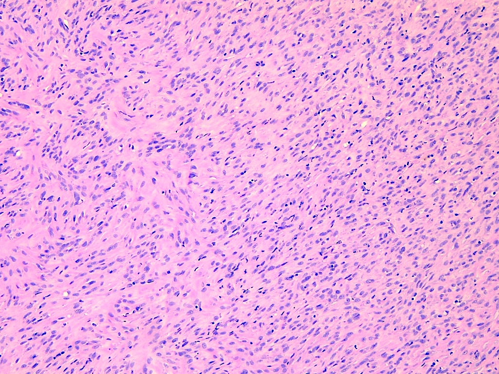
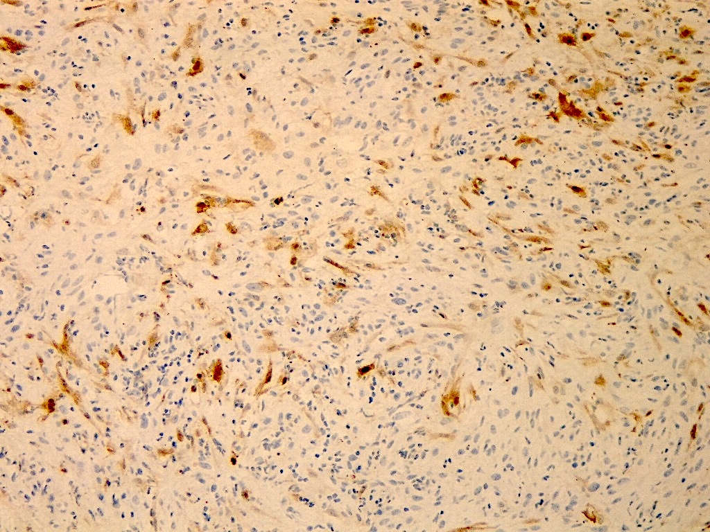
- Differentiate schwannoma (positive) from neurofibroma (negative or focal) (Am J Clin Pathol 2004;122:552)
Microscopic (histologic) images
Virtual slides
Positive staining - normal
- Mesothelial cells (Hum Pathol 2003;34:994)
- CNS (neurons, granular cells, astrocytes, Purkinje cells) (Hum Pathol 2003;34:994, Hum Pathol 2006;37:312)
- Leydig cells (Hum Pathol 2003;34:994)
- Ovary (theca cells, granulosa cells) (Hum Pathol 2003;34:994)
- Endometrial stromal cells (Hum Pathol 2003;34:994)
- Breast luminal cells (weak) (Hum Pathol 2003;34:994)
- Adrenal cortical cells and sustentacular cells (Hum Pathol 2003;34:994, Appl Immunohistochem Mol Morphol 2010;18:414)
- Salivary gland serous cells (weak) (Hum Pathol 2003;34:994)
- Colonic ganglion cells (Pediatr Dev Pathol 2021;24:19)
- Adipocytes (Hum Pathol 2006;37:312)
Positive staining - disease
- Mesothelioma (81 - 100%; 57% sarcomatoid) (Hum Pathol 2003;34:994, Mod Pathol 2007;20:248, Sci Rep 2021;11:12554, Indian J Pathol Microbiol 2021;64:655, Am J Clin Pathol 2008;130:771, Am J Surg Pathol 2007;31:1139, Hum Pathol 2019;92:48)
- Adrenal cortical lesions:
- Adenoma (100%) (Hum Pathol 2003;34:994, Appl Immunohistochem Mol Morphol 2010;18:414)
- Carcinoma (50 - 80%) (Hum Pathol 2003;34:994, Am J Surg Pathol 2011;35:678)
- Testicular sex cord stromal tumors:
- Leydig cell tumor (79 - 100%) (Hum Pathol 2003;34:994, Am J Surg Pathol 2021;45:1303)
- Juvenile granulosa cell tumors (86%) (Am J Surg Pathol 2021;45:1303)
- Large cell calcifying Sertoli cell tumors (50%) (Am J Surg Pathol 2021;45:1303)
- Adult granulosa cell tumors (50%) (Am J Surg Pathol 2021;45:1303)
- Sertoli cell tumors (44%) (Am J Surg Pathol 2021;45:1303)
- Ovarian sex cord stromal tumors:
- Adult granulosa cell tumor (90%) (Hum Pathol 2005;36:195)
- Sertoli-Leydig cell tumor (57%) (Hum Pathol 2005;36:195)
- Female adnexal tumor of probable Wolffian origin (40 - 57%) (Mod Pathol 2020;33:734, Arch Pathol Lab Med 2022;146:166)
- STK11 adnexal tumor (100%) (Am J Surg Pathol 2021;45:1061)
- Cervical mesonephric adenocarcinoma (50 - 88%) (Am J Surg Pathol 2018;42:1596, Am J Surg Pathol 2001;25:379)
- Adenomatoid tumors (100%) (Appl Immunohistochem Mol Morphol 2012;20:173)
- Ameloblastoma (90 - 94%) (Histopathology 2000;37:27, Appl Immunohistochem Mol Morphol 2014;22:762)
- Keratocystic odontogenic tumor (80%) (Appl Immunohistochem Mol Morphol 2014;22:762)
- Lipoma (96%) (Hum Pathol 2006;37:312)
- Liposarcoma (97%) (Hum Pathol 2006;37:312)
- Cardiac myxoma (74%) (Histol Histopathol 2001;16:1031)
- Colonic medullary carcinoma (73%) (Hum Pathol 2009;40:398)
- Schwannoma (96%) (Am J Clin Pathol 2004;122:552)
Negative staining
- Note: calretinin is usually negative in the below entities, with variable, generally rare, positive cases (see percentages below)
- Derm:
- Squamous cell carcinoma (19%) (Hum Pathol 2003;34:994)
- Malignant melanoma (14%) (Hum Pathol 2003;34:994)
- Head and neck:
- Laryngeal squamous cell carcinoma (23%) (Hum Pathol 2003;34:994)
- Oral cavity, squamous cell carcinoma (22%) (Hum Pathol 2003;34:994)
- Adenomatoid odontogenic tumor (0%) (Appl Immunohistochem Mol Morphol 2014;22:762)
- Thoracic:
- Lung small cell carcinoma (41 - 49%) (Hum Pathol 2003;34:994, Am J Surg Pathol 2003;27:150)
- Lung squamous cell carcinoma (22 - 34%) (Hum Pathol 2003;34:994, Am J Surg Pathol 2003;27:150)
- Lung adenocarcinoma (0 - 11%) (Am J Surg Pathol 2003;27:150, Hum Pathol 2003;34:1155)
- Thymic carcinoma (36%) (Hum Pathol 2003;34:1155)
- Thymoma (3%) (Hum Pathol 2003;34:1155)
- Breast:
- Invasive mammary carcinoma of no special type (ductal carcinoma) (4%) (Hum Pathol 2003;34:994)
- Medullary carcinoma (44%) (Hum Pathol 2003;34:994)
- Mucinous carcinoma (11%) (Hum Pathol 2003;34:994)
- Papillary carcinoma (14%) (Hum Pathol 2003;34:994)
- Phyllodes tumor (23%) (Hum Pathol 2003;34:994)
- Gynecologic:
- Ovarian endometrioid carcinoma (18 - 36%) (Hum Pathol 2003;34:994, Hum Pathol 2005;36:195)
- Ovarian serous carcinoma (10 - 45%) (Hum Pathol 2003;34:994, Am J Clin Pathol 2008;130:771, Am J Surg Pathol 2007;31:1139, Am J Surg Pathol 1998;22:1203)
- Ovarian microcystic stromal tumor (6%) (Am J Surg Pathol 2009;33:367)
- Ovarian fibroma - thecoma (14 - 33%) (Hum Pathol 2005;36:195)
- Squamous cell carcinoma (cervix, vulva, vagina) (10 - 18%) (Hum Pathol 2003;34:994)
- Endocervical adenocarcinoma (0%) (Am J Surg Pathol 2018;42:1596)
- Endometrial mesonephric-like carcinoma (0%) (Am J Surg Pathol 2018;42:1596)
- Endometrial endometrioid adenocarcinoma (7%) (Am J Surg Pathol 2018;42:1596)
- Endometrial serous carcinoma (9%) (Am J Surg Pathol 2018;42:1596)
- Endometrial clear cell carcinoma (14%) (Am J Surg Pathol 2018;42:1596)
- Endometrial carcinosarcoma (26%) (Am J Surg Pathol 2018;42:1596)
- Gastrointestinal:
- Hepatocellular carcinoma (2%) (Hum Pathol 2003;34:994)
- Pancreatic adenocarcinoma (5%) (Hum Pathol 2003;34:994)
- Gastric adenocarcinoma (6%) (Hum Pathol 2003;34:994)
- Small intestine, adenocarcinoma (11%) (Hum Pathol 2003;34:994)
- Colonic adenocarcinoma (5 - 6%) (Hum Pathol 2003;34:994, Pathol Oncol Res 2016;22:725)
- Genitourinary:
- Prostatic adenocarcinoma (7%) (Hum Pathol 2003;34:994)
- Renal cell carcinoma, clear cell type (3 - 10%) (Hum Pathol 2003;34:994, Appl Immunohistochem Mol Morphol 2010;18:414, Am J Surg Pathol 2011;35:678)
- Renal cell carcinoma, papillary type (10%) (Hum Pathol 2003;34:994)
- Urothelial carcinoma (5%) (Hum Pathol 2003;34:994)
- Testicular seminoma (9%) (Hum Pathol 2003;34:994)
- Testicular fibroma - thecoma (25%) (Am J Surg Pathol 2021;45:1303)
- Testicular myoid gonadal stromal tumor (0%) (Am J Surg Pathol 2021;45:1303)
- Rete testis adenocarcinoma (40%) (Am J Surg Pathol 2019;43:670)
- Neuroendocrine:
- Paraganglioma (16 - 27%) (Hum Pathol 2003;34:994, Appl Immunohistochem Mol Morphol 2010;18:414)
- Pheochromocytoma (3%) (Hum Pathol 2003;34:994)
- Thyroid medullary carcinoma (30%) (Hum Pathol 2003;34:994)
- CNS:
- Astrocytoma (28%) (Hum Pathol 2003;34:994)
- Ependymoma (7%) (Hum Pathol 2003;34:994)
- Glioblastoma multiforme (16%) (Hum Pathol 2003;34:994)
- Oligodendroglioma (17%) (Hum Pathol 2003;34:994)
- Other:
- Desmoplastic small round cell tumor (19%) (Mod Pathol 2003;16:229)
- Neurofibroma (7%) (Am J Clin Pathol 2004;122:552)
Sample pathology report
- Right pleura, core needle biopsy:
- Mesothelioma
- Immunohistochemistry performed shows the following staining profile in the lesional cells:
- Positive: calretinin, WT1
- Negative: TTF1, Claudin4
Board review style question #1
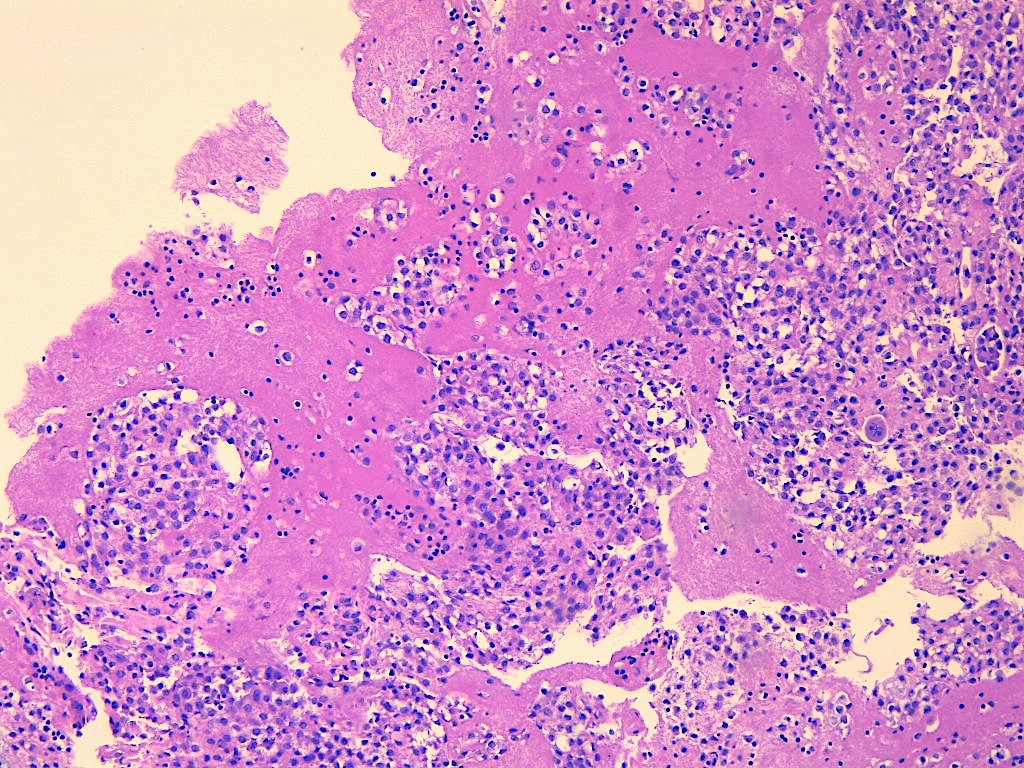
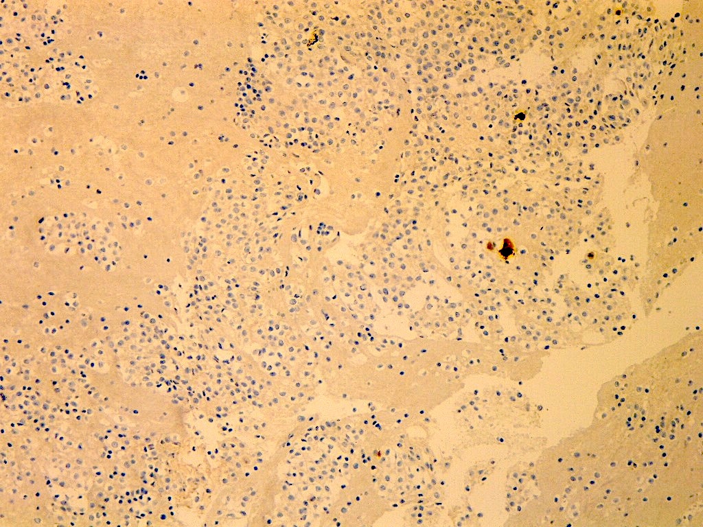
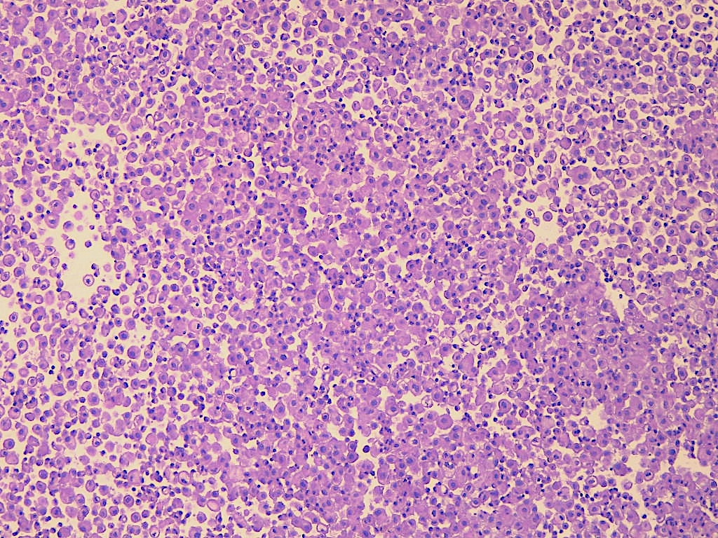
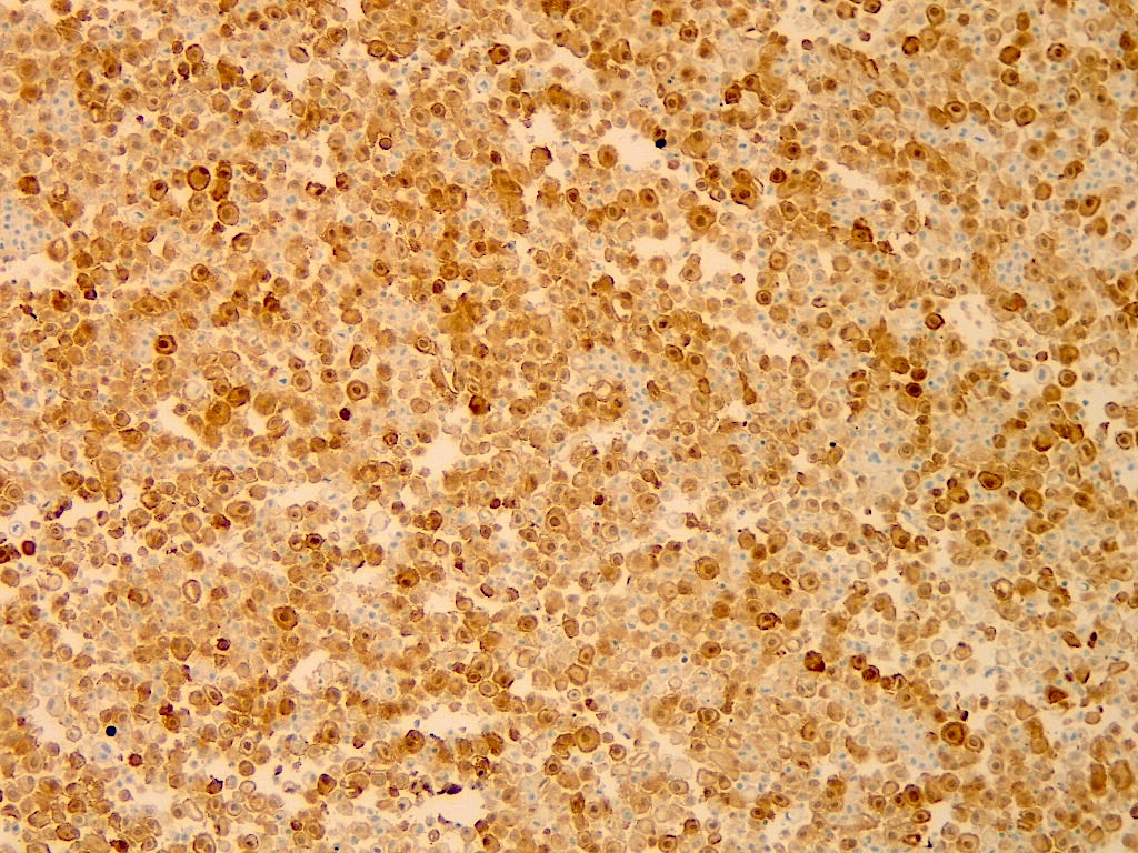
A pleural fluid specimen consists of abundant clusters of atypical cells. Pancytokeratin and WT1 stains are positive, while Claudin4 and BerEp4 are negative. The figure above shows a calretinin stain. What is the most appropriate statement?
- The biomarker result is noncontributory due to a lack of specificity
- The biomarker result is supportive of a lung adenocarcinoma
- The biomarker result is supportive of a lung squamous cell carcinoma
- The biomarker result is supportive of malignant mesothelioma
Board review style answer #1


