Table of Contents
Definition / general | Essential features | Interpretation | Uses by pathologists | Microscopic (histologic) images | Positive staining - normal | Positive staining - disease | Negative staining | Additional references | Board review style question #1 | Board review style answer #1 | Board review style question #2 | Board review style answer #2Cite this page: Khan AM, Topilow AA. S100. PathologyOutlines.com website. https://www.pathologyoutlines.com/topic/stainsS100.html. Accessed March 31st, 2025.
Definition / general
- Name is from its solubility in 100% saturated ammonium sulfate at neutral pH
- Family of multigenic group of nonubiquitous cytoplasmic homologous intracellular Ca2+ binding proteins with EF hand motifs and significant structural similarities at both protein and genomic levels
- Low molecular weights (9 -1 3 kDa) and are able to form heterodimers, homodimers and oligomeric assemblies
- S100 is composed of two main subunits, an alpha and a beta chain; most clinical stains use antibodies to the beta chain
- S100 protein family has 25 known members solely in vertebrates coded by unique genes
- There are 22 proteins (S100A1-A8, trichohylin, filaggrin and repetin) mapped onto chromosome locus 1q21, while the other proteins are located on chromosome loci 4p16 (S100P), 5q14 (S100Z), 21q22(S100B), Xp22(S100G), 5q13(S100Z), 7q22-q3(S100A11P) (Curr Mol Med 2013;13:24, Nat Rev Cancer 2015;15:96)
- Structurally similar to calmodulin and troponin-C but these proteins are cell specific and dependent upon environmental factors via cytokines, growth factors and toll-like receptors (TLR) ligands (Curr Mol Med 2013;13:24, Nat Rev Cancer 2015;15:96)
Essential features
- Family of multigenic group of nonubiquitous cytoplasmic homologous intracellular Ca2+ binding proteins with EF hand motifs and significant structural similarities at both protein and genomic levels
- Found in melanoma, nerve sheath tumors, clear cell sarcoma of soft tissue, various histiocytic tumors, glial tumors and myoepithelial tumors
- Aid in diagnosis of tumors of unknown origin and to show nerve sheath involvement
Interpretation
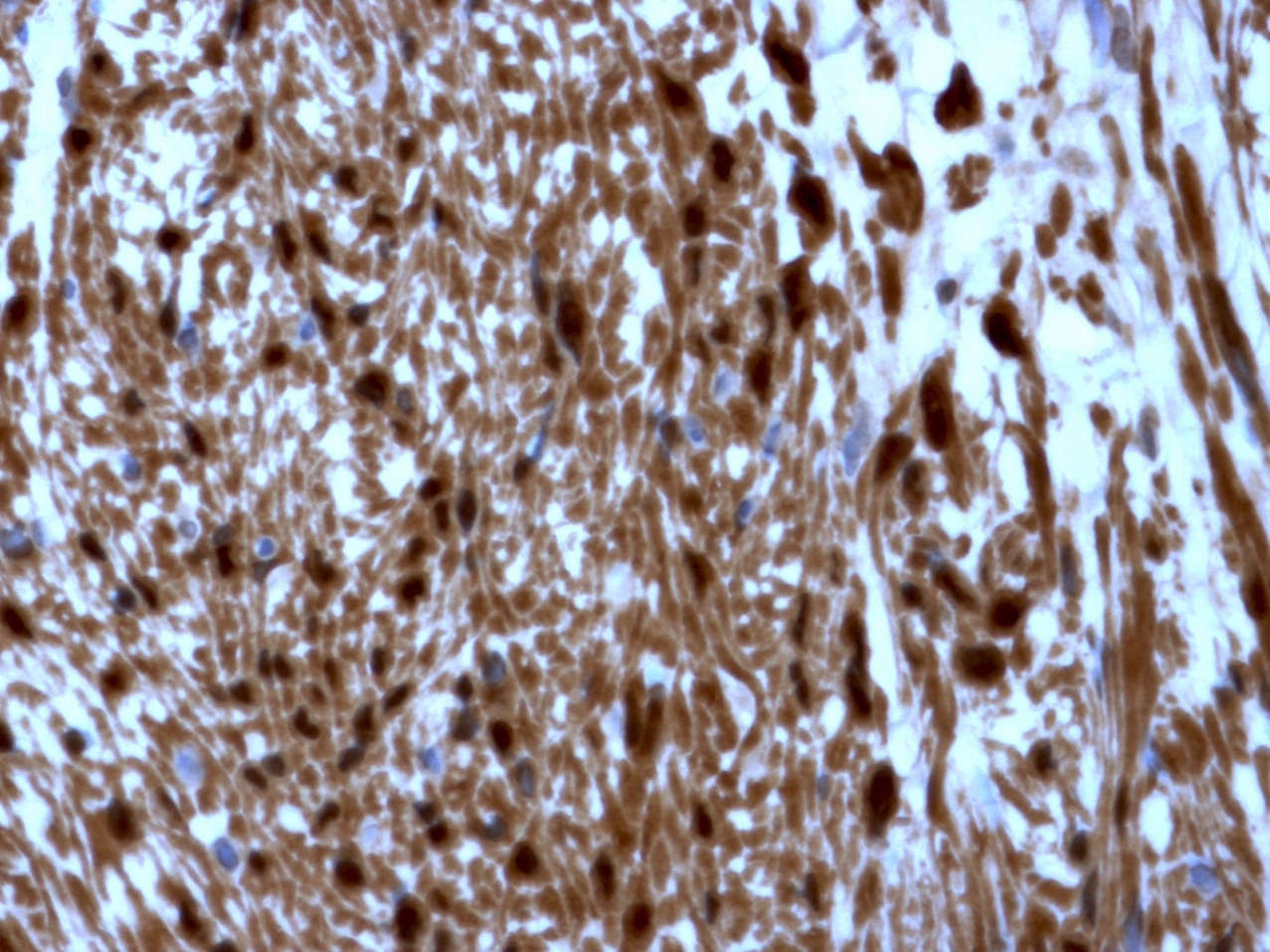
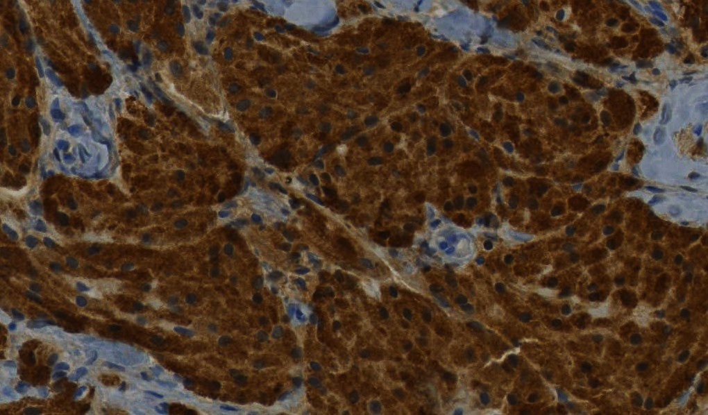
- Cytoplasmic and nuclear staining usually required to call positive
Uses by pathologists
- Marker of schwann cells and melanocytes; useful for evaluating nerve sheath tumors and melanoma
- Differentiate benign nerve sheath tumors (S100 strong and diffuse) from malignant peripheral nerve sheath tumors (typically weak or negative S100) (Am J Surg Pathol 2005;29:1042)
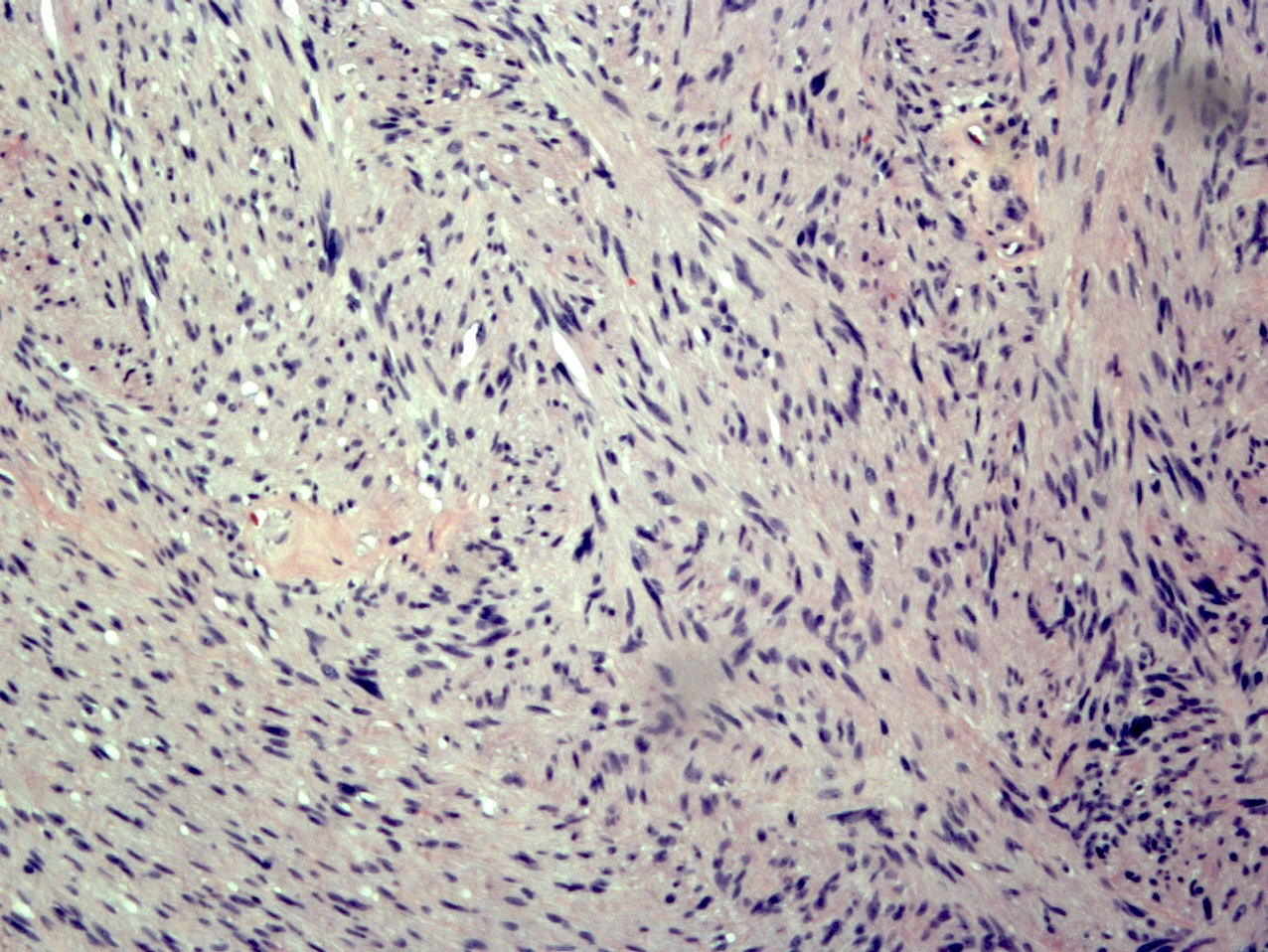
- Help differentiate between schwannoma (stains all cells) and neurofibroma (mixture of positive and negative cells)
- Distinguish Langerhans cell histiocytosis from other histiocytosis syndromes (Arch Pathol Lab Med 2015;139:1211)
- Identifying perineural involvement
- Myoepithelial cell marker and identification of tumors with myoepithelial cells
- MelanA and MART1 superior to S100 for evaluating sentinel lymph nodes for melanoma (Am J Surg Pathol 2001;25:1039)
Microscopic (histologic) images
Positive staining - normal
- Neurons, schwann cells, melanocytes, glial cells
- Myoepithelial cells
- Adipocytes
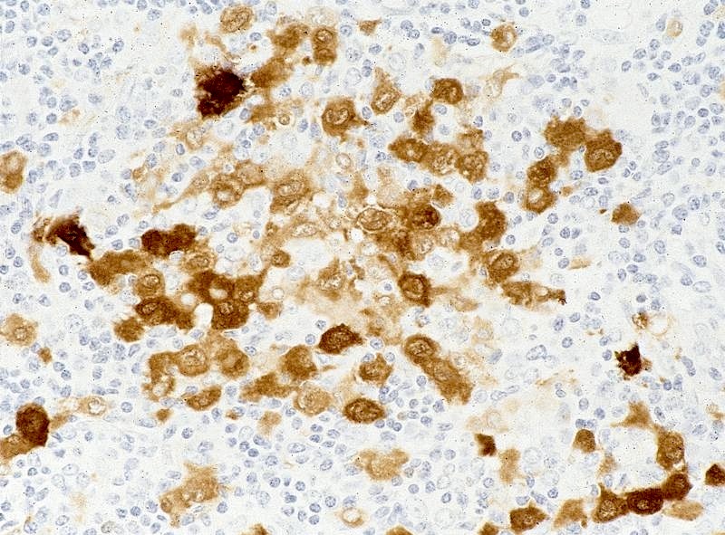
- Langerhans cells, tissue dendritic cells and interdigitating dendritic cells
- Chondrocytes and notochordal cells
Positive staining - disease
- Mesenchymal:
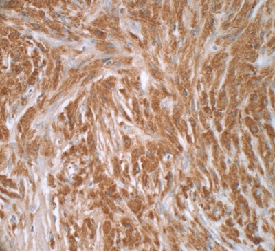
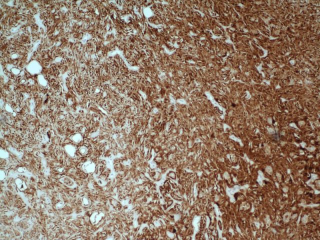
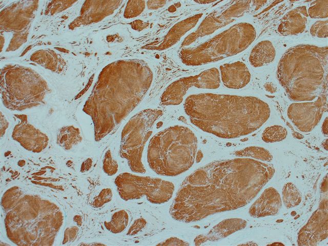
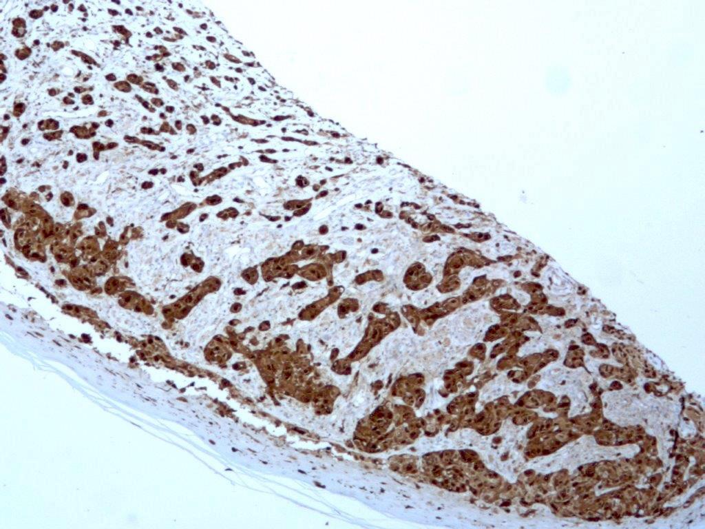
- Nerve sheath tumors: schwannoma (diffuse), ganglioneuroma (diffuse), dermal nerve sheath myxoma, neurofibroma (predominantly positive with negative nonschwannian component)
- Malignant peripheral nerve sheath tumor (30 - 67% show any staining; low grade lesions typically diffuse and conventional high grade examples are weak / patchy) (J Clin Pathol 2003 Nov;56:826)
- Adipocytic component of lipogenic tumors
- Chondroid tumors: enchondroma, osteochondroma, chondrosarcoma, chondromyxoid fibroma, chondroblastoma and mesenchymal chondrosarcoma (chondrocytic component only) (Arch Pathol Lab Med 2012;136:61)
- Notochordal tumors: benign notochordal cell tumor, conventional chordoma, chondroid chordoma (Mod Pathol 2008;21:1461)
- Clear cell sarcoma of soft tissue (Am J Surg Pathol 2008;32:452)
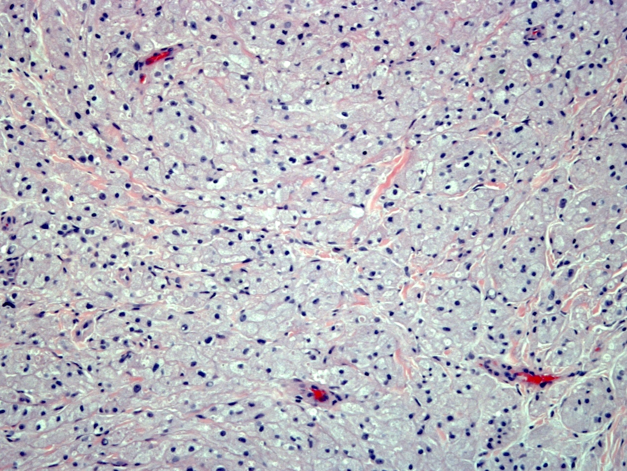
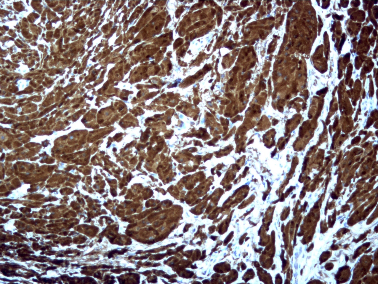
- Granular cell tumor (Arch Pathol Lab Med 2004;128:771)
- Biphenotypic sinonasal sarcoma (Arch Pathol Lab Med 2018;142:1196)
- Ossifying fibromyxoid tumor (Am J Surg Pathol 2008;32:996)
- Extraskeletal myxoid chondrosarcoma (31%) (Pathol Int 2005;55:453)
- Synovial sarcoma (34%) (Am J Clin Pathol 1999;112:641)
- Ewing sarcoma (variable) (Appl Immunohistochem Mol Morphol 2012;20:445)
- Perivascular epithelioid cell tumors (less commonly than other melanocytic markers; 33%) (Am J Surg Pathol 2005;29:1558)
- Mycobacterial spindle cell pseudotumor (Am J Surg Pathol 1992;16:276)
- Brain:
- Glial tumors: astrocytoma, oligodendroglioma, ependymoma
- Choroid plexus papilloma and carcinoma (Am J Surg Pathol 1986;10:394)
- Meningioma (Positive in ~70% of fibrous variant) (Appl Immunohistochem Mol Morphol 2015;23:215)
- Macrophage lineage:
- Interdigitating dendritic cell tumors (Am J Hematol 2007;82:725)
- Langerhans cell tumors (Am J Surg Pathol 2007;31:947)
- Rosai-Dorfman disease (Am J Surg Pathol 1992;16:122)
- Epithelial / myoepithelial:
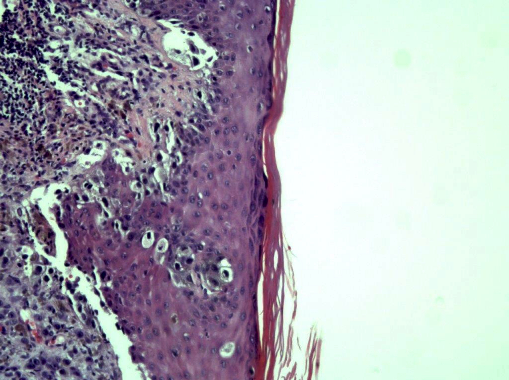
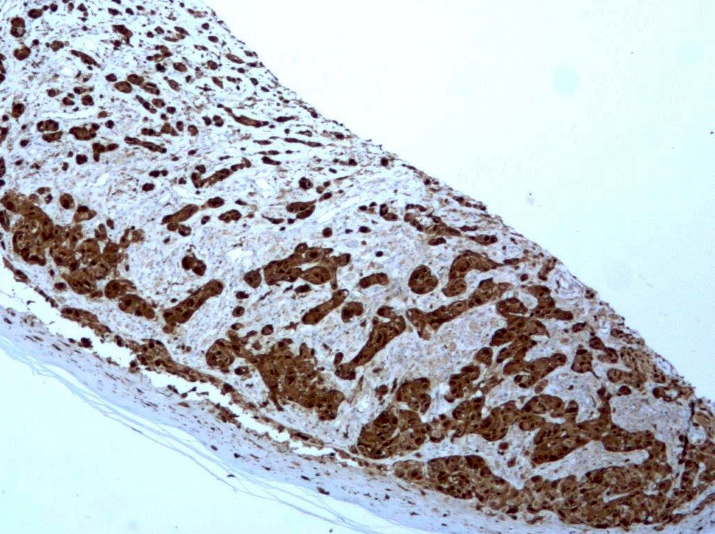
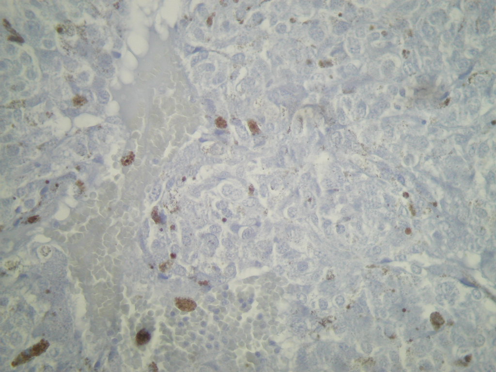
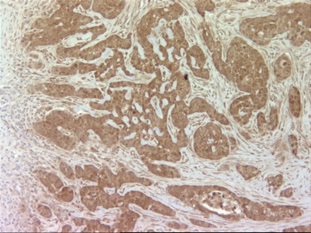
- Melanoma (95%, including desmoplastic and spindle cell tumors); S100- melanomas often had prior S100+ metastases, often are ocular (Hum Pathol 2005;36:1016)
- Invasive breast carcinoma (subset; particularly basal-like subtype) (Contemp Oncol (Pozn) 2016;20:436)
- Metaplastic breast carcinoma (Diagn Pathol 2014;9:139)
- Myoepithelial tumors: myoepithelioma, myoepithelial carcinoma
- Salivary gland tumors with myoepithelial differentiation: pleomorphic adenoma, myoepithelioma, basal cell adenoma, adenoid cystic carcinoma, polymorphous adenocarcinoma, epithelial-myoepithelial carcinoma, sialoblastoma (Acta Histochem Cytochem 2012;45:269)
- Mammary analogue secretory carcinoma (Head Neck Pathol 2015;9:85)
- Others:
- Paraganglioma / pheochromocytoma: sustentacular cells
- Neuroendocrine tumors (subset, often focal) (Am J Surg Pathol 2005;29:1194)
- Sex cord-stromal tumors (variable in any of this category) (Mod Pathol 1997;10:693)
Negative staining
- Mesenchymal:
- Perineurioma (Am J Surg Pathol 2005;29:845)
- Cellular neurothekeoma (Am J Surg Pathol 2007;31:329)
- Fibroblastic reticular cell tumor (Am J Surg Pathol 1998;22:1048)
- Alveolar soft part sarcoma (Arch Pathol Lab Med 2015;139:1459)
- Rhabdomyoma (may be focally positive)
- Sinonasal glomangiopericytoma (Pathol Res Pract 2019;215:983)
- Clear cell sarcoma of the kidney (Arch Pathol Lab Med 2019;143:1022)
- Solitary fibrous tumor (Iran J Pathol 2016;11:195)
- Mammary type myofibroblastoma (Am J Surg Pathol 2016;40:361)
- Cellular angiofibroma (Am J Surg Pathol 2004;28:1426)
- Gastrointestinal stromal tumor (Transl Gastroenterol Hepatol 2018;3:27)
- Benign fibrous histiocytoma (Am J Dermatopathol 2001;23:104)
- Atypical fibroxanthoma (Anticancer Res 2015;35:5717)
- Macrophage lineage:
- Follicular dendritic cell tumors (may show rare positive cases) (Am J Hematol 2007;82:725)
- Erdheim-Chester disease (Blood 2014;124:483)
- Juvenile xanthogranuloma (rare positive but generally negative) (Am J Surg Pathol 2005;29:21)
- Xanthoma
- Salivary gland tumors without myoepithelial differentiation: salivary duct carcinoma, mucoepidermoid carcinoma, squamous cell carcinoma, oncocytoma / oncocytic carcinoma, Warthin tumor (Arch Pathol Lab Med 2015;139:55)
- Squamous cell carcinoma
- Lymphoma
Additional references
Board review style question #1
Board review style answer #1
Board review style question #2
- A biopsied lung lesion is radiographically suspected to be a metastasis of unknown primary. Which staining profile suggests that the tumor is melanoma?
- S100 positive, CK5/6 positive, p63 negative, GFAP negative
- S100 negative, HMB45 positive, TTF1 negative, CK20 negative
- S100 positive, HMB45 positive, CK7 negative, p63 negative
- S100 negative, TTF1 positive, SOX10 positive, AE1/AE3 negative
- S100 positive, CD21 positive, CD35 positive, Clusterin positive
Board review style answer #2


