Stains & CD markers
LEF1

Copyright: 2020-2025, PathologyOutlines.com, Inc.
PubMed Search: LEF1 [title] pathology
LEF1
Author: Alessandra C. Schmitt, M.D.
Editorial Board Member: Kelly Magliocca, D.D.S., M.P.H.
Editor-in-Chief: Debra L. Zynger, M.D.
Last author update: 29 September 2020
Last staff update: 21 June 2021
Copyright: 2020-2025, PathologyOutlines.com, Inc.
PubMed Search: LEF1 [title] pathology
Table of Contents
Definition / general | Essential features | Interpretation | Uses by pathologists | Prognostic factors | Microscopic (histologic) description | Microscopic (histologic) images | Positive staining - normal | Positive staining - disease | Negative staining | Board review style question #1 | Board review style answer #1Cite this page: Schmitt AC. LEF1. PathologyOutlines.com website. https://www.pathologyoutlines.com/topic/stainsLEF1.html. Accessed March 31st, 2025.
Definition / general
- Lymphoid enhancer binding factor 1 (LEF1) is a transcription factor for B and T cells and is involved in the Wnt / CTNNB1 pathway which regulates normal development of airway submucosal glands, mammary glands, teeth and adnexal structures (Nat Rev Immunol 2005;5:21, Development 1999;126:4441)
Essential features
- Not specific
- Helpful when used as a panel
- Positive staining favors:
- Pleomorphic adenoma, basal cell adenoma or basal cell adenocarcinoma over adenoid cystic carcinoma
- Chronic lymphocytic B cell leukemia / small lymphocytic lymphoma over other small B cell lymphomas
- Cribriform morular variant of papillary thyroid carcinoma over other papillary thyroid carcinoma variants
- Solid pseudopapillary tumor over pancreatic neuroendocrine carcinoma or pancreatic adenocarcinoma
Interpretation
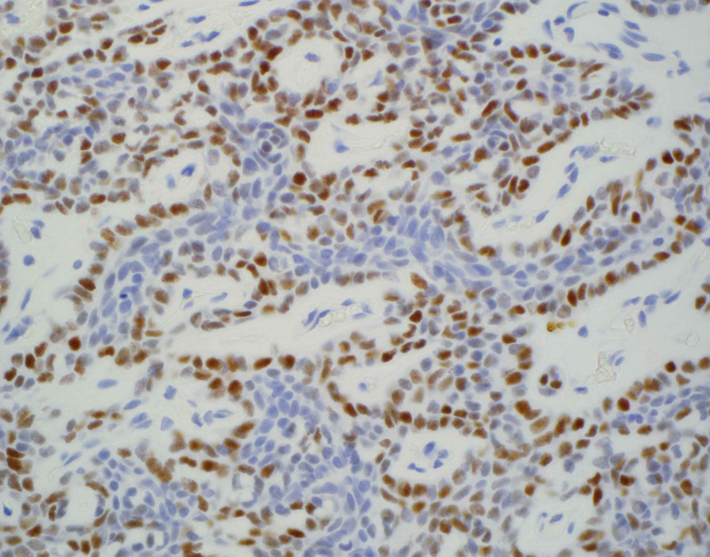
- Nuclear stain
Uses by pathologists
- Positive LEF1 weighs against diagnosis of adenoid cystic carcinoma in the setting of basaloid salivary gland neoplasm fine needle aspiration (Diagn Cytopathol 2017;45:1078)
- Identify cribriform morular variant of papillary thyroid carcinoma in the setting of papillary thyroid carcinoma neoplasm (Head Neck Pathol 2018;12:455)
- Diagnose chronic lymphocytic B cell leukemia (Am J Clin Pathol 2017;147:292)
- Diagnose solid pseudopapillary tumor (Oncotarget 2017;8:93404)
Prognostic factors
- Poor prognostic factor in colorectal carcinoma (Appl Immunohistochem Mol Morphol 2014;22:728)
- Poor prognostic factor in oral squamous cell carcinoma (J Formos Med Assoc 2014;113:934)
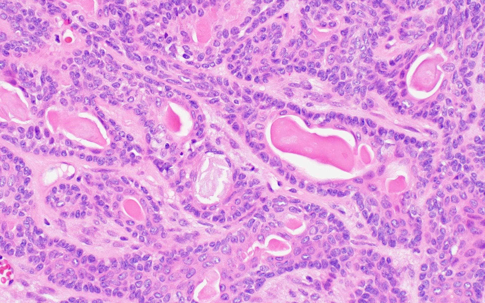
Microscopic (histologic) description
- Focal staining with peripheral accentuation possible in basal cell adenoma and pleomorphic adenoma (shown in figures below)
- Positive staining in pleomorphic adenoma is not specific
Microscopic (histologic) images
Positive staining - normal
- Benign thyroid (Head Neck Pathol 2018;12:455)
- Reactive T cells (Am J Clin Pathol 2017;147:292)
- Scar (Appl Immunohistochem Mol Morphol 2018;26:648)
Positive staining - disease
- Basal cell adenoma (86%) (Diagn Cytopathol 2017;45:1078)
- Basal cell adenocarcinoma (69%) (Hum Pathol 2015;46:255)
- Calcifying cystic odontogenic tumor (64%) (Hum Pathol 2015;46:255)
- Adamantinomatous craniopharyngioma (Histopathology 2004;45:573)
- Cutaneous lesions such as squamous cell carcinoma, cutaneous basal cell carcinoma and pilomatricoma (Br J Dermatol 2009;160:1353)
- Papillomavirus related multiphenotypic sinonasal carcinoma (Head Neck Pathol 2019;13:220)
- Cribriform morular variant of papillary thyroid carcinoma (86%) (Head Neck Pathol 2018;12:455)
- Chronic lymphocytic B cell leukemia / small lymphocytic lymphoma (96%) (Am J Clin Pathol 2017;147:292)
- Solid pseudopapillary tumor (93%) (Oncotarget 2017;8:93404)
- Basal cell carcinoma (96%) (Br J Dermatol 2009;160:1353)
- Desmoid type fibromatosis (72%) (Appl Immunohistochem Mol Morphol 2018;26:648)
Negative staining
- Parotid gland (Diagn Cytopathol 2017;45:1078)
- Adenoid cystic carcinoma (95%) (Diagn Cytopathol 2017;45:1078)
- Papillary thyroid carcinoma (other than morular variant)
- Follicular lymphoma (97%) (Am J Clin Pathol 2017;147:292)
- Marginal zone lymphoma
- Mantle cell lymphoma
- Pancreatic adenocarcinoma (Oncotarget 2017;8:93404)
- Pancreatic neuroendocrine tumor (Oncotarget 2017;8:93404)
Board review style question #1

A 50 year old woman presented with an infiltrative left submandibular mass. LEF1 stain was performed and was negative. The tumor is best diagnosed as
- Adenoid cystic carcinoma
- Basal cell adenocarcinoma
- Basal cell adenoma
- Cellular pleomorphic adenoma
Board review style answer #1
