Table of Contents
Definition / general | Uses by pathologists | Positive staining - normal | Positive staining - not malignant | Positive staining - malignant | Negative staining | Microscopic (histologic) images | Additional referencesCite this page: Pernick N. Cytokeratin 14 (CK14, K14). PathologyOutlines.com website. https://www.pathologyoutlines.com/topic/stainsCK14.html. Accessed April 2nd, 2025.
Definition / general
- CK14 is a type I acidic keratin expressed in mitotically active basal cells of stratified epithelium (Front Oncol 2020;10:623)
- Molecular weight of 50 kDa
- Partner is CK5
- May be detected by cytokeratin 34BE12
- CK5/6+ or CK14+ tumors define a basal subtype of DCIS (Mod Pathol 2006;19:1506) or invasive breast carcinoma; represents 9% of sporadic invasive ductal breast cancers, ER-, PR-, HER2-, high grade, poor prognosis (Mod Pathol 2005;18:1321, Eur J Cancer 2006;42:3149 but see Clin Cancer Res 2004;10:5988-not poor prognosis), associated with BRCA1 (Clin Cancer Res 2005;11:5175)
- In cervix, loss of expression is associated with high grade SIL and high risk HPV (Hum Pathol 2001;32:1351)
- Prostate tumors with distinct basal cells on H&E that are negative for 34BE12 are also negative for CK14 (Pathol Res Pract 2006;202:651)
- Mutations cause epidermolysis bullosa simplex (J Invest Dermatol 2006;126:773), Naegeli syndrome / dermatopathia pigmentosa reticularis (no fingerprints, OMIM 161000)
Uses by pathologists
- Distinguish parathyroid oxyphil adenoma (CK14+) from carcinoma (CK14-, Am J Surg Pathol 2002;26:344)
- Distinguish breast papilloma (stronger and more diffuse CK14 staining) from papillary DCIS (Am J Surg Pathol 2005;29:625)
- Distinguish sinonasal squamous cell carcinoma (poorly differentiated or nonkeratinizing, both CK14+) from sinonasal undifferentiated carcinoma or nasopharyngeal carcinoma (CK14-, Am J Surg Pathol 2002;26:1597)
Positive staining - normal
- Basal keratinocytes in stratified epithelium (various tissue/organs)
- Hair follicles (Br J Dermatol 2004;150:860)
- Myoepithelial cells (breast and salivary gland)
- Thyroid oncocytes
Positive staining - not malignant
- Breast papilloma (see above)
- Odontogenic neoplasms (Oral Dis 2003;9:1)
- Parathyroid oxyphil adenoma (see above)
- Pseudoepitheliomatous hyperplasia-spinous and superficial layers of oral mucosa with paracoccidioidomycosis (Med Mycol 2006;44:399)
- Renal and other oncocytoma (Histopathology 2001;39:455)
- Thymoma
- Trichoblastoma
Positive staining - malignant
- Basal cell (Am J Dermatopathol 2001;23:501)
- Breast-basal phenotype (see above)
- Salivary gland tumors except acinic cell carcinoma (Pathologica 2006;98:147)
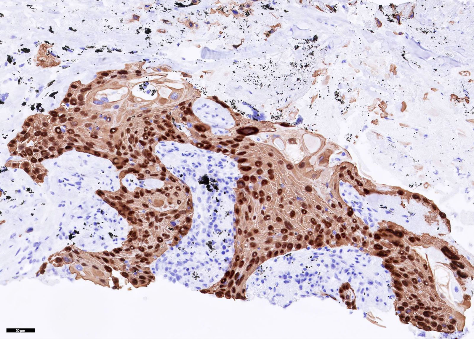
- Squamous cell carcinoma (esophagus-Nepal Med Coll J 2006;8:75 and other sites-Histopathology 2001;39:9)
- Squamous differentiation in urothelial (J Clin Pathol 1997;50:1032) and other tumors
Negative staining
- Normal oral mucosa, most renal cell carcinomas
Microscopic (histologic) images
Additional references


