Table of Contents
Definition / general | Terminology | Uses by pathologists | Microscopic (histologic) images | Positive staining - normal | Positive staining - disease | Negative stainingCite this page: Pernick N. CDX2. PathologyOutlines.com website. https://www.pathologyoutlines.com/topic/stainsCDX2.html. Accessed March 31st, 2025.
Definition / general
- Homeobox gene that encodes a nuclear transcription factor critical for intestinal embryonic development; relatively specific for intestinal epithelium (but see positive staining below)
Terminology
- Also called caudal-related homeobox gene 2, caudal type homeobox transcription factor 2
- Homologue of Drosophila melanogaster homeobox gene-caudal
Uses by pathologists
- Fairly specific marker of GI origin for adenocarcinomas, but also stains selected adenocarcinomas of other sites indicated below (Am J Surg Pathol 2003;27:303); can use to determine origin of metastatic adenocarcinoma as part of panel (Arch Pathol Lab Med 2007;131:1561)
- Distinguish primary and secondary colorectal adenocarcinomas (Arch Pathol Lab Med 2005;129:920)
- Distinguish primary bladder adenocarcinoma (CDX2-, villin-) from colorectal carcinoma extending / metastatic to bladder (CDX2+, villin+, Mod Pathol 2005;18:1217)
- Distinguish mucinous bronchioloalveolar adenocarcinoma (CDX2-) from metastatic mucinous colorectal adenocarcinoma (CDX2+, Am J Clin Pathol 2004;122:421)
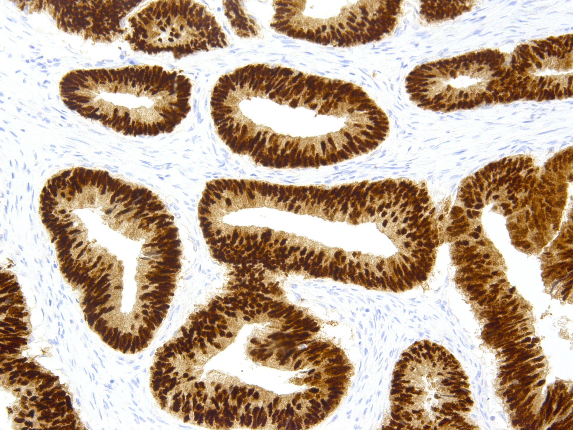
Microscopic (histologic) images
Positive staining - normal
- Nuclei of intestinal epithelium lining colonic villi and crypts, subset of epithelium of pancreas, stomach, esophagus
- Urachal remants of glandular type, bladder intestinal metaplasia
Positive staining - disease
- Bladder: bladder/urachal adenocarcinoma, urothelial carcinoma in situ with glandular differentiation (Hum Pathol 2011;42:1653), intestinal metaplasia (83%, Mod Pathol 2006;19:1395), noninvasive urachal mucinous cystic tumor (Am J Surg Pathol 2011;35:787)
- Colon: hyperplastic polyps (Am J Clin Pathol 2008;129:416), colorectal adenocarcinomas (86-100%, less if poorly differentiated)
- Endometrium: lesions with squamous differentiation, especially morular-type differentiation (Hum Pathol 2008;39:1072)
- Esophagus: intestinal metaplasia in Barrett esophagus (100%, Am J Clin Pathol 2008;129:571, metaplastic esophageal columnar epithelium without goblet cells (43%, Am J Surg Pathol 2009;33:1006)
- Extrahepatic bile duct carcinoma: 37% (Am J Clin Pathol 2005;124:361)
- Small intestinal carcinomas: 60%, usually diffuse (Am J Clin Pathol 2007;128:808), ampulla of Vater carcinomas: intestinal subtype-22%, mucinous-75% (Am J Clin Pathol 2011;135:202), intra-ampullary papillary-tubular neoplasm (Am J Surg Pathol 2010;34:1731)
- Stomach: adenocarcinoma (36-70%, with variable intensity, Hum Pathol 2011;42:1777)
- Carcinoid tumors: GI (varies by location, Am J Clin Pathol 2005;123:394); neuroendocrine tumors (appendix, ileum, pancreas, Am J Surg Pathol 2008;32:420)
- Mucinous adenocarcinomas: lung and ovary; cervical adenocarcinoma (endometrioid > endocervical, Am J Surg Pathol 2008;32:1608); rarely other carcinomas
- Signet ring cell adenocarcinoma: colon (strong, diffuse) and stomach (weak, patchy, Am J Clin Pathol 2004;121:884)
- Teratoma: metastatic testicular-100% (Am J Clin Pathol 2008;130:265)
Negative staining
- Anal glands; bladder urothelium, gastric mucosa
- Carcinomas of anal gland (Arch Pathol Lab Med 2007;131:1304), breast and prostate (Am J Surg Pathol 2007;31:1323, but see Hum Pathol 2007;38:72) and urothelial carcinoma, sinonasal high grade adenocarcinoma (Am J Surg Pathol 2011;35:971)


