Table of Contents
Definition / general | Pathophysiology | Diagrams / tables | Interpretation | Uses by pathologists | Microscopic (histologic) images | Positive staining - normal | Positive staining - disease | Negative staining | Board review style question #1 | Board review style answer #1Cite this page: Pernick N. BCL2. PathologyOutlines.com website. https://www.pathologyoutlines.com/topic/stainsBCL2.html. Accessed April 2nd, 2025.
Definition / general
- "b cell lymphoma #2"
- Proto-oncogene at 18q21.3
- Encodes 25 kDa protein, mainly localized to inner mitochondrial membrane; also endoplasmic reticulum and nuclear envelope
Pathophysiology
- Prevents cells from undergoing apoptosis
- BCL2 overexpression increase lifespan of B cells; may maintain memory B cells, plasma cells and neurons by prolonging life span without cell division
- May participate in ion channel formation and alteration of membrane permeability, necessary for initiation of apoptosis
- Non-phosphorylated BCL2 inhibits apoptosis, and Bax homodimers normally cause apoptosis; Bax can bind to and inhibit non-phosphorylated BCL2, promoting apoptosis
- Epstein-Barr virus latent membrane protein-1 (LMP-1) interacts with BCL2 promoter, leading to prolonged lifespan for B cells
- Surprisingly, BCL2 overexpression in osteoblasts causes osteocyte apoptosis (PLoS One 2011;6:e27487)
Diagrams / tables
Interpretation
- Nuclear and cytoplasmic staining
Uses by pathologists
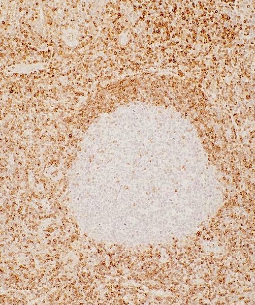
- (1) Distinguish follicular hyperplasia of lymph node (germinal centers are BCL2 negative) from follicular lymphoma (germinal centers are BCL2+); BCL2 usually overexpressed in follicular lymphoma due to t(14,18)(q32;q21), which brings BCL2 gene adjacent to active immunoglobulin heavy chain (IgH) gene; however, some follicular lymphomas are BCL2 negative (Am J Surg Pathol 2011;35:1691, Am J Surg Pathol 2009;33:591)
- (2) Detect immature enteric ganglion cells in pediatric intestinal pseudo-obstruction (Am J Surg Pathol 2005;29:1017)
- (3) Diffuse large cell lymphoma: adverse prognostic factor in some studies (Mod Pathol 2005;18:1113, Clin Cancer Res 2011;15:7785)
- (4) Myelodysplastic syndrome: increased expression associated with progression
- (5) May have prognostic value in early stage breast cancer (Br J Cancer 2010;103:668)
Microscopic (histologic) images
Positive staining - normal
- Lymph node: small B lymphocytes in mantle zone and cells within T cell areas
- Adrenal cortex, melanocyties, thymus-medullary cells, thyroid gland solid cell nests
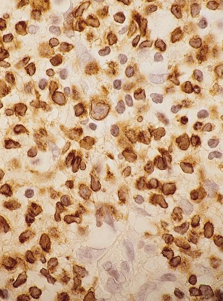
- Immature (but not mature) small ganglion cells
Positive staining - disease
- Hematopoietic: follicular lymphoma (germinal centers stain); also acute lymphoblastic leukemia, angiotropic / intravascular lymphoma, Burkitt lymphoma (occasionally, Indian J Pathol Microbiol 2011;54:290), diffuse large B cell lymphoma (variable), extranodal NK/T-cell lymphoma, nasal-type: 39% (Am J Clin Pathol 2008;130:343), Hodgkin lymphoma (classical): 27% (Hum Pathol 2007;38:103), mantle cell lymphoma, marginal zone lymphoma (variable), persistent polyclonal lymphocytosis, plasmablastic lymphoma, SLL
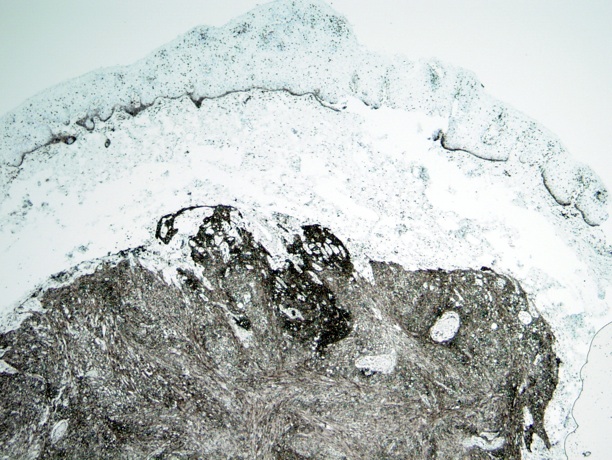
- Other: adrenal cortical adenoma and carcinoma (Mod Pathol 1998;11:716), atrioventricular node tumor of heart, basal cell carcinoma (skin and prostate, Am J Surg Pathol 2007;31:697), breast-benign stromal spindle cell tumor, breast-columnar cell lesions, breast-micropapillary carcinoma, breast-phyllodes tumor (stromal cells), cervix-mesonephric remnants / rests, cervix-tuboendometrial metaplasia, collecting duct carcinoma-kidney, colorectal adenomas / carcinomas, dermatofibrosarcoma protuberans
- Giant cell angiofibroma, hemangiopericytoma, large cell neuroendocrine carcinoma (salivary glands), low grade fibromyxoid sarcoma of soft tissue, lymphoepithelioma-like carcinoma, melanoma (uvea, Arch Pathol Lab Med 1996;120:497), myofibroblastoma, pheochromocytoma (variable)
- Sclerosing epithelioid fibrosarcoma, small cell carcinoma (breast), solitary fibrous tumor, spindle cell epithelioma of vagina (Arch Pathol Lab Med 2001;125:547), squamous cell carcinoma (uterus), squamous papilloma (conjunctiva, Ann NY Acad Sci 2004;1030:419), synovial sarcoma (Hum Pathol 1996;27:1060), thyroid gland-CASTLE
Negative staining
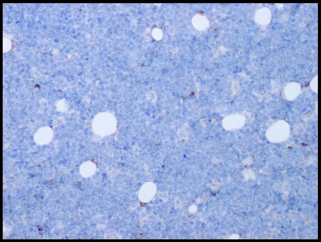
- Anaplastic lymphoma, apocrine benign / malignant lesions, benign fibrous histiocytoma, lymphoid hyperplasia (marginal zone cells in hyperplastic areas are BCL2+, germinal centers are BCL2-), lymphoplasmayctic lymphoma
Board review style question #1
The translocation that puts the IgH locus on chromosome 14 and the BCL2 locus on chromosome 18 together, is a trademark of which disease?
- Anaplastic large cell lymphoma
- Burkitt lymphoma
- Follicular lymphoma
- Splenic marginal cell leukemia
- Hodgkin lymphoma
Board review style answer #1
C. Follicular lymphoma; more than 90% of FL is associated with t(14;18).
Citation: Kumar: Robbins & Cotran Pathologic Basis of Disease, Eighth Edition, 2009, McPherson: Henry's Clinical Diagnosis and Management by Laboratory Methods, Twenty Second Edition, 2011 (Chapter 33)
Source: BoardVitals, 2/5/2014
- Incorrect: anaplastic large cell lymphoma is primarily associated with t(2;5).
- Incorrect: Burkitt lymphoma is primarily associated with t(8;14), also t(2;8) and t(8;22)
- Incorrect: Hodgkin lymphoma is not associated with a specific translocation (although BCL6 translocations may be seen in L&H cells).
- Incorrect: splenic marginal cell leukemia is not associated with a specific translocation.
Citation: Kumar: Robbins & Cotran Pathologic Basis of Disease, Eighth Edition, 2009, McPherson: Henry's Clinical Diagnosis and Management by Laboratory Methods, Twenty Second Edition, 2011 (Chapter 33)
Source: BoardVitals, 2/5/2014


