Table of Contents
Definition / general | Uses by pathologists | Clinical features | Microscopic (histologic) images | Positive staining - normal | Positive staining - disease | Negative stainingCite this page: Pernick N. Alcian blue. PathologyOutlines.com website. https://www.pathologyoutlines.com/topic/stainsAlcianblue.html. Accessed December 4th, 2024.
Definition / general
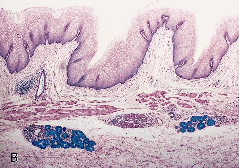
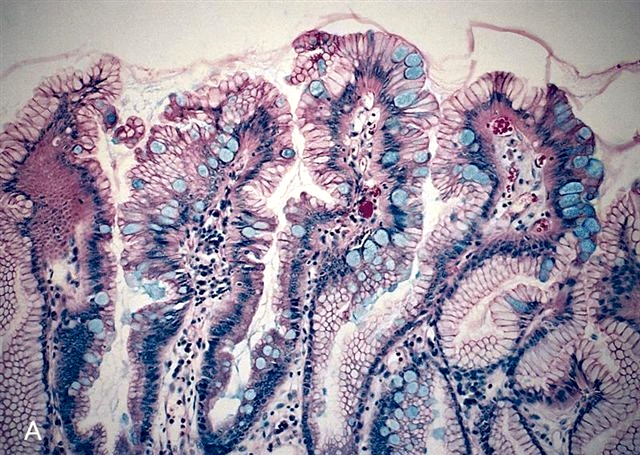
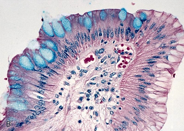
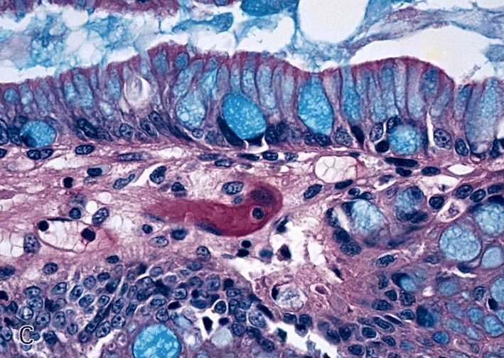
- Common "routine" stain (not an immunohistochemical stain) to detect mucins (Wikipedia: Alcian Blue Stain [Accessed 8 August 2018])
- At pH 2.5, detects acidic mucins
- At pH 1.0, detects highly acidic mucins
- Stained parts are blue to bluish green
- Note: all references below are to pH 2.5 unless otherwise indicated
Uses by pathologists
- Stains acid-simple, nonsulfated and acid-simple mesenchymal mucins at pH 2.5, acid-complex sulfated mucins at pH 1.0 and acid-complex connective tissue mucins at pH 0.5; does NOT stain neutral mucins
- PAS-Alcian blue may be best pan mucin combination; PAS also stains glycogen, but predigestion with diastase will remove the glycogen
- Alcian blue-high iron diamine detects sulfomucins (brown) and sialomucins (blue)
Clinical features
- Procedure (University of Utah)
- May be useful in FNA diagnosis of salivary gland pleomorphic adenoma (J Cytol 2012;29:221)
- Stains glycosaminoglycan deposits in macular corneal dystrophy (Korean J Ophthalmol 2013;27:454)
Microscopic (histologic) images
AFIP images and Cases #78, 94 and 110
Images hosted other servers:
Positive staining - normal
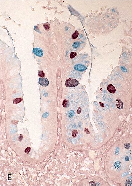
- Colloid of thyroid gland, goblet cells and mucous glands
Positive staining - disease
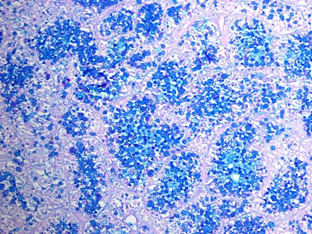
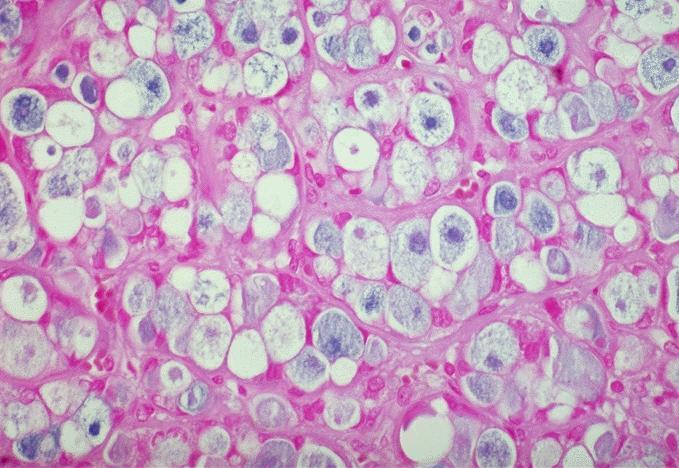
- Adenocarcinoma, adenosquamous carcinoma, mast cell leukemia (Acta Med Croatica 2013;67:61)
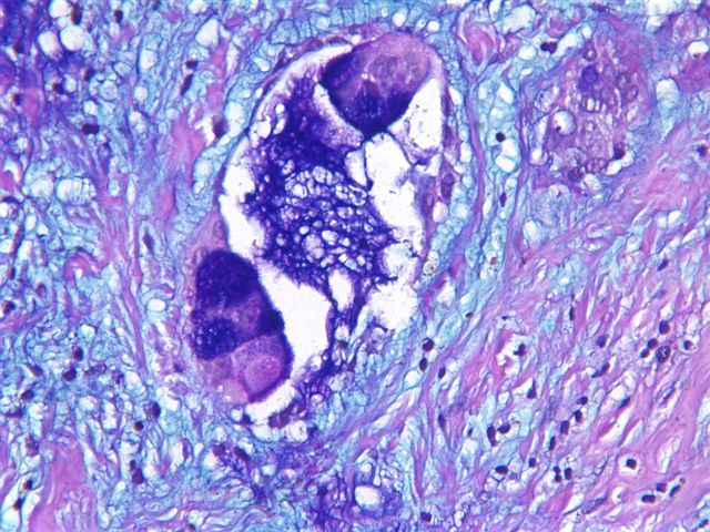
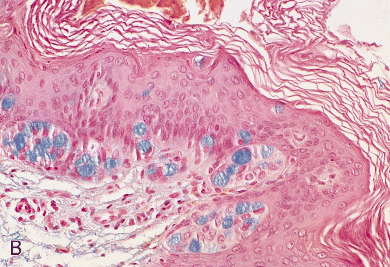
- Mucinous tumors, myxedema (dermal mucin), myxoma (mucoid matrix), nodular mucinosis (breast, other) and Paget disease of scrotum
Negative staining
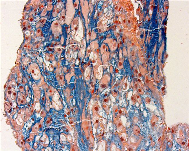
- Lipids / lipid entities (lipoma, liposarcoma), Paget disease of esophagus, squamous cell carcinoma (acantholytic variant-breast & other sites, pseudoglandular variant-penis & other sites), xanthelasma


