Table of Contents
Definition / general | Case reports | Gross description | Gross images | Microscopic (histologic) description | Microscopic (histologic) images | Positive stains | Differential diagnosisCite this page: Mansouri J. Epithelial cyst. PathologyOutlines.com website. https://www.pathologyoutlines.com/topic/spleenepithelialcysts.html. Accessed January 10th, 2025.
Definition / general
- Usually children or young adults
- Solitary or multiple; may be associated with accessory spleen
- May mimic mucinous cystic neoplasm if in intrapancreatic accessory spleen
- Called "epithelioid" / "epidermoid" if squamous lining
- Origin unknown; may derive from metaplasia in mesothelial cysts (Am J Surg Pathol 1988;12:275)
- Often large and requires splenectomy
Case reports
- 62 year old man with epidermoid cyst in intrapancreatic accessory spleen (JOP 2011;12:279)
Gross description
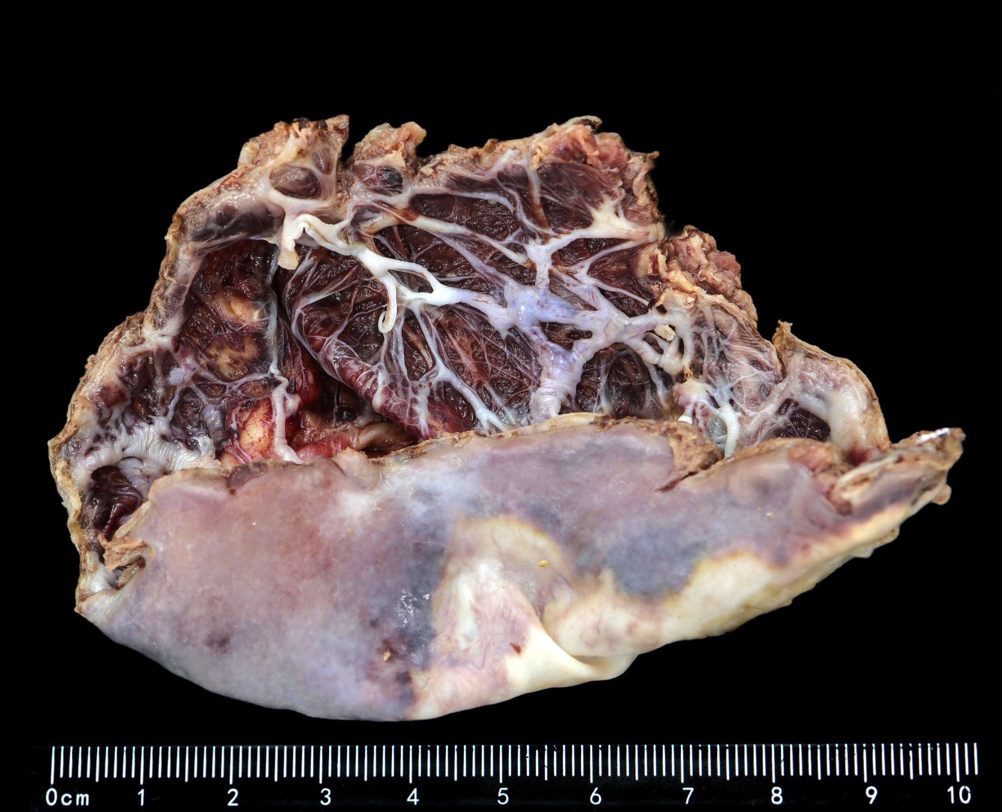
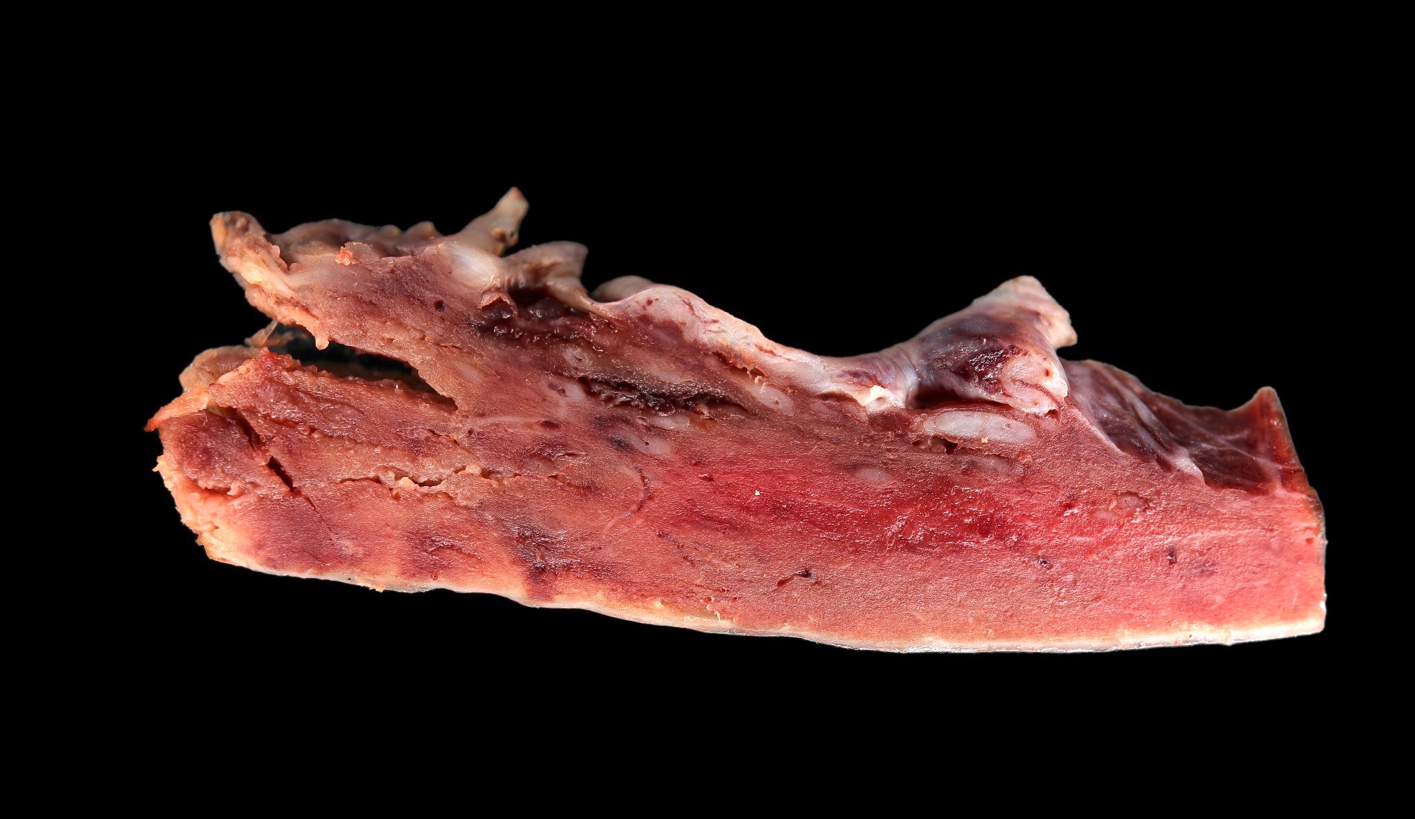
- Glistening inner surface with marked trabeculation
Gross images
Microscopic (histologic) description
- Lined by squamous, columnar, cuboidal or mesothelial-like epithelium
- No skin adnexae
- Rarely mucinous and associated with pseudomyxoma peritonei
Microscopic (histologic) images
Positive stains
- CEA, CA19-9 (Am J Surg Pathol 1998;22:704)
Differential diagnosis
- Mucinous / cystic neoplasm
- Pseudocyst


