Page views in 2024: 2,769
Page views in 2025 to date: 462
Cite this page: Alexiev BA. Staging-GIST. PathologyOutlines.com website. https://www.pathologyoutlines.com/topic/softtissuestaginggist.html. Accessed March 30th, 2025.
Definition / general
- This staging protocol should be used for all resection specimens from patients with gastrointestinal stromal tumor (GIST)
- This staging protocol is not required for the following procedures: biopsy, local excision, primary resection specimen with no residual tumor (e.g., following neoadjuvant therapy), cytologic specimens
Essential features
- AJCC 7th edition staging was sunset on December 31, 2017; as of January 1, 2018, use of the 8th edition is mandatory
- The TNM staging system applies to all GISTs of the esophagus, gastroesophageal junction, stomach, small intestine (duodenum, jejunum, ileum, Meckel diverticulum, small intestine, NOS), appendix, Ileocecal valve, large intestine (cecum, ascending colon, hepatic flexure of colon, transverse colon, descending colon, sigmoid colon, rectosigmoid junction, rectum, large intestine, NOS), retroperitoneum and peritoneum / abdomen
- See CAP for additional information
Terminology
- Reporting of pT (tumor), pN (lymph node) and when applicable, pM (metastasis) categories is based on information available to the pathologist at the time the report is issued; as per the AJCC, it is the managing physician's responsibility to establish the final pathologic stage based upon all pertinent information (Amin: AJCC Cancer Staging Manual, 8th Edition, 2018)
- Tumor (T):
- Based on the tumor size, as measured grossly or with imaging
- Largest dimension in any plane
- Best to provide 3 dimensional size if possible
- Nodes (N):
- Lymph node status needs to be determined both clinically and microscopically
- Metastasis (M):
- Prefix:
- If the lesion was previously treated, use the prefix y (ypTNM)
- If the lesion is recurrent, use the prefix r (rpTNM)
ICD coding
- ICD-O: 8936/3 - gastrointestinal stromal tumor
- ICD-11: 2B5B & XH9HQ1 - gastrointestinal stromal tumor, primary site & gastrointestinal stromal sarcoma
Diagrams / tables
Table 1: Guidelines for risk assessment of primary gastrointestinal stromal tumor (GIST)
| Tumor parameters
| Risk of progressive disease (%)
|
| Mitotic rate
| Size
| Stomach
| Duodenum
| Jejunum / ileum
| Rectum
|
≤ 5 per 5 mm2
| ≤ 2 cm
| None
| None
| None
| None
|
| > 2 - ≤ 5 cm
| Very low
| Low
| Low
| Low
|
| > 5 - ≤ 10 cm
| Low
| Insufficient data
| Moderate
| Insufficient data
|
| > 10 cm
| Moderate
| High
| High
| High
|
> 5 per 5 mm2
| ≤ 2 cm
| None
| Insufficient data
| High
| High
|
| > 2 - ≤ 5 cm
| Moderate
| High
| High
| High
|
| > 5 - ≤ 10 cm
| High
| Insufficient data
| High
| Insufficient data
|
| > 10 cm
| High
| High
| High
| High
|
Primary tumor (pT)
- pT not assigned (cannot be determined based on available pathological information)
- pT0: no evidence of primary tumor
- pT1: tumor ≤ 2 cm
- pT2: tumor > 2 cm but not > 5 cm
- pT3: tumor > 5 cm but not > 10 cm
- pT4: tumor > 10 cm in greatest dimension
Regional lymph nodes (pN)
- pN not assigned (no nodes submitted or found)
- pN not assigned (cannot be determined based on available pathological information)
- pN0: no regional lymph node metastasis
- pN1: regional lymph node metastasis
Notes:
- When no lymph nodes are present (as is often the case with resection for GIST), the pathologic N category is not assigned (pNX is not used for GIST) and should not be reported
Prefixes
- Not applicable
- m: multiple primary synchronous tumors in a single organ
- y: preoperative radiotherapy or chemotherapy
- r: recurrent tumor stage
Risk assessment
- None
- Very low risk
- Low risk
- Moderate risk
- High risk
- Overtly malignant / metastatic
- Cannot be determined: ___________
Notes:
Histologic grade (G)
- GX: cannot be assessed
- G1: low grade (mitotic rate ≤ 5 per 5 mm2)
- G2: high grade (mitotic rate > 5 per 5 mm2)
- Other (specify): ___________
Notes:
- Mitotic count should be initiated in an area that reveals the highest level of mitotic activity on screening magnification and then should be performed as consecutive high power fields (HPF)
- Mitoses should be counted in 5 mm2 of tumor
- With the use of older model microscopes, 50 HPF is equivalent to 5 mm2
- Most modern microscopes with wider fields require ~20 - 25 HPF to encompass 5 mm2
- See CAP for additional information
Histopathologic type
- Gastrointestinal stromal tumor, spindle cell type
- Gastrointestinal stromal tumor, epithelioid type
- Gastrointestinal stromal tumor, mixed
- Gastrointestinal stromal tumor, other (specify): ___________
Gross images
Contributed by Borislav A. Alexiev, M.D.
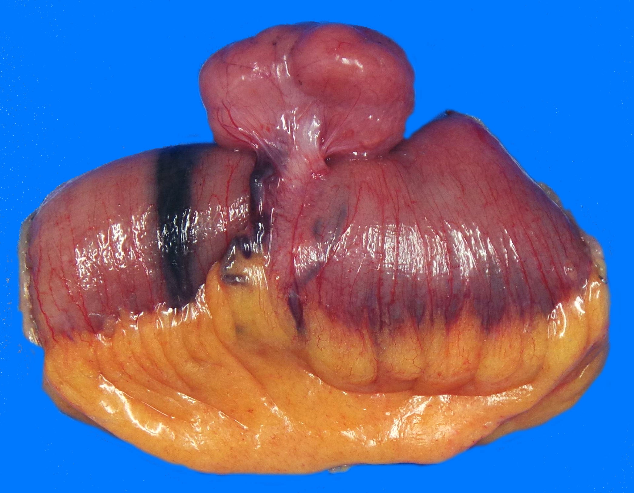
Small bowel mass

Macroscopic appearance
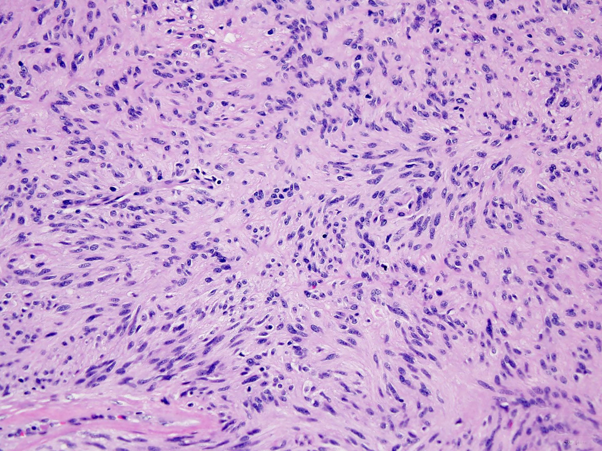
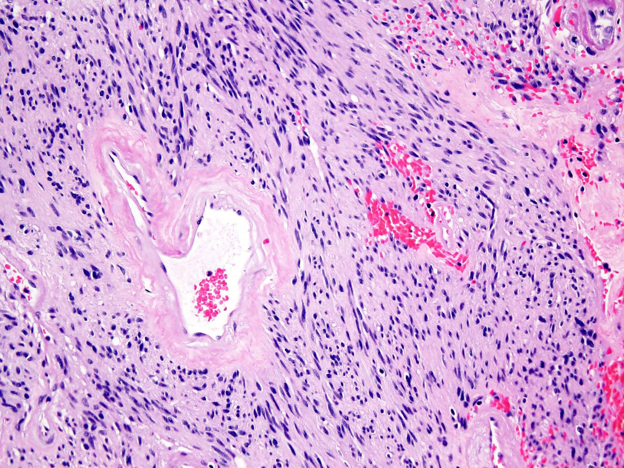
Microscopic (histologic) images
Contributed by Borislav A. Alexiev, M.D.
Spindle cell tumor
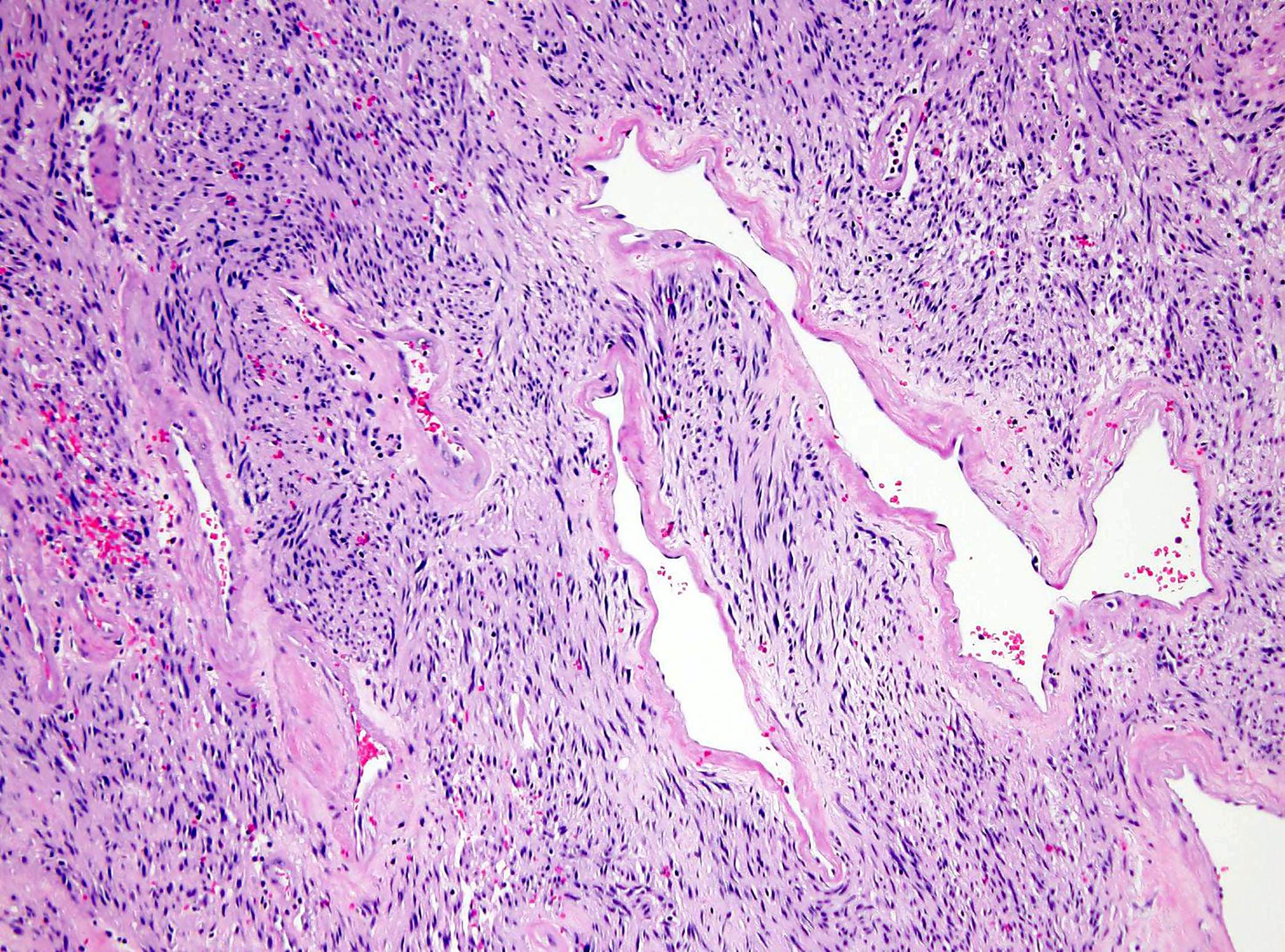
Regressive vascular changes
Board review style question #1
The gastrointestinal stromal tumor (GIST) staging protocol is required for which of the following procedures?
- Biopsy
- Cytologic specimen
- Local excision
- Primary resection specimen with no residual tumor
- Resection
Board review style answer #1
E. Resection. The staging protocol should be used for all resection specimens from patients with GIST, except primary resection specimen with no residual tumor.
Comment Here
Reference:
Staging-GIST Board review style question #2
Which is the correct pT for a gastric gastrointestinal stromal tumor (GIST) that is 12 cm?
- pT0
- pT1
- pT2
- pT3
- pT4
Board review style answer #2
E. pT4. pT4 is defined as a GIST that is > 10 cm in its greatest dimension.
Comment Here
Reference:
Staging-GIST

Back to top