Table of Contents
Definition / general | Etiology | Clinical features | Case reports | Treatment | Gross description | Gross images | Microscopic (histologic) description | Microscopic (histologic) images | Negative stainsCite this page: Shankar V. Myospherulosis. PathologyOutlines.com website. https://www.pathologyoutlines.com/topic/softtissuemyospherulosis.html. Accessed January 6th, 2025.
Definition / general
- Iatrogenic benign mass composed of fungi-like spherules that are actually erythrocytes damaged by endogenous and exogenous fat
Etiology
- Due to erythrocyte damage from endogenous and exogenous fat
- Also due to endogenous membranocystic degeneration of fat that occurs in lupus erythematosus and in membranous lipodystrophy with dermal atrophy due to local application of steroid ointment (Arch Dermatol 1991;127:88)
- In the gluteal region, this entity is described in relation to old injections of petrolatum based hormones and penicillin (Diagn Cytopathol 1988;4:137, J Am Acad Dermatol 1989;21:400)
History:
- First described by McClatchie (E Afr Med J 1969;46:625), who reported 7 patients from Kenya with unusual soft tissue nodules in arm, legs and subcutaneous tissue of buttock
- Called myospherulosis due to the involvement of skeletal muscle in some patients (Am J Clin Pathol 1969;51:699)
- Five patients were subsequently reported by Hutt in Uganda (Trans R Soc Trop Med Hyg 1971;65:182)
- Initially these structures were thought to be a fungus, but the usual stains for fungi were negative
- Kyriakos (Am J Clin Pathol 1977;67:118) reported non African cases in paranasal sinuses, nasal cavity and middle ear; most patients had undergone surgery and the surgical wound was packed with gauze impregnated with petrolatum and tetracycline ointment, suggesting an iatrogenic etiology
- De Schriver and Kyriakos confirmed this etiology by inducing similar lesions in experimental animals (Am J Pathol 1977;87:33)
- Rosai (Am J Clin Pathol 1978;69:475) and Wheeler (Arch Otolaryngol 1980;106:272) demonstrated that the spherules were erythrocytes damaged by endogenous and exogenous fat
- Travis (Arch Pathol Lab Med 1986;110:763) and Shimada (Am J Surg Pathol 1988;12:427) confirmed the presence of damaged erythrocytes by immunostaining for hemoglobin
- Kakizaki (Am J Clin Pathol 1993;99:249) demonstrated that the wall of the spherules was due to the physical emulsion phenomenon that occurs between lipid containing materials and blood
- The damaged erythrocytes are enclosed by a lipid membrane and later phagocytosed by histiocytes as part of the lipogranulomatous reaction that takes place in adipose tissue
Clinical features
- May occur with aspergillosis of the maxillary sinus (Oral Sur Oral Med Oral Pathol 1987;63:582, Arch Pathol Lab Med 2005;129:e84)
Case reports
- 39 year old man with large buttock tumor (Case of the Week #173)
- 60 year old man with retroperitoneal tumor (Asian J Neurosurg 2010;5:91)
Treatment
Treatment and prognosis:
- Benign process; no treatment needed other than for symptomatic relief
Gross description
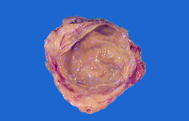
- Large saccular cyst-like lesion, surrounded by fat
- May contain oily substance with a yellow color
Microscopic (histologic) description
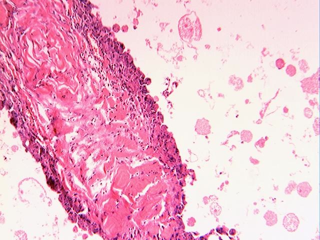
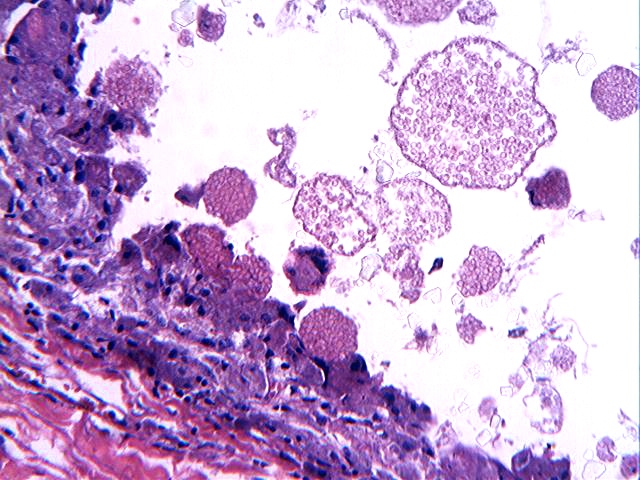
- Cyst composed of spherules which are damaged erythrocytes
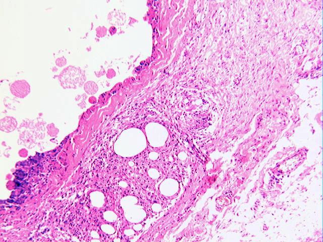
- Wall of the cyst made of fibrous tissue, accompanied by a lipogranulomatous reaction
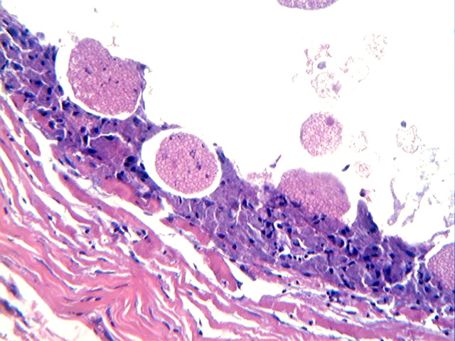
- Many eosinophilic spherules containing red blood cells are within histiocytes lining the cyst wall
- Some larger spherules resemble a bag of marbles
- Dermal nodules are cystic cavities with a fibrous wall lined by histiocytes and multinucleated foreign body giant cells, with lipogranulomatous inflammation in the adipose tissue adjacent to the cavities


