Table of Contents
Definition / general | Case reports | Clinical images | Gross images | Microscopic (histologic) description | Microscopic (histologic) images | Differential diagnosisCite this page: Shankar V. Extraskeletal osteosarcoma. PathologyOutlines.com website. https://www.pathologyoutlines.com/topic/softtissueeskosteo.html. Accessed December 28th, 2024.
Definition / general
- Adults, extremities
- May occur after Xray exposure
- 60% mortality, worse than chondrosarcoma
- Subtypes: osteoblastic, chondroblastic, fibroblastic, MFH-like, telangiectactic, well-differentiated (parosteal)
Case reports
- 30 year old man with chest wall mass (BMC Cancer 2010;10:645)
- 56 year old man with cutaneous tumor on scar of previous bone graft (Ann Dermatol 2011;23:S160)
- 62 year old woman with well-differentiated tumor arising from retroperitoneum that recurred as anaplastic spindle cell sarcoma (Case Rep Med 2010;2010:327591)
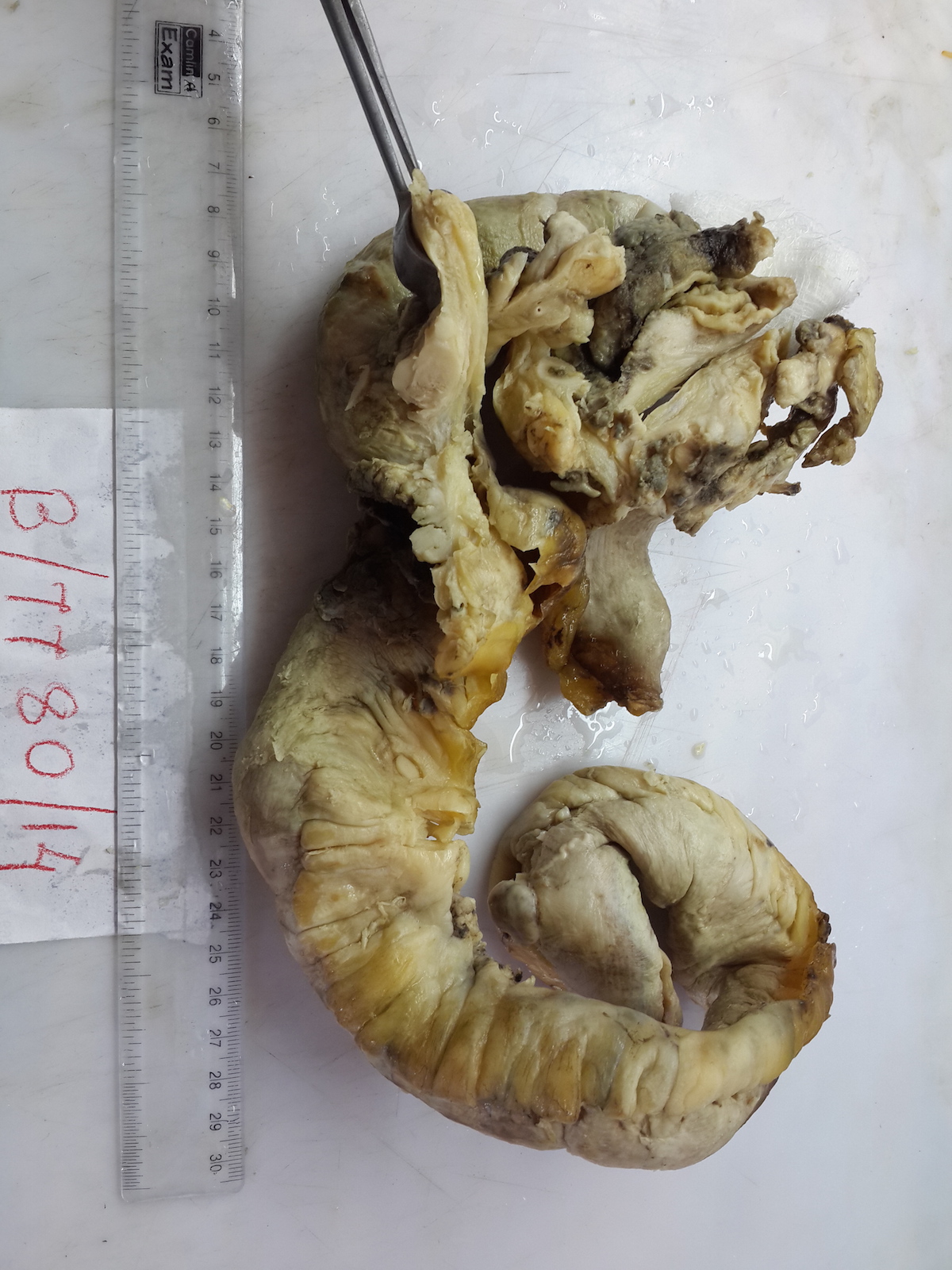
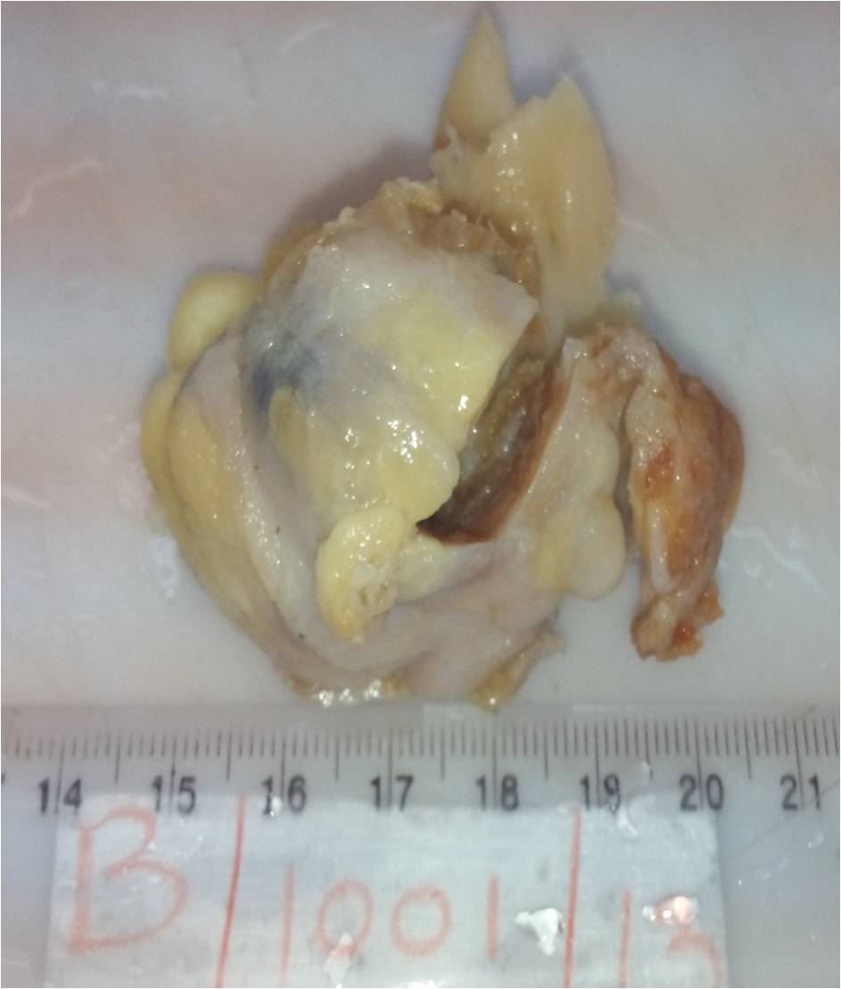
- 75 year old man with acute intestinal perforation and 55 year old woman with gluteal mass (Case #370)
- 79 year old man with upper arm tumor (Oncol Lett 2011;2:75)
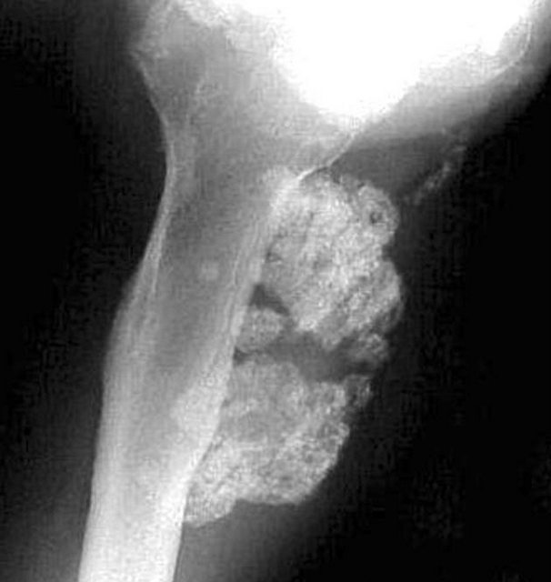
Clinical images
Gross images
Microscopic (histologic) description
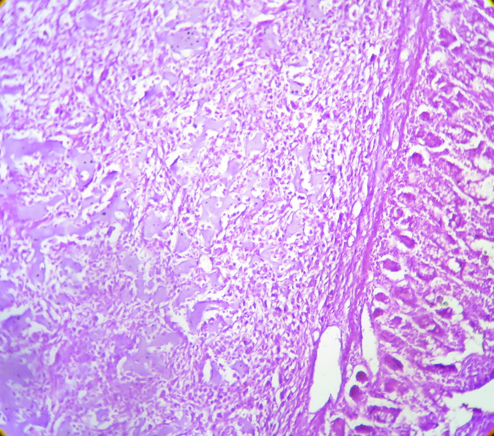
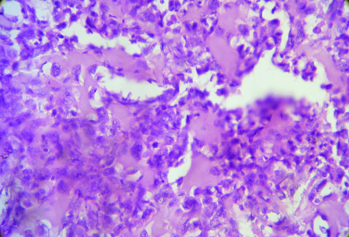
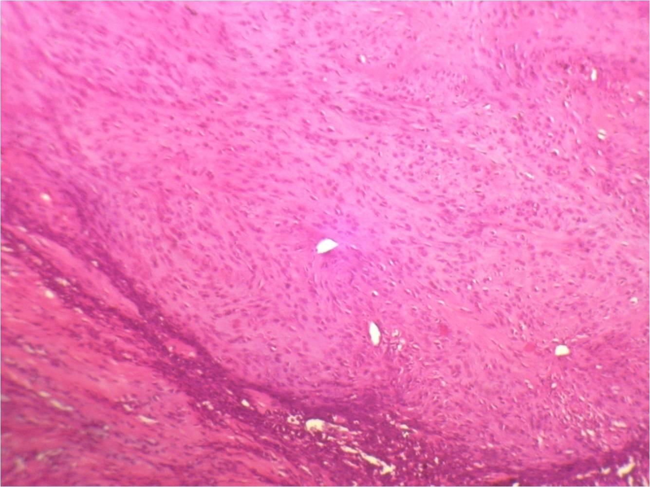
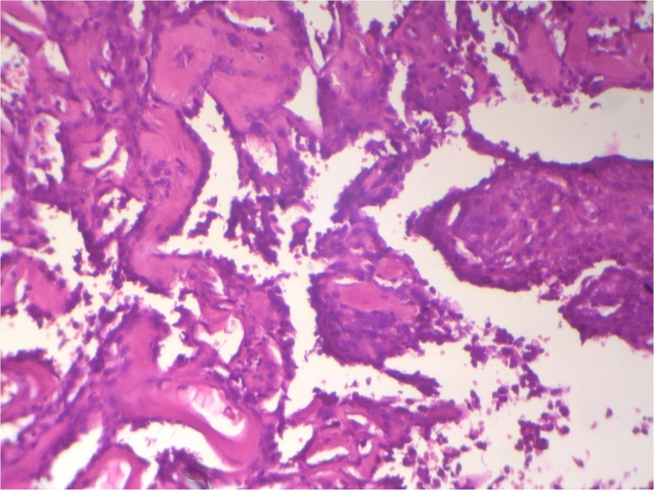
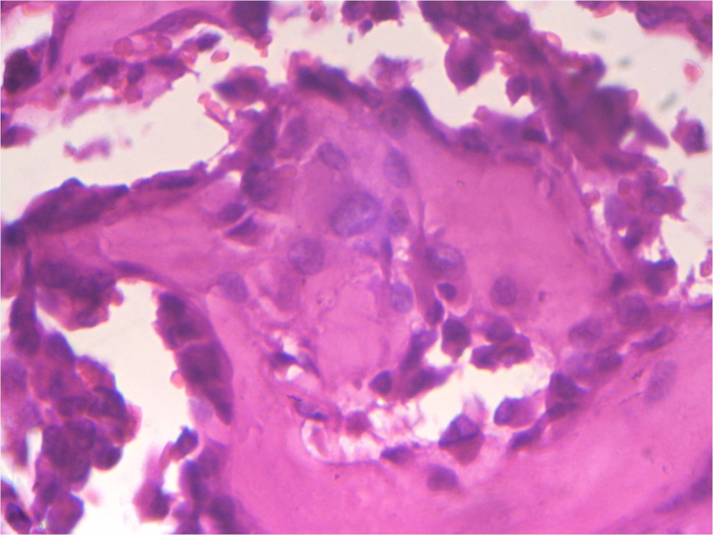
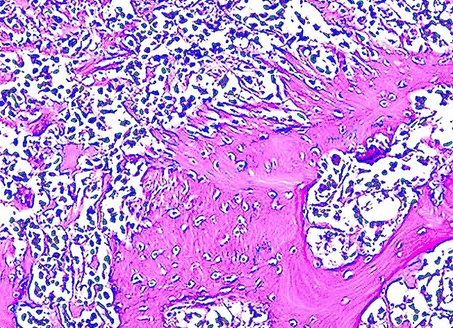
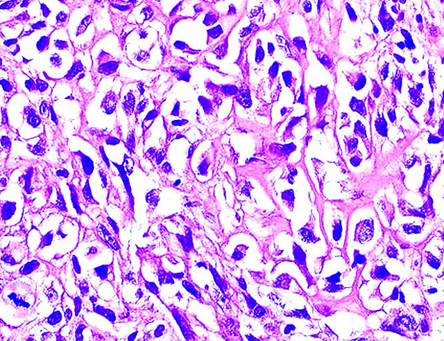
- Osteoid and bone formation produced by tumor cells, without interposition of cartilage
Microscopic (histologic) images
Differential diagnosis
- Myositis ossificans: no nuclear atypia, zonal
- Other sarcomas producing metaplastic bone: MFH, synovial sarcoma, fibrosarcoma


