
Small intestine & ampulla
Carcinoma
Neuroendocrine carcinoma
Author: Hanni Gulwani, M.B.B.S.

Last author update: 1 August 2012
Last staff update: 19 November 2020
Copyright: 2003-2025, PathologyOutlines.com, Inc.
PubMed Search: Neuroendocrine carcinoma[TIAB] small bowel


Table of Contents
Definition / general | Case reports | Clinical features | Microscopic (histologic) description | Microscopic (histologic) images | Electron microscopy descriptionCite this page: Gulwani H. Neuroendocrine carcinoma. PathologyOutlines.com website. https://www.pathologyoutlines.com/topic/smallbowelNECarcinoma.html. Accessed March 31st, 2025.
Definition / general
- Usually fatal
Case reports
- 50 year old man with obstructive jaundice with an ampullary mass: collision tumor-mixed adenoneuroendocrine carcinoma (Case #322)
- 52 year old man with collision tumor of primary adenocarcinoma and neuroendocrine carcinoma of duodenum (Rare Tumors 2012;4:e20)
- 55 year old woman with large cell neuroendocrine carcinoma with glandular differentiation (J Clin Pathol 2004;57:1098)
- 73 year old woman with large cell neuroendocrine carcinoma with squamous cell and glandular components (Jpn J Clin Oncol 2011;41:434)
- 74 year old man with small cell neuroendocrine carcinoma with villous adenoma (World J Gastroenterol 2008;14:4709)
- 74 year old with large cell neuroendocrine carcinoma (Arch Pathol Lab Med 2003;127:221)
Clinical features
- Ampullary tumors are rare, present with progressing jaundice
- Aggressive with poor prognosis (Hepatobiliary Pancreat Dis Int 2008;7:422)
- Small cell carcinoma is rare; in few cases reported, prognosis better than for small cell lung tumors (Arch Pathol Lab Med 2003;127:e357, J Clin Oncol 2004;22:2730, J Clin Pathol 2010;63:620)
Microscopic (histologic) description
- Marked pleomorphism, large irregular hyperchromatic nuclei, prominent nucleoli, tumor necrosis, frequent mitotic figures
- Large cell carcinoma:
- Islands and trabeculae of large cells with brisk mitotic activity and extensive necrosis
- Cells have more cytoplasm than small cell carcinoma, irregular chromatin, frequent nucleoli
- Small cell carcinoma:
- Sheets and nests of small, round cells with scanty cytoplasm, hyperchromatic nuclei, stippled chromatin, indistinct nucleoli, numerous mitotic figures and apoptotic cells
- Foci of necrosis and vascular invasion common
- Resembles pulmonary tumor
- Pure or mixed with adenocarcinoma
Microscopic (histologic) images
Case #322
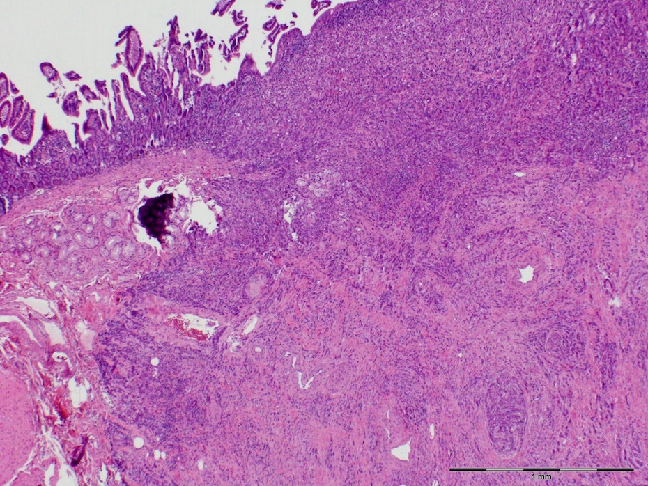

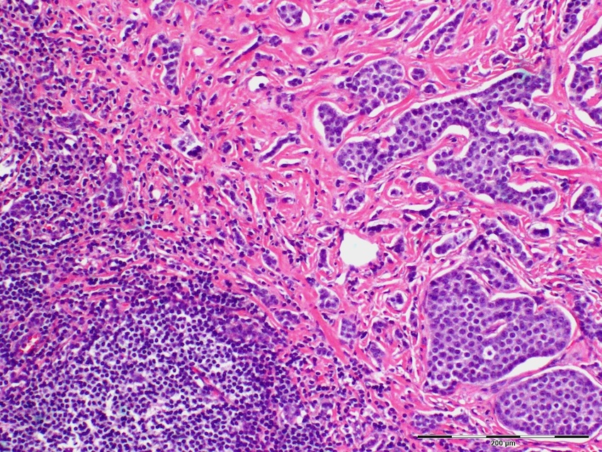
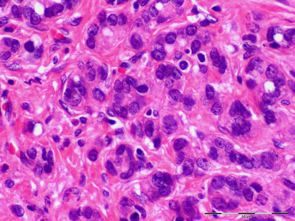


Mixed adenoneuroendocrine carcinoma
Line 2: CDX2 (left); chromogranin (middle); synaptophysin (right)
Images hosted on other servers:


Large cell neuroendocrine carcinoma

Small cell neuroendocrine carcinoma

Nests of small cells
with uniform round
nuclei with classic salt
and pepper chromatin
Electron microscopy description
- Small cell carcinoma: membrane bound dense core granules
