
Skin nonmelanocytic tumor
Adnexal tumors
Follicular derived
Trichofolliculoma
Editorial Board Member: Jonathan D. Ho, M.B.B.S., D.Sc.


Last author update: 8 February 2023
Last staff update: 8 February 2023
Copyright: 2002-2025, PathologyOutlines.com, Inc.
PubMed Search: Trichofolliculoma [title]

Table of Contents
Definition / general | Essential features | ICD coding | Epidemiology | Sites | Pathophysiology | Clinical features | Diagnosis | Prognostic factors | Case reports | Treatment | Clinical images | Microscopic (histologic) description | Microscopic (histologic) images | Virtual slides | Positive stains | Negative stains | Molecular / cytogenetics description | Videos | Sample pathology report | Differential diagnosis | Additional references | Board review style question #1 | Board review style answer #1 | Board review style question #2 | Board review style answer #2Cite this page: Asadbeigi SN, Nguyen C. Trichofolliculoma. PathologyOutlines.com website. https://www.pathologyoutlines.com/topic/skintumornonmelanocytictrichofolliculoma.html. Accessed April 1st, 2025.
Definition / general
- Benign adnexal hamartomatous follicular tumor
- Histologically shows multiple follicles in various stages spreading from a central cystic follicle
Essential features
- Solitary papule in adults in the head and neck region
- Central dilated primary follicle with secondary follicles budding from the primary follicle
- Can show a spectrum of morphology depending on the hair follicle cycle
ICD coding
Epidemiology
- Mostly solitary papule or nodule
- Occurs in adulthood with no definitive racial or gender predilection (An Bras Dermatol 2015;90:519)
- Rarely occurs as a congenital lesion (Int J Mol Sci 2021;22:4759)
- No proven association with any dermatological or systemic diseases (Am J Pathol 1976;85:479)
Sites
- Face, with nose as the most common site (Am J Dermatopathol 2010;32:35)
Pathophysiology
- Repeated development of hair follicles with disordered hair cycle; defective sonic hedgehog polarization (J Dermatol 2017;44:1050, Am J Dermatopathol 2009;31:248)
- Distorted ability to control the size of hair follicles
- Trichofolliculoma with sebaceous differentiation: follicular and sebaceous components have independent cycles
- Primary follicle: (J Dermatol 2017;44:1050)
- Primary infundibular cystic structure which shows a thin wall with radiating secondary follicles
- Secondary follicles:
- Follicles radiating from the primary follicle
- Most follicles are in anagen phase
- Tertiary follicles:
- Regression of secondary follicles to tertiary follicles
- Shift of anagen hair to catagen phase
- Variation in size of hair from vellus hair to thick terminal hair
- Quaternary follicles:
- Regression of tertiary follicles to quaternary follicles
Clinical features
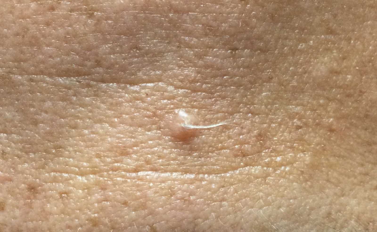
- Solitary, skin colored papule or nodule (approximately 0.2 - 1.5 cm in diameter) with central depression (An Bras Dermatol 2015;90:780, Arch Dermatol 1960;81:922)
- Multiple tufts of vellus and thin hairs emerging from the central section
Diagnosis
- Clinical: central primary follicle with multiple tufts of vellus hair (Am J Pathol 1976;85:479)
- If the hair is plucked, trichofolliculoma can be clinically misdiagnosed as basal cell carcinoma, molluscum contagiosum, keratoacanthoma, milium, trichoepithelioma, syringoma or sebaceous hyperplasia (J Eur Acad Dermatol Venereol 2017;31:e123)
- Dermoscopy: shows troll hair sign - tight plumes of white and thin hairs, similar to children's troll dolls (Australas J Dermatol 2021;62:90)
- Biopsy and histological examination
Prognostic factors
- Excellent prognosis (Dermatol Online J 2013;19:19264)
- Rarely recurs at the primary site (Arch Dermatol 1979;115:1003)
- Rarely coexists with basal cell carcinoma (Australas J Dermatol 2007;48:127)
- Malignant transformation with perineural invasion has been reported in a single case report (Arch Dermatol 1979;115:1003)
Case reports
- 15 year old girl with a hairy papule on nose since birth (Dermatol Online J 2020;26:13030)
- 21 year old woman with multiple skin colored lesions on face (Indian J Dermatol 2015;60:214)
- 52 year old woman with a collision tumor of trichofolliculoma and basal cell carcinoma (Australas J Dermatol 2007;48:127)
- 54 year old woman with multiple soft lesions in zosteriform distribution (Indian J Dermatol 2013;58:330)
- 57 year old man with a tender nodule on canthus (Arch Dermatol 1979;115:1003)
Treatment
- Typically no need for treatment but rarely excision is recommended (Int J Mol Sci 2021;22:4759)
Clinical images
Contributed by Sepideh Nikki Asadbeigi, M.D.
Trichofolliculoma
Images hosted on other servers:

Papule with white vellus hair
Microscopic (histologic) description
- Dilated central cystic follicle with surrounding multiple fully formed vellus or terminal follicles
- The central cystic follicle shows connection / opening to epidermis
- Early lesion: a mildly dilated infundibulum and radiating secondary curved vellus follicles; cystic dilatation may be absent
- Late lesion: thin walled primary infundibular cystic structure and radiating vellus or terminal follicles that are mostly in the anagen phase
- Trichofolliculoma demonstrates outer root sheath differentiation
- Central follicle shows stratified squamous cell epithelium with a granular layer with dilation or cystic changes and contains keratinous material and may have vellus hairs
- Primary follicle has keratinized stratified epithelium with keratohyaline granules
- Branched follicles may show varying degree of maturation, including rudimentary structures or epithelial cords and anagen, catagen or telogen hair in older lesions
- Secondary follicles are small with many epithelial strands and abortive pilar formation
- Sebaceous differentiation may be present
- Trichofolliculoma is usually surrounded by well developed connective tissue, which is frequently cellular
- Each follicle is surrounded by an individual perifollicular sheath
- Late stage can show a solid pattern as the regressing secondary follicles and developing tertiary follicles coalesce
- Sebaceous trichofolliculoma variant: sebaceous gland attached to radiating follicles (J Cutan Pathol 1980;7:394)
Microscopic (histologic) images
Contributed by Sepideh Nikki Asadbeigi, M.D.
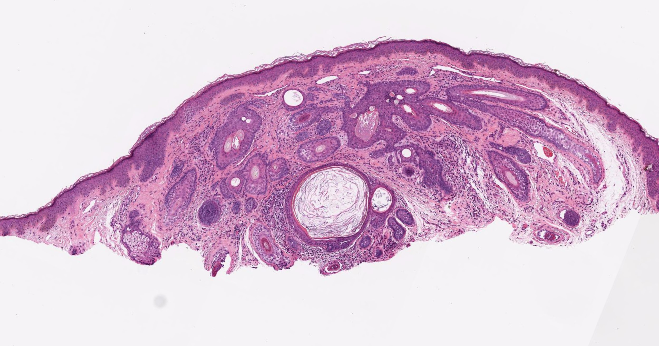
Primary and secondary follicles
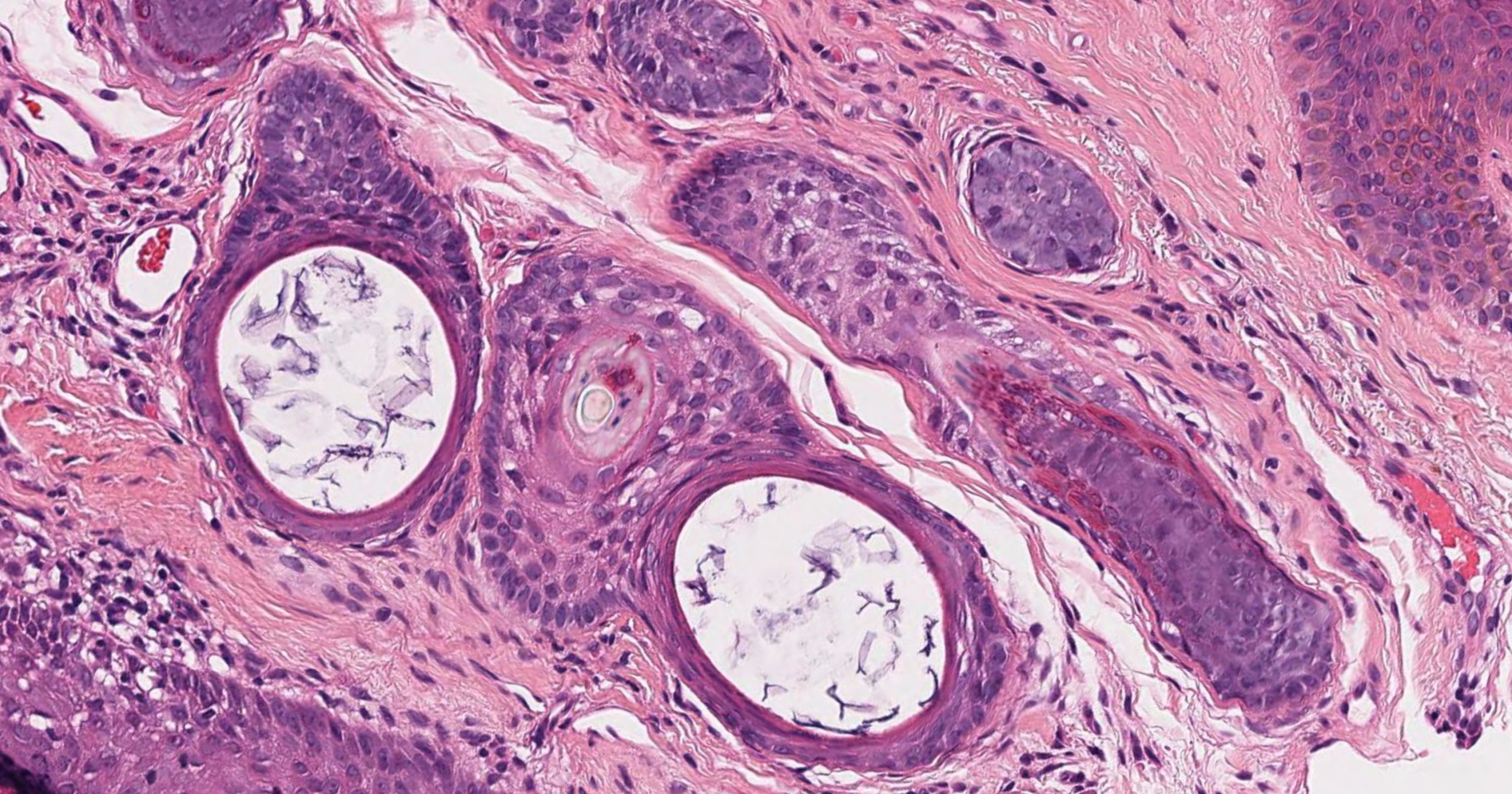
Secondary follicles
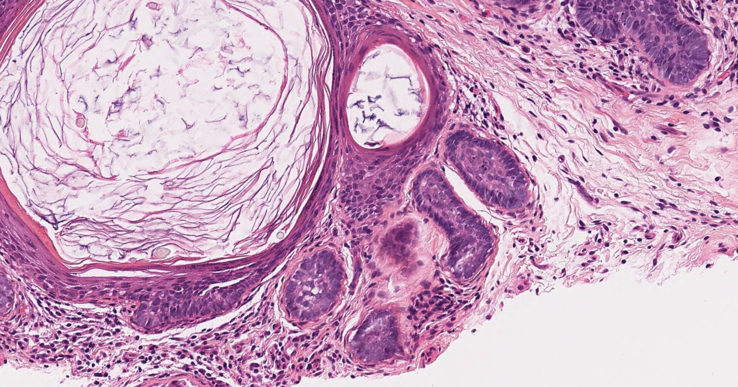
Primary cystic follicle
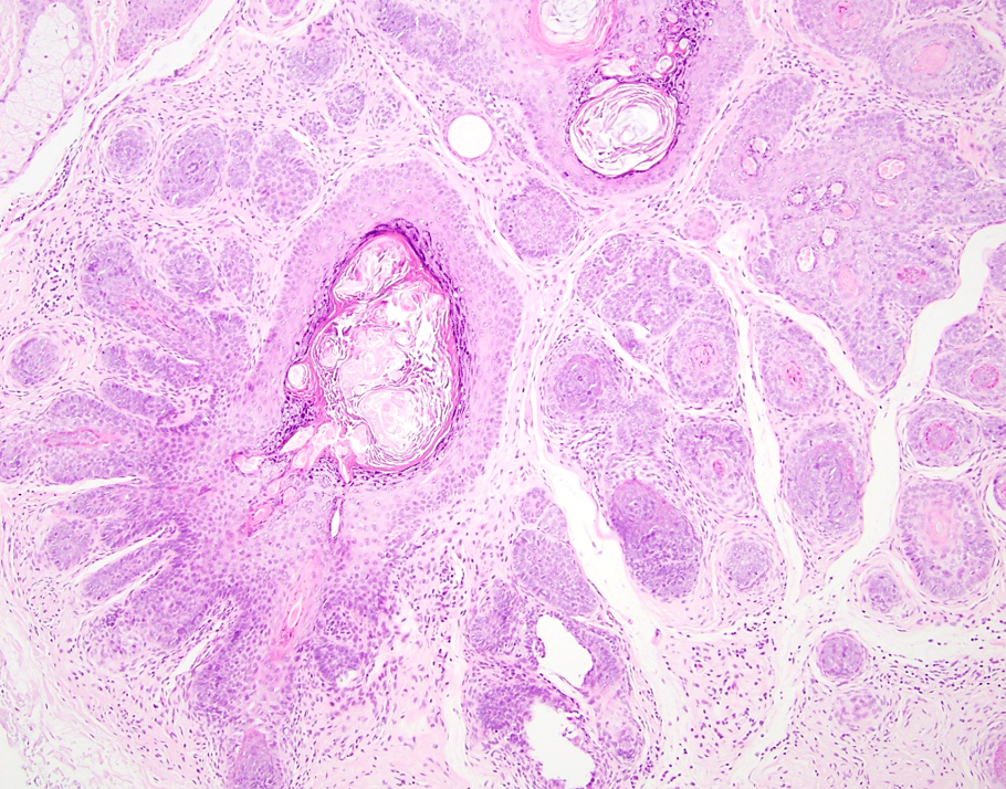
Dilated hair follicle
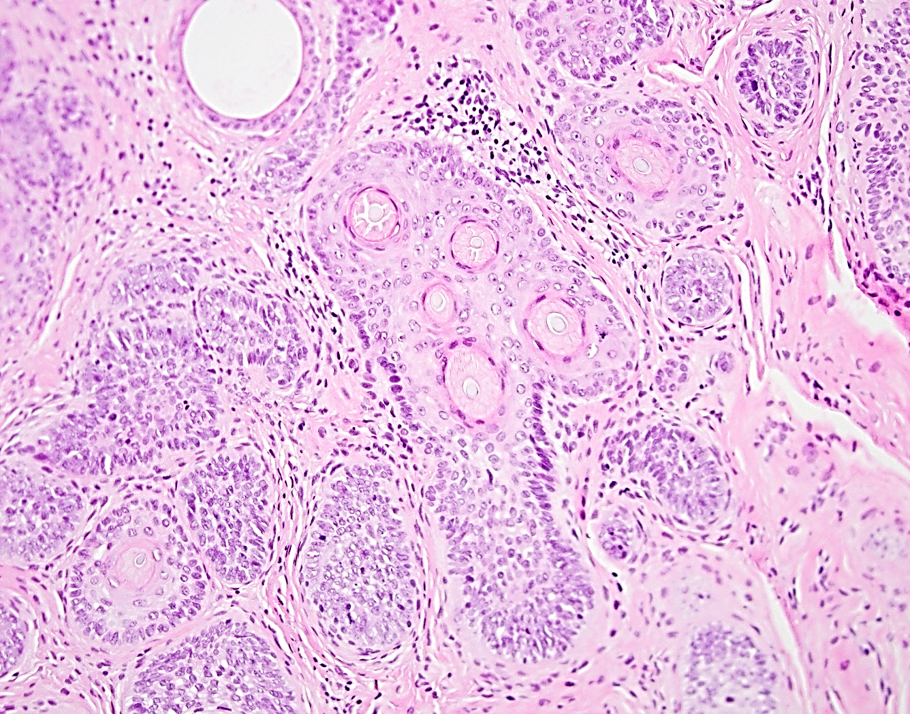
Secondary hair follicle
Virtual slides
Images hosted on other servers:

Trichofolliculoma with sebaceous differentiation
Positive stains
- Immunohistochemistry is not usually needed for diagnosis
- CK15 in the basal cells of the secondary follicles (Br J Dermatol 2003;148:597)
- CK16 and CK17 in the suprabasal cells of the immature secondary hair follicles (Br J Dermatol 2003;148:597)
- BerEP4 expression in basaloid germ-like structures but weak staining in the secondary or tertiary follicles compared to the normal hair (Am J Dermatopathol 2010;32:35)
- CD34 in basal cells of the outer root sheath in the thick terminal follicles (Am J Dermatopathol 2010;32:35)
Negative stains
Molecular / cytogenetics description
- BMP and PYGO2 signaling pathway in experimental studies; not used in clinical setting (Int J Mol Sci 2021;22:4759)
Videos
Trichofolliculoma clinical and pathology by Dr. Michael Lee
Trichofolliculoma histopathology by Dr. Jerad Gardner
Sample pathology report
- Forehead skin, shave biopsy:
- Trichofolliculoma (see comment)
- Comment: Atypia is not identified.
- Microscopic description: Sections demonstrate a centrally dilated follicular unit with multiple radiating follicular units. At the center of the cystic structure there is keratotic debris and immature hair shafts (trichoids). Surrounding the structure is a mantle of well organized connective tissue. Atypical features were not noted.
Differential diagnosis
- Trichoadenoma:
- Multiple multilayered squamous epithelial islands with a central cystic cavity containing keratinous material
- Does not contain hair shafts
- Solitary trichoepithelioma:
- Islands of basaloid cells are present, which is not a feature of trichofolliculoma
- Fibrofolliculoma:
- Both show a large central follicle with multiple arising epithelial attachments
- Fibrofolliculoma only has strands of follicular epithelium and does not contain any hair shafts
- Dilated pore of Winer:
- Widened follicular infundibulum
- Unlike trichofolliculoma, it does not show the radiating secondary follicles
- Hair follicle nevus:
- Both lesions show multiple hair follicles with vellus hairs but trichofolliculoma has a central cystic component
- Follicular infundibulum cyst:
- Does not contain the secondary or tertiary follicular structures
- Pilar sheath acanthoma:
- Less florid pattern than trichofolliculoma
- Dilated central follicle with cystic features and radiating acanthotic epithelium
- Unlike trichofolliculoma, pilar sheath acanthoma does not have hair shafts in the peripheral buds
- Folliculosebaceous cystic hamartoma:
- Folliculosebaceous structures with surrounding stroma with various mesenchymal elements
- Sebaceous component is the more prominent component
- Although it has been suggested that this is a late stage of sebaceous trichofolliculoma, reports of congenital folliculosebaceous cystic hamartoma suggest that this idea is incorrect (Am J Dermatopathol 2008;30:500, J Cutan Pathol 2008;35:843)
Additional references
Board review style question #1
A 50 year old Caucasian woman presents with a solitary flesh colored papule with a central depression. What is the diagnosis?
- Dilated pore of Winer
- Pilar sheath acanthoma
- Trichoepithelioma
- Trichofolliculoma
Board review style answer #1
Board review style question #2
The biopsy of a skin colored papule on the face of a 34 year old man shows a central dilated follicle with smaller radiating follicular units containing hair shafts. What is the most accurate diagnosis?
- Hair follicle nevus
- Pilar sheath acanthoma
- Trichoadenoma
- Trichofolliculoma
Board review style answer #2
