Table of Contents
Definition / general | Case reports | Treatment | Clinical images | Gross description | Microscopic (histologic) description | Microscopic (histologic) images | Positive stains | Additional referencesCite this page: Hale CS. Leukemia cutis. PathologyOutlines.com website. https://www.pathologyoutlines.com/topic/skintumornonmelanocyticleukemias.html. Accessed April 3rd, 2025.
Definition / general
- Skin involvement ("leukemia cutis") occurs in 5% with CML, 8% with CLL, 10% with monocytic leukemia
- Myeloid leukemia with monocytic differentiation more commonly involves the skin than other types of myeloid leukemia
- Usually is abnormal peripheral blood count at diagnosis
- Skin involvement is rarely initial manifestation of recurrence (Am J Clin Pathol 2008;129:130)
- May also have accompanying vasculitis (Am J Clin Pathol 1997;107:637)
- Aggressive behavior and short survival (J Am Acad Dermatol 1999;40:966)
Case reports
- 68 year old woman with history of AML (Case of Week #140)
Treatment
- Systemic chemotherapy directed at eradicating the leukemic clone
Gross description
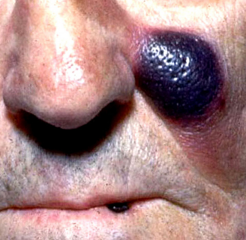
- Multiple nodules / papules
Microscopic (histologic) description
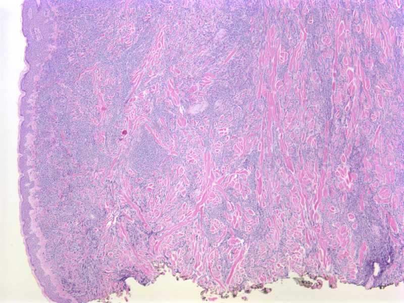
- In CLL, may be perivascular, periadnexal, nodular or band-like dermal infiltrate
- Infiltrate in leukemic patients is often NOT neoplastic, but reactive
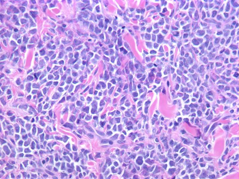
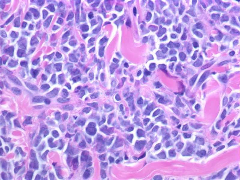
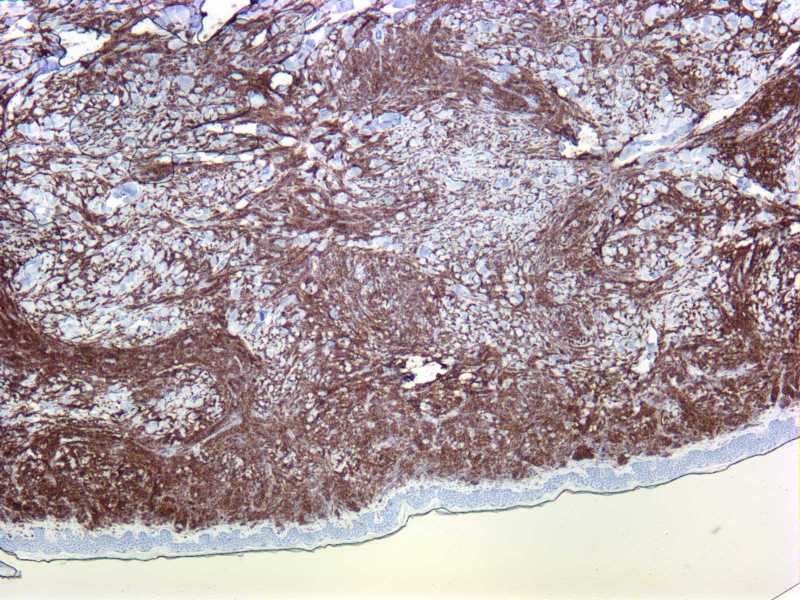
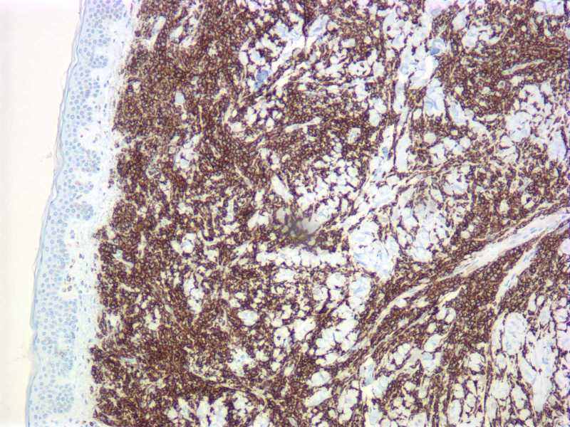
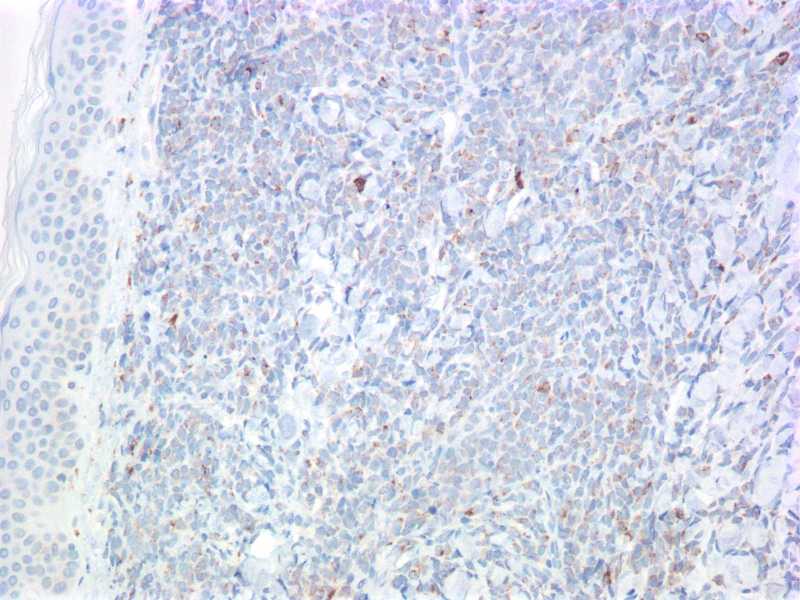
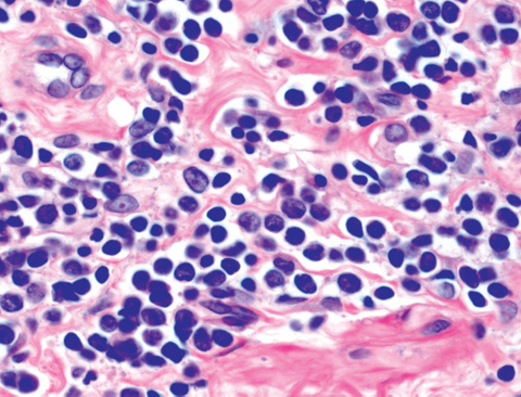
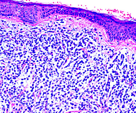
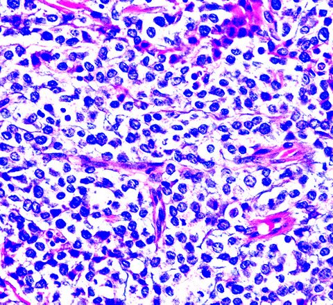
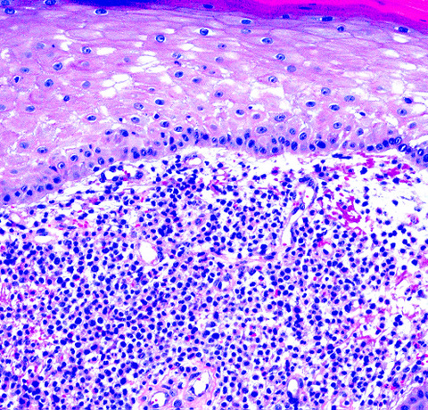
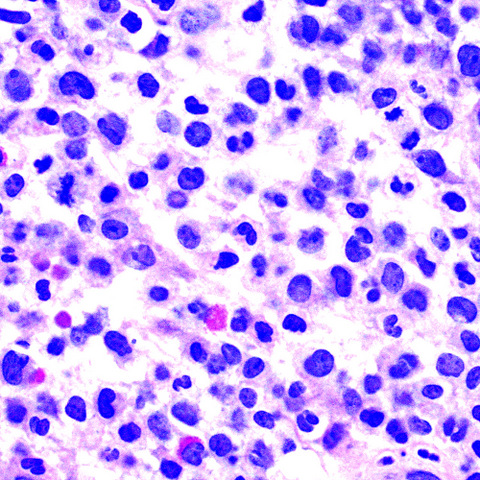
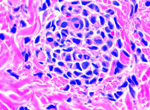
- AML: dermis and superficial subcutaneous fat are diffusely infiltrated by a monotonous population of large cells with a high nuclear to cytoplasmic ratio, round to slightly irregular nuclear contours, finely dispersed chromatin and prominent nucleoli
Microscopic (histologic) images
Positive stains
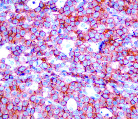
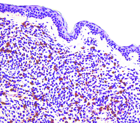
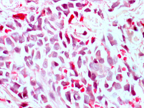
- Myeloblasts: chloroacetate esterase (Leder stain), myeloperoxidase
Additional references


