
Skin nonmelanocytic tumor
Benign (nonmelanocytic) epidermal tumors or tumor-like lesions
Large cell acanthoma
Board of reviewers: Caroline I. Mullins, M.D.

Last author update: 21 November 2024
Last staff update: 13 December 2024
Copyright: 2002-2025, PathologyOutlines.com, Inc.
PubMed Search: Large cell acanthoma

Table of Contents
Definition / general | Essential features | ICD coding | Epidemiology | Sites | Pathophysiology | Etiology | Clinical features | Diagnosis | Prognostic factors | Case reports | Clinical images | Microscopic (histologic) description | Microscopic (histologic) images | Virtual slides | Videos | Sample pathology report | Differential diagnosis | Additional references | Board review style question #1 | Board review style answer #1 | Board review style question #2 | Board review style answer #2Cite this page: Almohamedi RS, Saeed Kamil Z. Large cell acanthoma. PathologyOutlines.com website. https://www.pathologyoutlines.com/topic/skintumornonmelanocyticlargecellacanthoma.html. Accessed April 2nd, 2025.
Definition / general
- Large cell acanthoma is a benign, intraepidermal proliferation of enlarged keratinocytes (J Cutan Pathol 2014;41:733)
- First reported by Pinkus in 1970 (Dermatol Pract Concept 2014;4:43)
Essential features
- Sharply demarcated lesion from adjacent normal skin
- Enlarged keratinocytes (2 times larger than adjacent normal keratinocytes) with no cytologic atypia
- Basal hyperpigmentation with pigment incontinence in superficial dermis
- Large cell acanthoma morphologically overlaps with solar lentigo (J Cutan Pathol 2014;41:733)
- Classification of large cell acanthoma as a variant of solar lentigo with cellular hypertrophy is debated (J Cutan Pathol 2014;41:733)
ICD coding
- ICD-O: 8072/0 - large cell acanthoma
- ICD-11: 2F21.Y & XH7AQ2 - other specified benign keratinocytic acanthoma & large cell acanthoma
Epidemiology
- More common in fair skinned individuals (Fitzpatrick skin types I - III) (Int J Dermatol 2003;42:36)
- Mainly affects middle aged and elderly adults (J Cutan Pathol 2014;41:733)
Sites
- Usually sun exposed skin is affected (Ocul Oncol Pathol 2019;5:312)
- Predilection for the head / neck and extremities (J Cutan Pathol 2014;41:733, Ocul Oncol Pathol 2019;5:312)
Pathophysiology
- There is significant overlap between large cell acanthoma and solar lentigo in terms of keratin expression as well as anti-apoptotic and proliferative proteins (J Cutan Pathol 2014;41:733)
Etiology
- Large cell acanthoma is associated with chronic ultraviolet (UV) exposure
Clinical features
- Clinically presents as a slow growing, well circumscribed, tan to brown macule / plaque measuring < 10 mm (Ocul Oncol Pathol 2019;5:312, Dermatol Pract Concept 2014;4:43)
Diagnosis
- Biopsy
Prognostic factors
- Benign tumor with no risk for recurrence or metastasis
- No malignant transformation has been reported to date (Ocul Oncol Pathol 2019;5:312)
Case reports
- 10 year old boy with large cell acanthoma (Int J Dermatol Venereol 2023;10:1097)
- 49 year old man with a conjunctival lesion with similar histological features of large cell acanthoma of the skin (J Cutan Pathol 2010;37:1103)
- 62 year old man with multiple disseminated large cell acanthoma of the skin associated with HPV6 (J Am Acad Dermatol 2005;53:335)
- 70 year old Asian woman with a growing white spot inside of the lower eyelid (Ocul Oncol Pathol 2019;5:312)
- 76 year old man with an elevated, flat skin lesion on left shoulder blade (Dermatology Review 2024;111:140)
Clinical images
Images hosted on other servers:

Sharply demarcated brown macule on the arm

Sharply demarcated brown macule on the cheek
Microscopic (histologic) description
- Sharply demarcated lesion from adjacent normal epidermis (J Cutan Pathol 2014;41:733)
- Large cell acanthoma is characterized by
- Mild acanthosis (Am J Dermatopathol 1992;14:140)
- Epidermal flattening
- Basketweave hyperkeratosis
- Enlarged polygonal keratinocytes (Ocul Oncol Pathol 2019;5:312)
- Hypergranulosis (Am J Dermatopathol 1992;14:140)
- Characteristics of the nucleus of the enlarged keratinocytes
- Twice the size of a normal keratinocyte (J Cutan Pathol 2014;41:733)
- Smooth and round nuclear contours (Am J Dermatopathol 1992;14:140)
- Prominent nucleoli (Ocul Oncol Pathol 2019;5:312)
- Lack of nuclear pleomorphism
- Rare mitotic figures
- Basal layer hyperpigmentation is frequently observed (Ocul Oncol Pathol 2019;5:312)
Microscopic (histologic) images
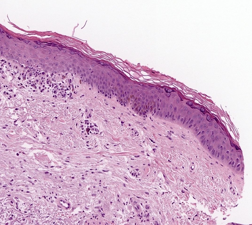
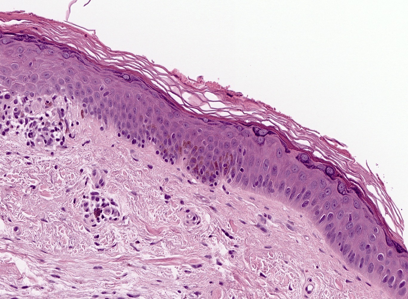
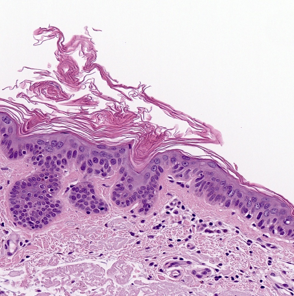
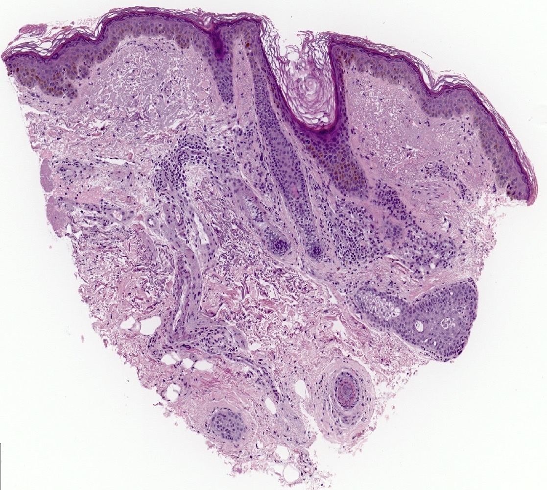
Contributed by Zaid Kamil, M.D. and Razan Saleh Almohamedi, M.D.
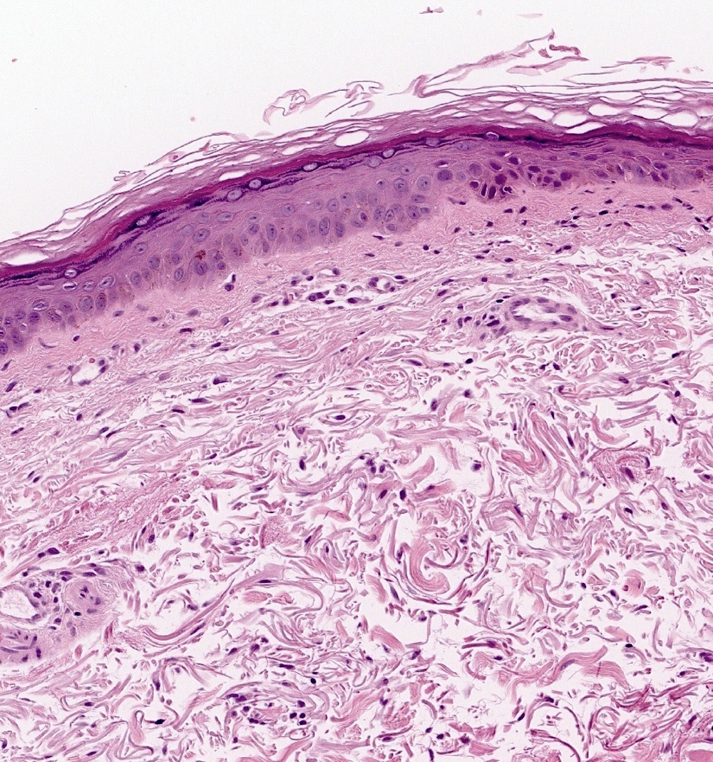
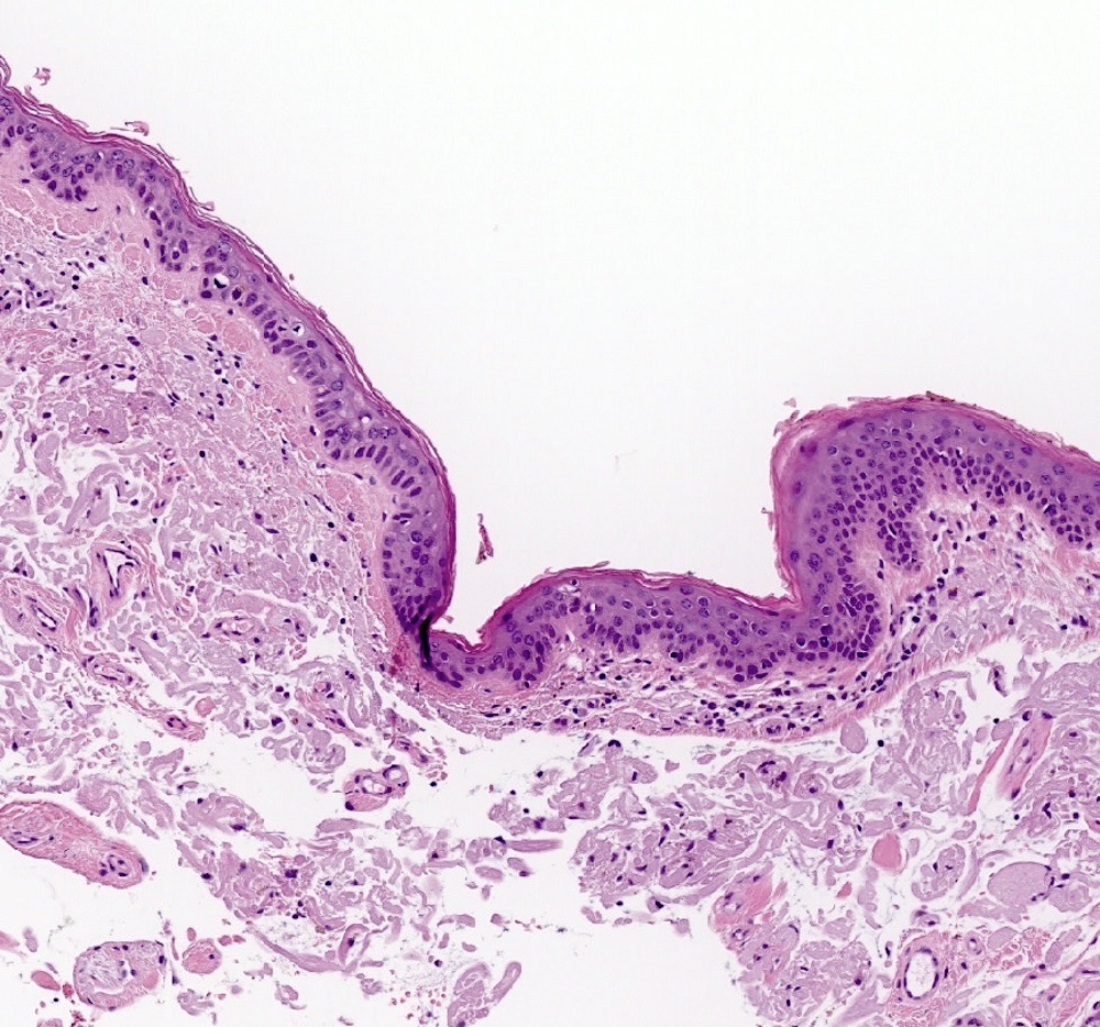
Abrupt large cell transition
Abrupt large cell transition
Large cells with hyperkeratosis
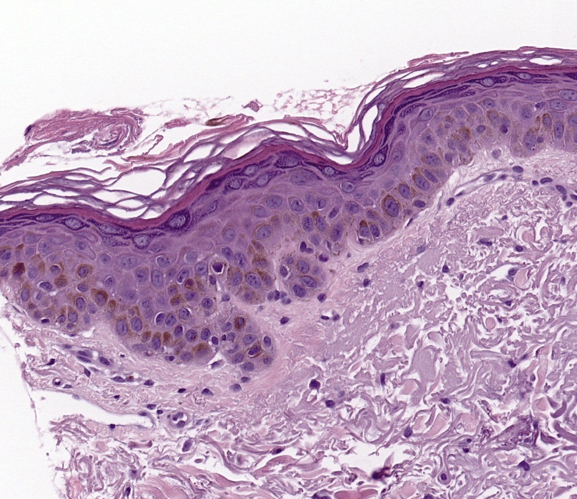
Large cells with basal hyperpigmentation
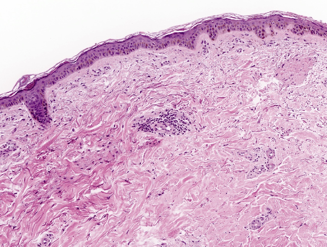
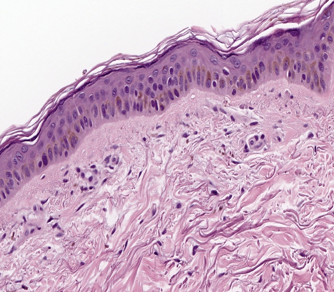
Large cells with basal hyperpigmentation
Virtual slides
Contributed by Zaid Kamil, M.D. and Razan Saleh Almohamedi, M.D.


Enlarged keratinocytes and basal hyperpigmentation
Videos
Large cell acanthoma by Dr. Jerad Gardner
Sample pathology report
- Skin, punch biopsy, left forearm:
- Large cell acanthoma (see comment)
- Comment: Sections show demarcated intraepidermal proliferation with mild acanthosis, hypergranulosis and enlarged keratinocytes. Basal pigmentation is seen. There is no evidence of atypical mitosis, nuclear pleomorphism or cytologic atypia. The overall features are of large cell acanthoma.
Differential diagnosis
- Clear cell acanthoma:
- Characterized by psoriasiform epidermal hyperplasia
- Keratinocytes have clear or pale cytoplasm that contain glycogen (StatPearls: Clear Cell Acanthoma [Accessed 16 October 2024])
- Keratinocytes are PAS positive (StatPearls: Clear Cell Acanthoma [Accessed 16 October 2024])
- Neutrophils within stratum corneum and dermal papillae
- Solar lentigo:
- Associated with basal hyperpigmentation, pigment incontinence and solar elastosis (J Cutan Pathol 2014;41:733)
- Elongated rete ridges
- Seborrheic keratosis:
- Characterized by pseudohorn cysts (J Dtsch Dermatol Ges 2023;21:265)
- Keratinocytes are small and without cytological atypia
- Actinic keratosis:
- Shows partial thickness keratinocyte atypia limited to the lower two - thirds of the epidermis
- Associated with parakeratosis and nuclear crowding (J Cutan Pathol 2014;41:733)
- Bowen's disease / squamous cell carcinoma in situ:
- Shows full thickness involvement of the epidermis by dysplastic squamous cells
- Associated with nuclear pleomorphism (J Cutan Pathol 2014;41:733)
Additional references
Board review style question #1
A 45 year old woman presented with a well circumscribed brown macule on the right forearm measuring 7 mm. A skin punch biopsy showed a demarcated intraepidermal proliferation with mild acanthosis, hypergranulosis and enlarged keratinocytes with nuclei twice the size of adjacent normal keratinocytes. No evidence of atypical mitosis, nuclear pleomorphism or squamous dysplasia. Based on the microscopic description and clinical presentation, what is the diagnosis?
- Actinic keratosis
- Clear cell acanthoma
- Large cell acanthoma
- Seborrheic keratosis
- Solar lentigo
Board review style answer #1
C. Large cell acanthoma is characterized by intraepidermal proliferation with mild acanthosis, hypergranulosis and enlarged keratinocytes with nuclei twice the size of adjacent normal keratinocytes. Answer E is incorrect because solar lentigo is associated with basal hyperpigmentation and dermal solar elastosis. Answer A is incorrect because actinic keratosis is characterized by atypia of the basal keratinocytes involving up to the mid epidermis. Answer D is incorrect because seborrheic keratosis is associated with acanthosis, papillomatosis, hyperkeratosis, keratin cysts and keratin pseudocysts. Answer B is incorrect because clear cell acanthoma is characterized by psoriasiform epidermal hyperplasia and keratinocytes with clear or pale cytoplasm containing glycogen, which is highlighted by PAS special stain.
Comment Here
Reference: Large cell acanthoma
Comment Here
Reference: Large cell acanthoma
Board review style question #2
Which of the following microscopic features is characteristic of large cell acanthoma?
- Basal hyperpigmentation and solar elastosis
- Full thickness involvement of the epidermis by dysplastic squamous cells
- Keratinocyte atypia of the lower and mid epidermis
- Keratinocyte nuclei that are twice the size as normal
- Psuedohorn cysts
Board review style answer #2
D. Keratinocyte nuclei that are twice the size as normal. Large cell acanthoma is characterized by large keratinocytes with a nucleus twice the size of a normal keratinocyte. Answer E is incorrect because psuedohorn cysts are a characteristic microscopic feature of seborrheic keratosis. Answer C is incorrect because keratinocyte atypia of the lower and mid epidermis is usually seen in actinic keratosis. There is no atypia or dysplasia seen in large cell acanthoma. Answer A is incorrect because basal hyperpigmentation and solar elastosis are typically seen in solar lentigo. Large cell acanthoma will only exhibit basal hyperpigmentation. Answer B is incorrect because full thickness involvement of the epidermis by dysplastic squamous cells is a characteristic microscopic feature of squamous carcinoma in situ.
Comment Here
Reference: Large cell acanthoma
Comment Here
Reference: Large cell acanthoma
