
Skin nonmelanocytic tumor
Vascular tumors
Tufted angioma
Authors: Joel Tjarks, M.D., Sara C. Shalin, M.D., Ph.D.


Last author update: 1 August 2016
Last staff update: 20 September 2024
Copyright: 2002-2025, PathologyOutlines.com, Inc.
PubMed Search: Acquired tufted angioma

Table of Contents
Definition / general | Essential features | Terminology | Epidemiology | Sites | Clinical features | Case reports | Treatment | Clinical images | Microscopic (histologic) description | Microscopic (histologic) images | Positive stains | Negative stains | Differential diagnosis | Additional referencesCite this page: Tjarks J, Shalin SC. Tufted angioma. PathologyOutlines.com website. https://www.pathologyoutlines.com/topic/skintumornonmelanocyticacquiredangioma.html. Accessed April 2nd, 2025.
Definition / general
- Benign, acquired vascular tumor most often arising in infancy or early childhood
- Rare; approximately 200 cases reported in literature to date
Essential features
- Tufts of capillaries infiltrating the dermis and subcutaneous adipose tissue in a “cannonball” or lobular pattern
- Considered to be on the same neoplastic spectrum as kaposiform hemangioendothelioma; some consider it to be the same entity
Terminology
- Synonyms: Tufted angioma, Nakagawa’s angioblastoma, progressive capillary hemangioma, tufted hemangioma
Epidemiology
- Most commonly affects children and young adults with no gender predilection
- Rare cases have been reported in adults
- May be seen in association with Kasabach-Merritt syndrome
- Some cases have been associated with liver transplantation, pregnancy, healed herpes zoster sites, or vaccination sites
Sites
- Commonly found on neck, shoulders and trunk
- Cranial and facial lesions are uncommon
Clinical features
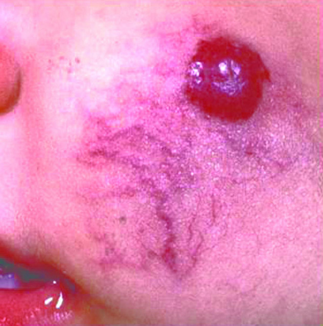
- Erythematous, poorly defined mottled macules and plaques typically ranging from 2 - 5 cm
- Usually solitary
- May have overlying hypertrichosis
- Slowly growing, spreading lesion
- Spontaneous regression rarely occurs; most lesions persist
Case reports
- 15 day old boy with palm sized erythematous patch (Ann Dermatol 2013;25:129)
- 2 month old girl with widespread disseminated red papules (J Cutan Pathol 2013;40:405)
- 47 year old woman with red, infiltrated, solitary plaque (An Bras Dermatol 2015;90:16)
- 72 year old man with 2 asymptomatic dusky red papules (Am J Dermatopathol 2015;37:162)
- Tufted angioma arising at a site of BCG vaccination (Eur J Dermatol 2013;23:102)
Treatment
- Surgical excision for small lesions; recurrences are common
- Low dose aspirin
- High dose steroids (intralesional or systemic)
- Pulsed dye laser
- Chemotherapy(vincristine)
Clinical images
Contributed by Mark R. Wick, M.D.
Images hosted on other servers:

Subcutaneous
nodule
Microscopic (histologic) description
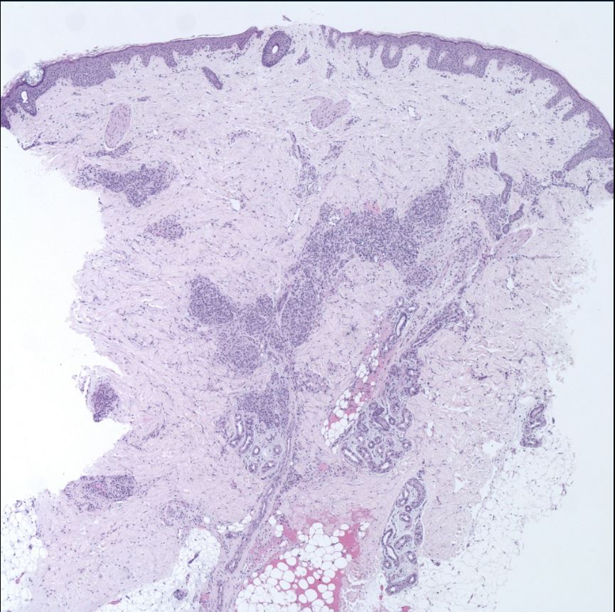
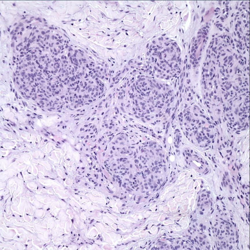
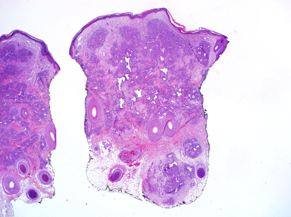
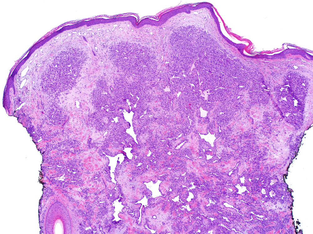
- Multiple, scattered lobules of small capillary type vessels with small oval to spindle shaped cells throughout the dermis and subcutaneous tissue imparting a “cannonball” or glomerular appearance
- May have variable mitoses without nuclear atypia
- Hemosiderin may be present; in contrast to pyogenic granuloma, inflammation is typically absent
- In contrast to Kaposi sarcoma, no slit-like vascular spaces and no plasma cells
- In contrast to kaposiform hemangioendothelioma, confined to skin and less infiltrative
Microscopic (histologic) images
Contributed by Manuel Valdebran, M.D., Phil LeBoit, M.D. and Joel Tjarks, M.D.

Images hosted on other servers:

Cannon ball distribution
Differential diagnosis
- Glomeruloid hemangioma
- Kaposiform hemangioendothelioma
- Kaposi sarcoma: tufted angioma lacks slit-like vessels and plasma cells; HHV8 negative
- Lobular capillary hemangioma (pyogenic granuloma): tufted angioma more plaque-like; lacks epidermal collarette and inflammation
Additional references
