
Skin nonmelanocytic tumor
Carcinoma (nonadnexal)
Lymphoepithelioma-like carcinoma
Authors: Ghassan A. Tranesh, M.D., Hong Qu, M.D.
Editorial Board Member: Sara C. Shalin, M.D., Ph.D.


Last author update: 1 November 2014
Last staff update: 15 February 2024
Copyright: 2003-2025, PathologyOutlines.com, Inc.
PubMed Search: Lymphoepithelioma-like carcinoma [title] skin

Table of Contents
Definition / general | Epidemiology | Sites | Pathophysiology | Etiology | Clinical features | Case reports | Treatment | Clinical images | Microscopic (histologic) description | Microscopic (histologic) images | Positive stains | Negative stains | Differential diagnosis | Additional referencesCite this page: Tranesh G. Lymphoepithelioma-like carcinoma. PathologyOutlines.com website. https://www.pathologyoutlines.com/topic/skintumornonmelanocyticLEL.html. Accessed April 2nd, 2025.
Definition / general
- Exceptionally rare and poorly differentiated cutaneous carcinoma with prominent reactive inflammatory infiltrate
- Mimics undifferentiated nasopharyngeal carcinoma (Rare Tumors 2013;5:e47)
- WHO defines these tumors (in nasopharynx/sinonasal cavity) as lymphoepithelial carcinoma
- Importantly, cutaneous LELC is NOT associated with Epstein Barr virus (EBV) infection
Epidemiology
- Older to elderly adult patients (Dermatol Online J 2008;14:12)
- Approximately equal incidence of females and males
Sites
- Head and neck most common site
Pathophysiology
- In skin, no apparent relationship with EBV (Dermatol Online J 2008;14:12, Am J Dermatopathol 1996;18:478)
- This is in contrast to consistent EBV association with similar tumors from lung, salivary glands, stomach, thymus, nasopharynx and sinonasal cavity
Etiology
- Uncertain origin (Dermatol Online J 2008;14:12)
- Unclear whether a poorly / undifferentiated squamous cell carcinoma versus poorly differentiated adnexal neoplasm
Clinical features
- Usually firm, variably colored, face or neck plaque, papule or nodule (Dermatol Online J 2008;14:12)
Case reports
- 73 year old woman with vulvar tumor (Diagn Pathol 2011 Jan 10;6:4)
- 85 and 87 year old men (Dermatol Online J 2012;18:7, Rare Tumors 2013;5:e47)
- 89 year old woman (Dermatol Online J 2008;14:12)
Treatment
- Wide local excision or Moh's microsurgery to ensure complete removal
- Radiation reserved for recurrence or lymph node involvement (Case Rep Oncol Med 2012;2012:241816)
- Generally low recurrence and metastatic potential with complete excision (in contrast to nasopharyngeal counterpart)
- Rare lymph node metastasis (Am J Dermatopathol 2006;28:211) or death from disease
Clinical images
Images hosted on other servers:


Forehead tumor (pre- and post-excision)
Microscopic (histologic) description
- With Diagnostic criteria:
- Well circumscribed lobules, nests or small aggregates of large, cohesive, epithelioid cells closely associated with a dense, mixed T and B lymphocytic and plasmacytic infiltrate (Rare Tumors 2013;5:e47)
- Biphasic nature may be more apparent at high magnification
- The epithelioid component generally lacks connection with the epidermis
- Cells are often polygonal and display poorly defined eosinophilic cytoplasm, vesicular nuclei with prominent nucleoli and increased mitotic activity, including atypical mitotic figures (Dermatol Online J 2008;14:12)
- Focal ductular differentiation or trichilemmal keratinization rarely reported (J Cutan Pathol 1991;18:93)
- Mandatory dense inflammatory infiltrate (polymorphous and polytypic) surrounding and intermingling with epithelial tumor cells
- Lymphoid infiltrate may obscure the epithelial component
- When inflammation predominates, may mimic lymphoproliferative disorder
- Well circumscribed lobules, nests or small aggregates of large, cohesive, epithelioid cells closely associated with a dense, mixed T and B lymphocytic and plasmacytic infiltrate (Rare Tumors 2013;5:e47)
Microscopic (histologic) images
Contributed by Sara Shalin, M.D., Ph.D.
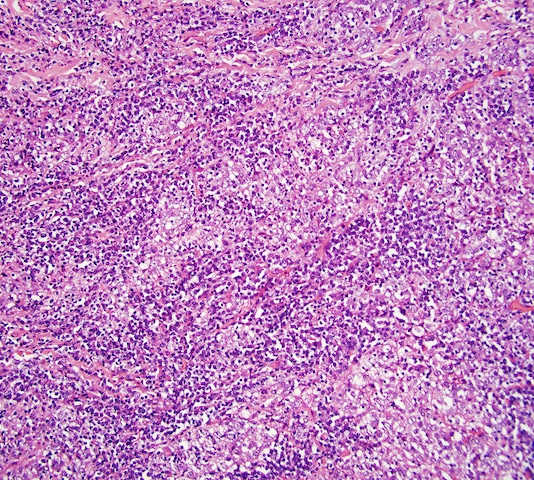
LELC
Images hosted on other servers:

Aggregates of atypical epithelial cells


Vulvar infiltrate with nodal micrometastasis

No epidermal infiltration


Atypical, epithelioid cells in syncytial pattern

Round to polygonal cells with eosinophilic cytoplasm


Proliferation of epithelial tumor cells and numerous small lymphocytes

H&E, CK5/6, EBV


AE1/AE3, EMA


CD20, CD3

CK5/6+
Positive stains
Negative stains
- Melanoma markers: S100 and HMB45
- Neuroendocrine markers: chromogranin, synaptophysin and CD56
- EBER (EBV encoded RNA in situ hybridization)
Differential diagnosis
- Cutaneous lymphadenoma
- Lymphoma
- Melanoma
- Merkel cell carcinoma
- Metastatic disease from nasopharyngeal carcinoma
- Poorly differentiated inflamed carcinoma
Additional references
