Table of Contents
Definition / general | Terminology | Epidemiology | Sites | Clinical features | Treatment | Clinical images | Microscopic (histologic) description | Microscopic (histologic) images | Differential diagnosisCite this page: Hale CS. Halo nevus. PathologyOutlines.com website. https://www.pathologyoutlines.com/topic/skintumormelanocytichalonevus.html. Accessed March 30th, 2025.
Definition / general
- Nevus surrounded by zone of hypopigmented skin (eMedicine: Halo nevus)
Terminology
- Also called leukoderma acquisitum centrifugum, Sutton nevus (Am J Dermatopathol 2003;25:349)
Epidemiology
- 1% of population before adulthood, no racial or gender predilection (J Am Acad Dermatol 2012;67:582)
Sites
- Back, followed by head / neck are most common sites (J Am Acad Dermatol 2012;67:582)
Clinical features
- Single or multiple
- Usually due to regression caused by cell mediated immunity, or less commonly humoral immunity or granulomatous inflammation (Am J Dermatopathol 2008;30:233)
- No fibrosis, in contrast to melanoma (J Cutan Pathol 2007;34:301)
- Seen in 18% of Turner syndrome patients (J Am Acad Dermatol 2004;51:354)
- May persist for decades (J Am Acad Dermatol 2012;67:582)
- Note: classify based on nevus cell population because halo phenomenon occurs in various nevus types (J Cutan Pathol 1995;22:342)
Treatment
- Observation, excision (for cosmetic reasons) or laser for facial lesions to induce repigmentation (J Cosmet Laser Ther 2007;9:245)
Clinical images
Microscopic (histologic) description
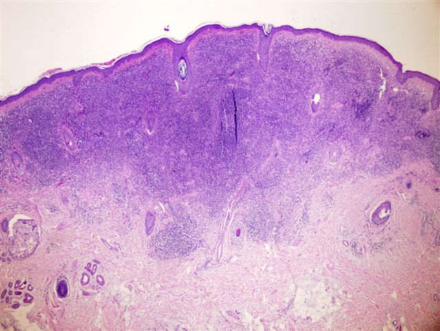
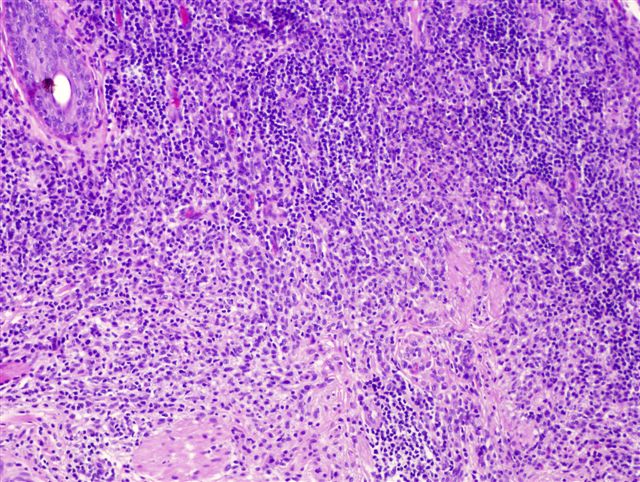
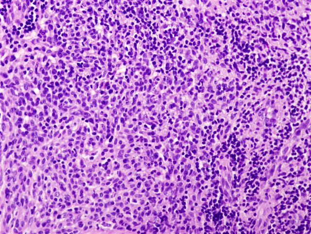
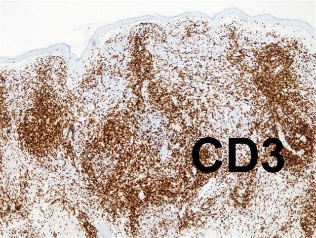
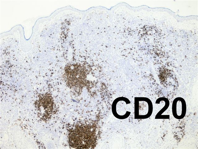
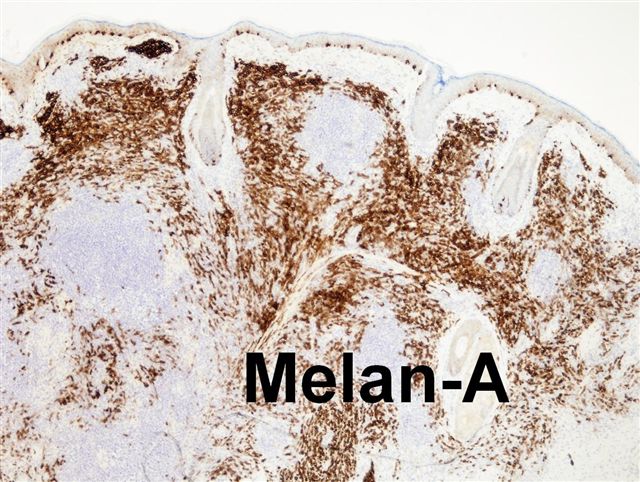
- Residual melanocytes with heavy infiltration by lymphocytes and histiocytes that destroy pigment containing cells
Microscopic (histologic) images


