
Skin nontumor
Keratinization disorders
Acanthosis nigricans
Last author update: 1 July 2011
Last staff update: 16 April 2021
Copyright: 2002-2025, PathologyOutlines.com, Inc.
PubMed Search: Acanthosis nigricans [title]


Table of Contents
Definition / general | Etiology | Clinical features | Clinical images | Microscopic (histologic) description | Microscopic (histologic) images | Differential diagnosis | Additional referencesCite this page: Hamodat M. Acanthosis nigricans. PathologyOutlines.com website. https://www.pathologyoutlines.com/topic/skinnontumoracanthosisnigricans.html. Accessed April 2nd, 2025.
Definition / general
- Brown, velvety and verrucous plaques in axillae, back of neck and other skin folds, associated with visceral malignancies, endocrine diseases and congenital disorders
Etiology
- Cutaneous manifestation of a diverse group of diseases
- May occur as inherited disorder, with Down syndrome or after ingestion of drugs
- 80% are "benign" type, either autosomal dominant or associated with tissue resistance to insulin, including diabetes, obesity and Cushing's disease
- 20% are associated with GI or other internal malignancies
- Usually age 40+ years
Clinical features
- Brown, velvety, and verrucous plaques in axillae, back of neck and other skin folds
- Oral mucosa (lips and tongue) affected in 25% of cases; rare involvement of esophagus
- Hyperkeratotic lesions may develop on the palms, soles and knuckles
Clinical images
Contributed by Mark R. Wick, M.D.

Breast
Images hosted on other servers:

Hyperpigmented, brownish, velvety lesions

Right neck
Microscopic (histologic) description
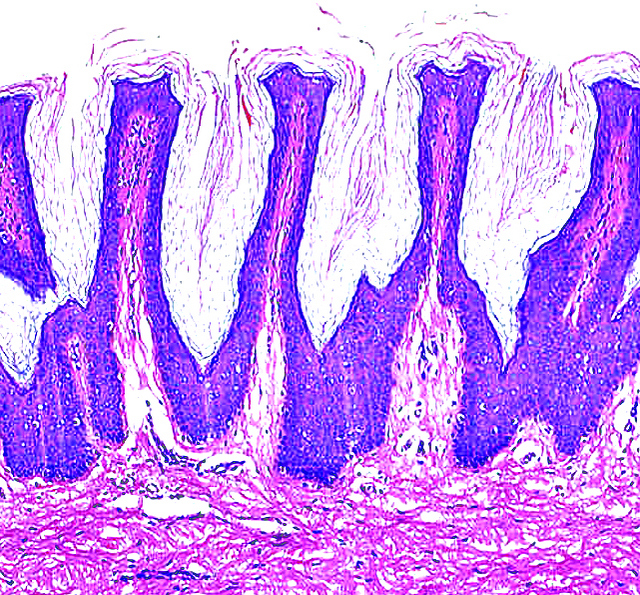
- Orthokeratotic hyperkeratosis (not actually acanthosis) and papillomatosis of stratum spinosum
- Hyperpigmentation of basal cell layer, but no melanocytic hyperplasia
- Usually no dermal inflammation
Microscopic (histologic) images
Contributed by Mark R. Wick, M.D.
Breast
Images hosted on other servers:

Papillomatosis and hyperkeratosis, H&E
Differential diagnosis
- Condyloma acuminata: oral lesions may resemble
- Epidermal nevus
- Seborrheic keratosis
Additional references
