
Salivary glands
Nonneoplastic tumors / tumor-like conditions
Lymphoepithelial cyst
Author: Zahra Maleki, M.D.
Editorial Board Member: Lisa Rooper, M.D.
Deputy Editor-in-Chief: Kelly Magliocca, D.D.S., M.P.H.

Last author update: 12 August 2022
Last staff update: 12 August 2022
Copyright: 2002-2025, PathologyOutlines.com, Inc.
PubMed Search: Benign lymphoepithelial cysts salivary

Table of Contents
Definition / general | Essential features | Terminology | ICD coding | Epidemiology | Sites | Pathophysiology | Etiology | Clinical features | Diagnosis | Radiology description | Radiology images | Prognostic factors | Case reports | Treatment | Clinical images | Gross description | Gross images | Frozen section description | Microscopic (histologic) description | Microscopic (histologic) images | Cytology description | Cytology images | Positive stains | Sample pathology report | Sample cytopathology report | Differential diagnosis | Differential diagnosis in cytology | Additional references | Board review style question #1 | Board review style answer #1 | Board review style question #2 | Board review style answer #2Cite this page: Maleki Z. Lymphoepithelial cyst. PathologyOutlines.com website. https://www.pathologyoutlines.com/topic/salivaryglandsb9lymphoepithelialcyst.html. Accessed March 30th, 2025.
Definition / general
- Unilocular cysts that involve the parotid gland
- Frequently but not always HIV related
- Can be associated with autoimmune disease (e.g., Sjögren syndrome) (Int J Surg Pathol 2009;17:421)
Essential features
- Well defined cysts on imaging (Laryngoscope 2007;117:106)
- Simple cyst lined by low stratified squamous epithelium surrounded by polymorphous lymphocytes with prominent germinal centers (Diagn Cytopathol 2012;40:684)
Terminology
- Salivary type lymphoepithelial cyst
- HIV associated cystic lymphoid hyperplasia (Laryngoscope 2007;117:106)
ICD coding
- ICD-10: K11.8 - other diseases of salivary glands
Epidemiology
- HIV
- Autoimmune disease (e.g., Sjögren syndrome)
Sites
- Almost all arise in parotid gland
- Very rare in submandibular gland
Pathophysiology
- Sporadic lymphoepithelial cyst may result from cystic dilation of ducts within intraparotid or periparotid lymph node or branchial cleft remnants
- HIV associated lymphoepithelial cyst likely forms due to hyperplasia of intra-salivary gland lymph nodes and associated ductal obstruction (J Int Assoc Provid AIDS Care 2017;16:120)
- Sjögren syndrome associated lymphoepithelial cysts arise secondary to infiltration of B cells into the ductal epithelium and their expansion within the striated ducts and subsequent basal ductal cell hyperplasia (Nat Rev Rheumatol 2021;17:333)
Etiology
- Cystic dilation of the salivary gland ducts
- HIV associated
- Autoimmune disease (e.g., Sjögren syndrome) associated
Clinical features
- Presents as a painless unilocular mass near or within the salivary gland
- Sporadic and autoimmune related cysts are generally unilateral
- HIV associated cysts can be bilateral, with an overall incidence of 3 - 5% in HIV patients
Diagnosis
- Unilateral, painless cystic salivary gland mass
- Unilateral or bilateral painless cystic salivary gland mass in HIV patients, with or without cervical lymphadenopathy
Radiology description
- Well defined, thin, smooth walled unilocular cystic mass (Int J Surg Case Rep 2017;41:383)
Radiology images
Contributed by Michael Kraut, M.D.
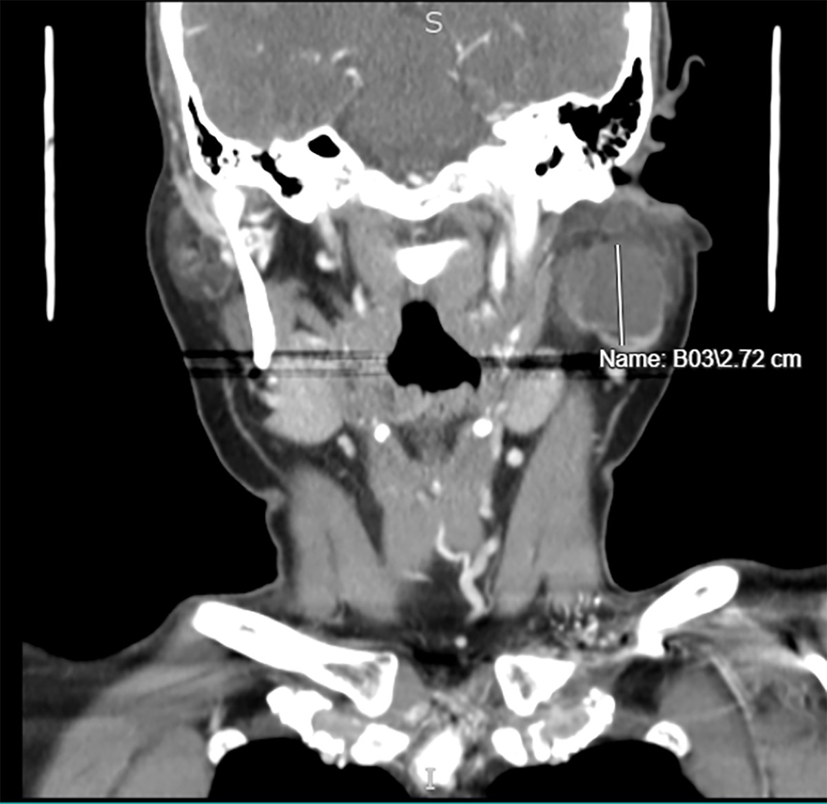
Lymphoepithelial cyst on coronal section
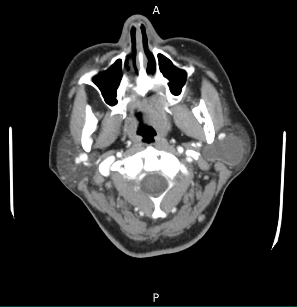
Lymphoepithelial cyst on axial section
Images hosted on other servers:

Well defined mass
Prognostic factors
- Excellent prognosis
- Low to null recurrence rate for all types of salivary gland lymphoepithelial cysts (J Surg Case Rep 2020;2020:rjaa300)
Case reports
- 32 year old woman presented with painless swelling of the left side of the neck for 8 months (J Pharm Bioallied Sci 2014;6:S185)
- 35 year old man with a soft, nontender swelling of the left parotid gland for 9 - 10 months (J Oral Maxillofac Pathol 2018;22:S91)
- 37 year old woman with a history of HIV and Hodgkin lymphoma presented with right sided facial swelling for 4 days; 47 year old man with a history of HIV / AIDS and Mycobacterium avium complex (MAC) infection presented with right sided jaw mass for 2 months (J Int Assoc Provid AIDS Care 2017;16:120)
Treatment
- Conservative therapy, with institution of highly active antiretroviral therapy medication in HIV related cases (HIV AIDS (Auckl) 2012;4:81)
- Surgical treatment not indicated for HIV associated lymphoepithelial cysts unless there is doubt about the diagnosis or there are cosmetic considerations (Head Neck 2018;40:1073)
- Repeated fine needle aspiration and drainage, sclerotherapy, radiotherapy, surgery (Head Neck 2018;40:1073)
Clinical images
Images hosted on other servers:

Diffuse swelling involving the left parotid gland
Gross description
- Cystic structure containing a serous clear watery straw colored fluid with smooth and glistening inner lining
Gross images
Images hosted on other servers:

Unilocular cyst with straw colored fluid
Frozen section description
- Benign lymphoepithelial cyst
Microscopic (histologic) description
- Most cases show a unilocular cyst with a thin stratified squamous lining
- Ciliated, cuboidal or columnar epithelial lining is seen in rare cases
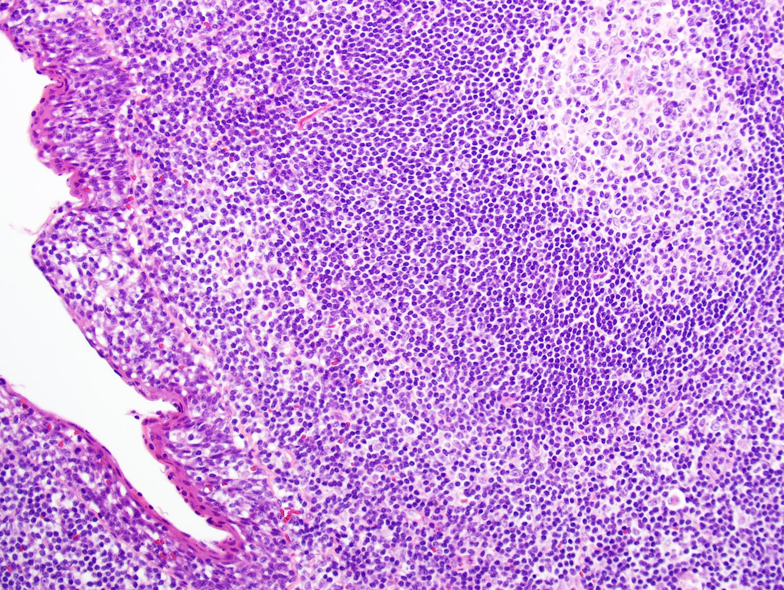
- Epithelium is surrounded by dense polymorphous lymphoid tissue with germinal centers and sinusoidal spaces
- Lymphocytes frequently permeate the epithelial cyst lining cells
Microscopic (histologic) images
Contributed by Zahra Maleki, M.D.
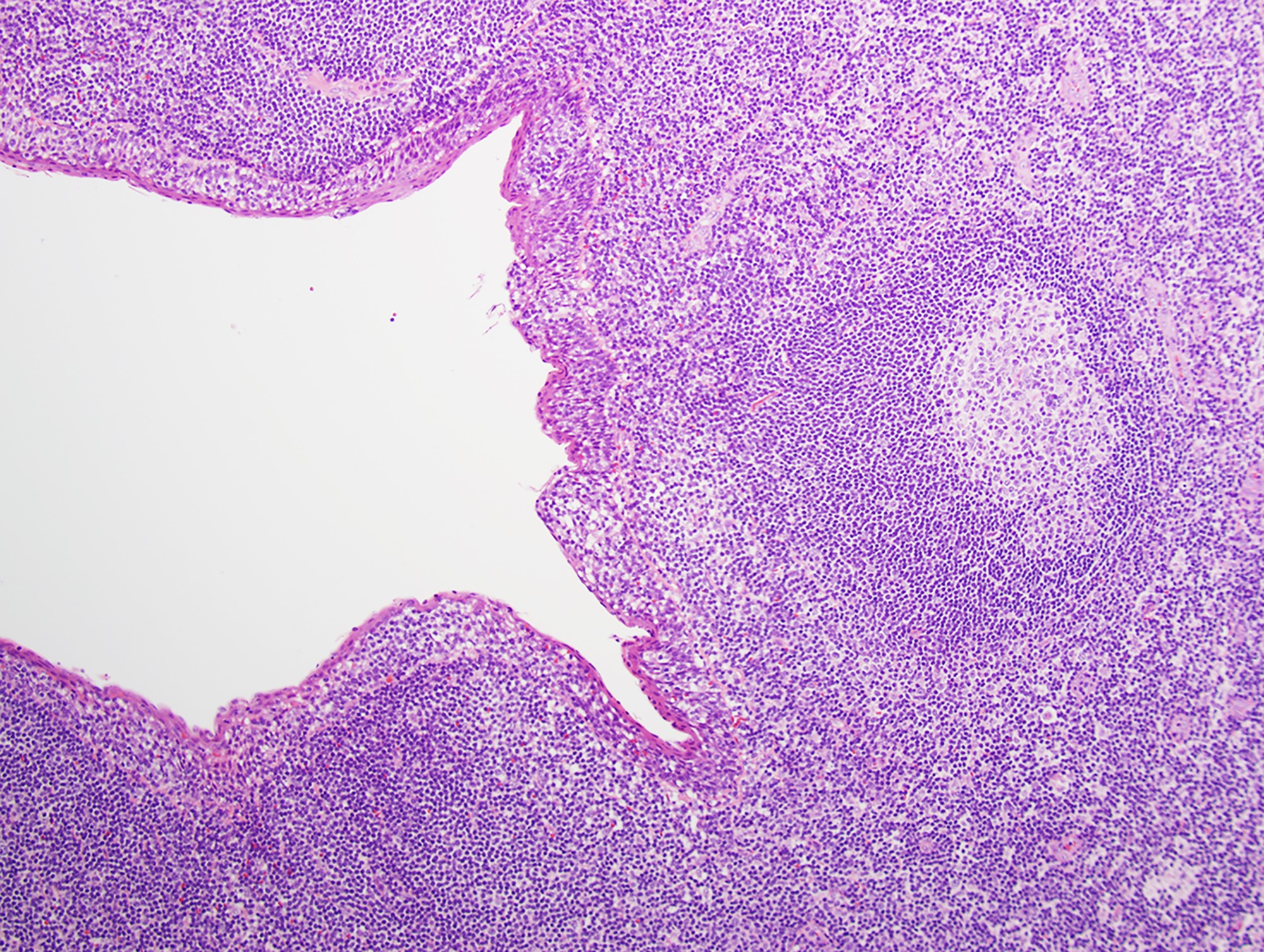
Epithelial and lymphoid component
Squamous lining and lymphoid tissue
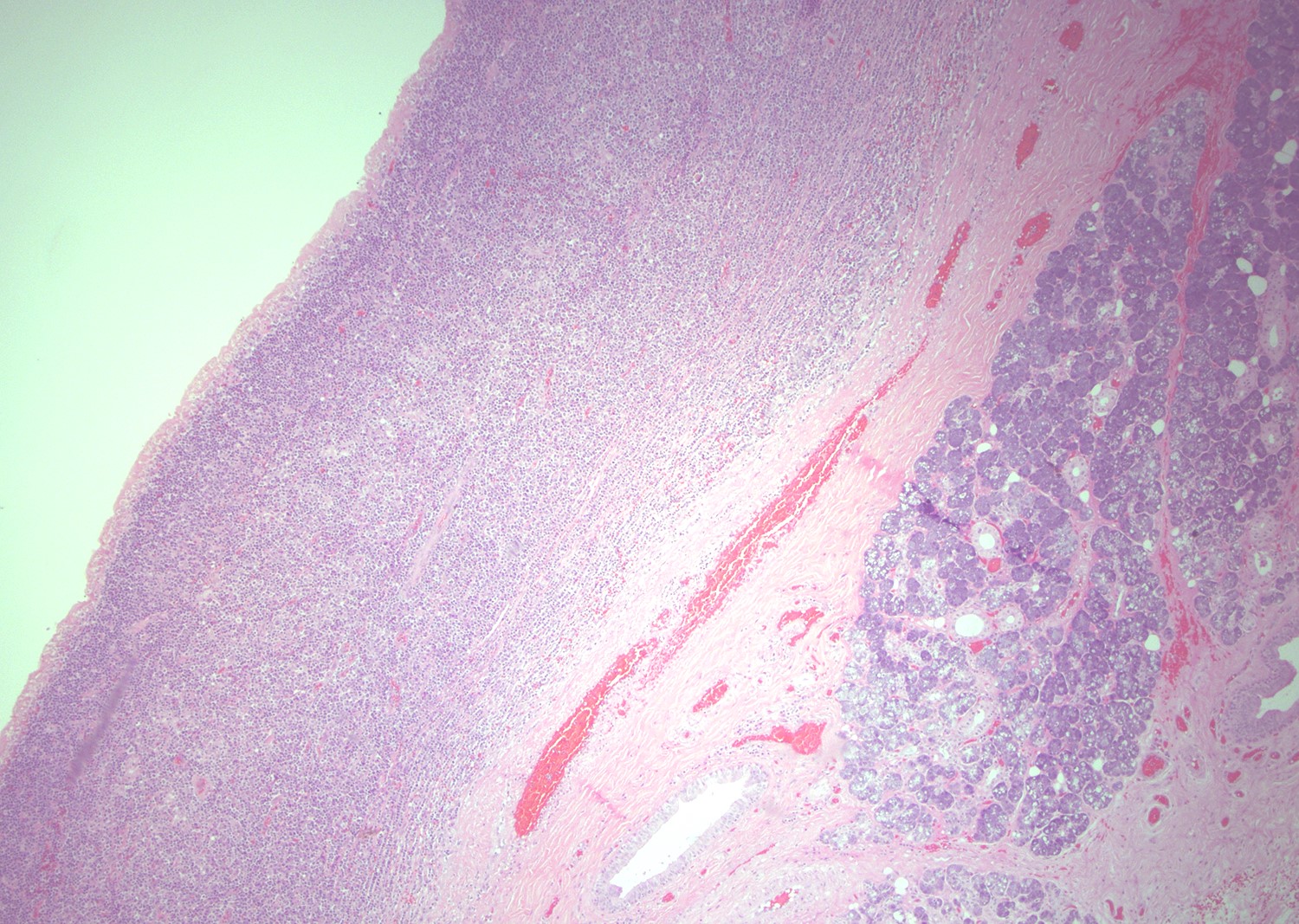
Epithelium surrounded by inflammatory cells
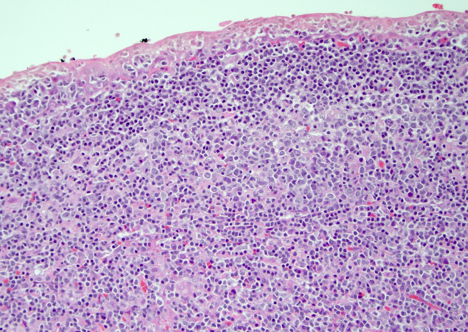
Epithelial and inflammatory cells
Cytology description
- Mature nucleated squamous cells with variable reactive atypia, anuclear cells and squamous epithelium (Int J Surg Case Rep 2017;41:383, Diagn Cytopathol 2012;40:684)
- Polymorphous lymphocytes and aggregates of epithelioid histiocytes
- Proteinaceous background
- Variable presence of acute inflammation, and bland appearing mucinous ductal cells and ciliated columnar cells
- Scant cellularity, abundant proteinaceous background, lack of squamous cells and epithelial cells, marked atypia of epithelial cells, abundance of lymphocytes pose diagnostic challenges
Cytology images
Contributed by Zahra Maleki, M.D.
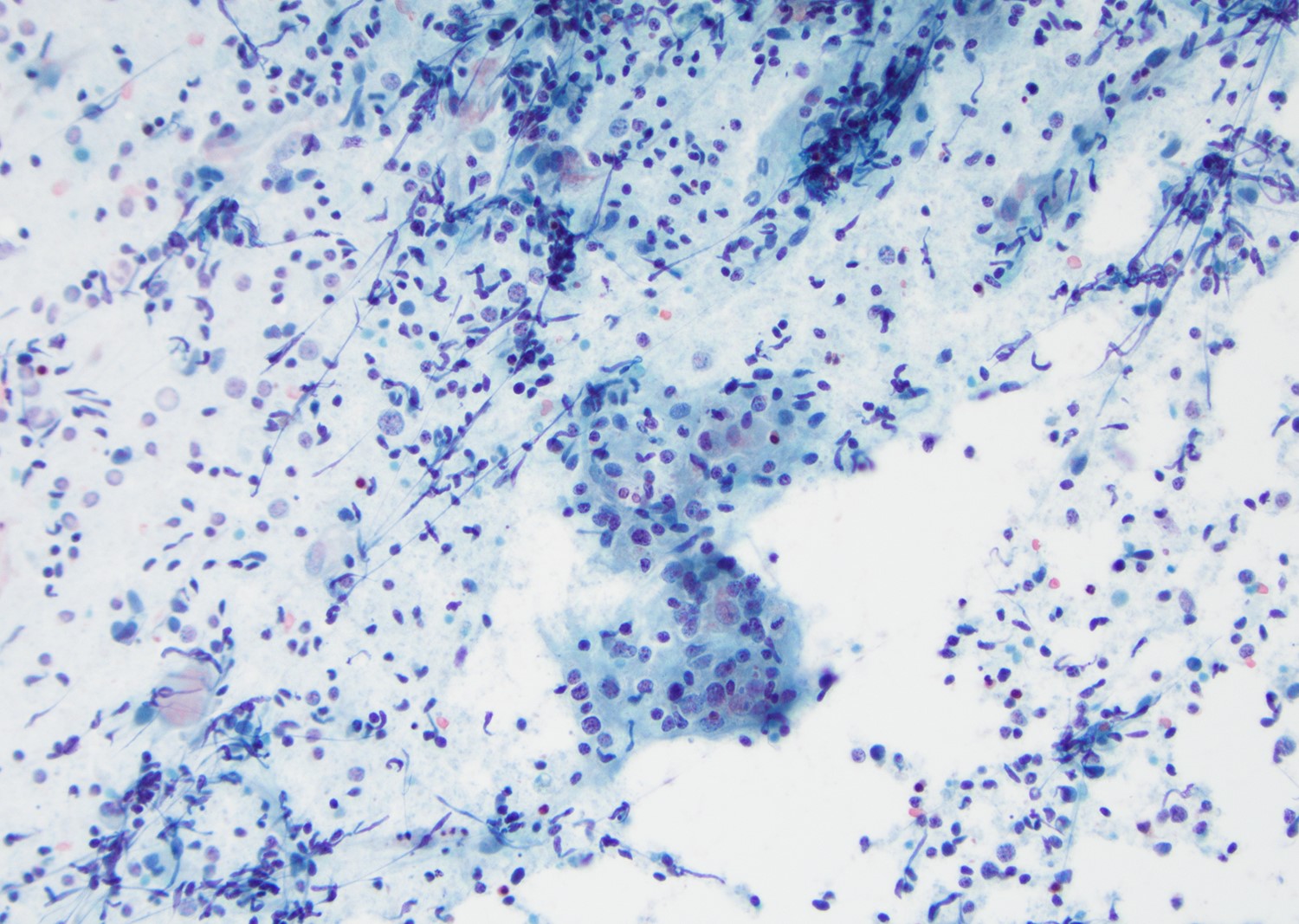
Numerous lymphocytes and histiocytes
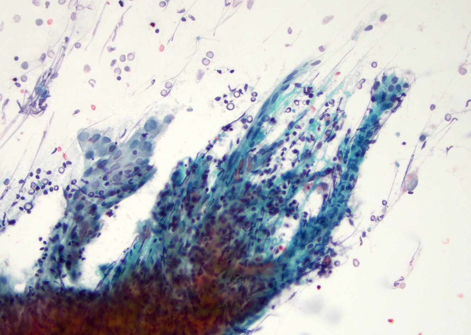
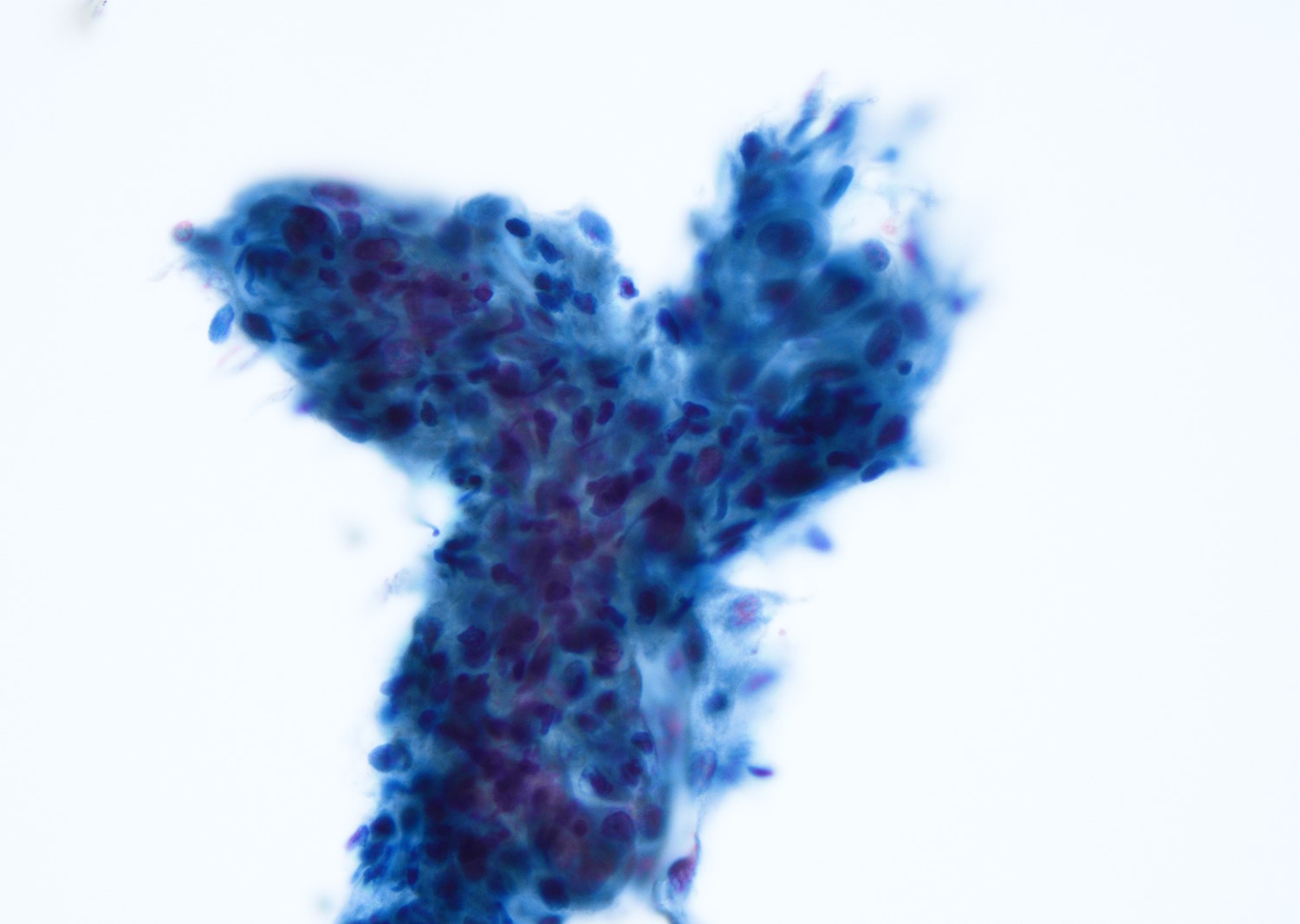
Large epithelial fragment
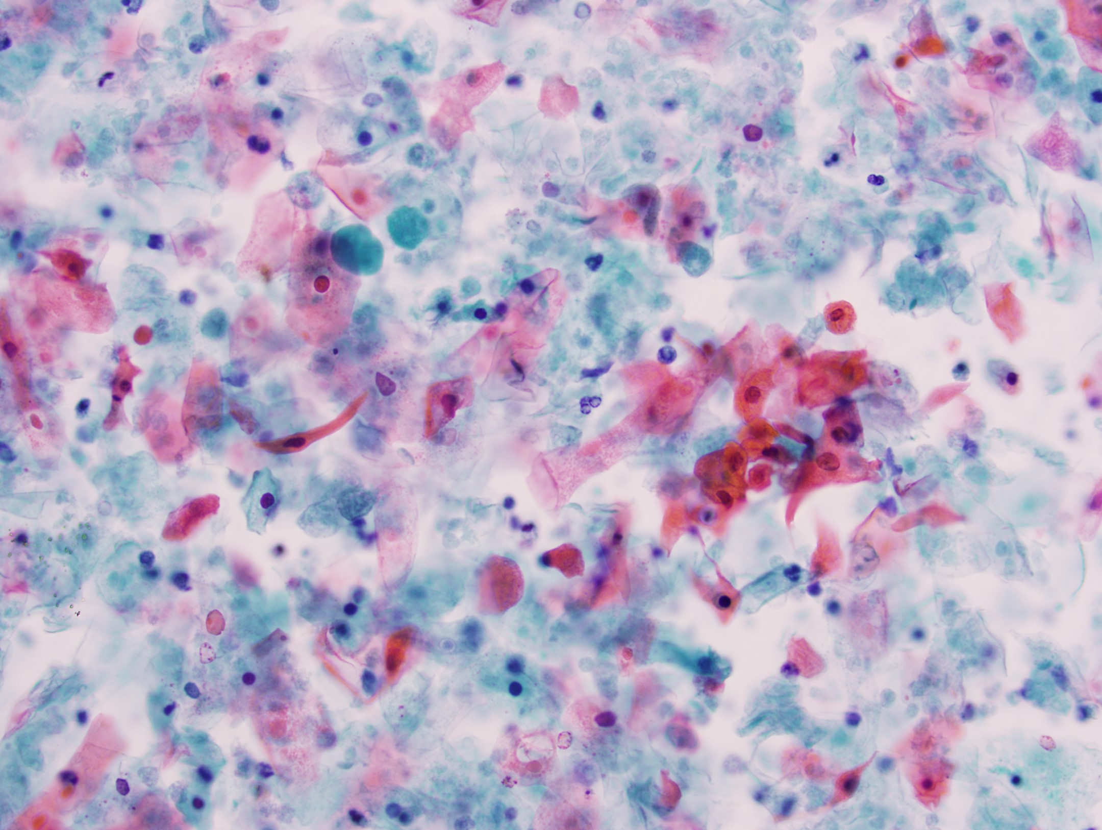
Dispersed squamous cells
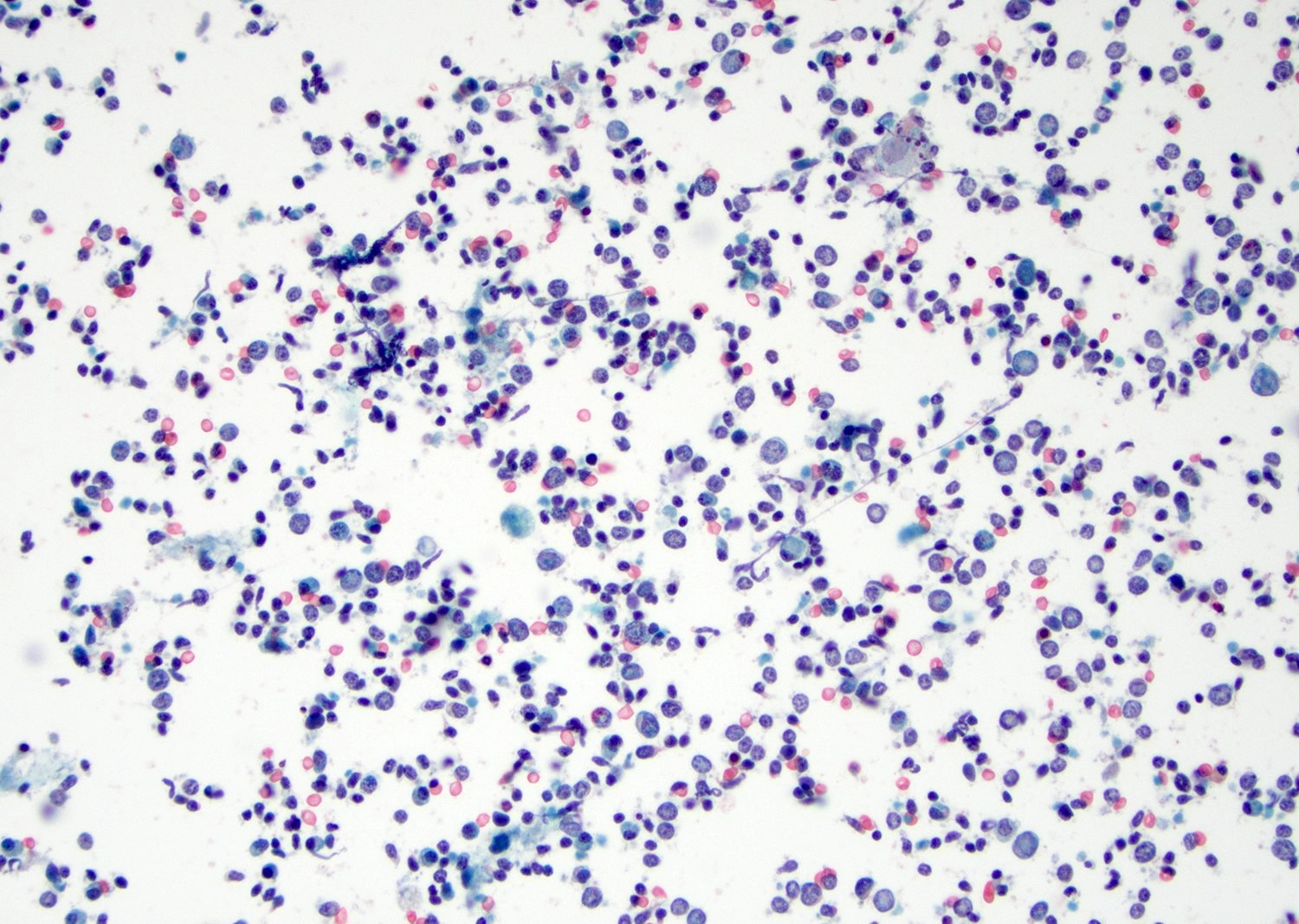
Polymorphous lymphocytes
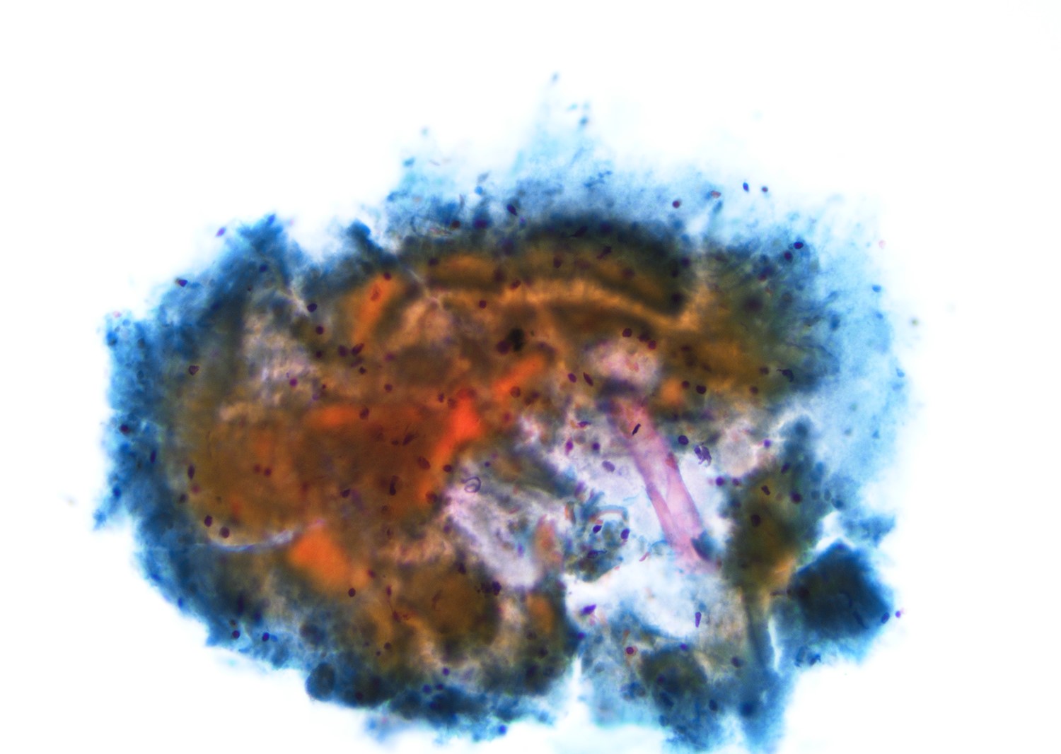
Cyst content
Positive stains
- AE1 / AE3 in squamous or ciliated columnar cells (Int J Surg Pathol 2021;29:78)
- Numerous intrafollicular CD8+ lymphocytes in HIV associated lymphoepithelial cysts (Oral Surg Oral Med Oral Pathol Oral Radiol 2019;128:52)
Sample pathology report
- Parotid mass, right, resection:
- Benign lymphoepithelial cyst with reactive follicular hyperplasia
Sample cytopathology report
- Parotid, left, fine needle aspiration:
- Fragments of squamous epithelium, lymphocytes and cyst content (see comment)
- Comment: The differential diagnosis includes benign lymphoepithelial cyst, cystic lymphoid hyperplasia, lymphoepithelial sialadenitis, benign cyst with lymph node sampling or a Warthin tumor.
- Recommend: Clinical and radiographic correlation.
Differential diagnosis
- Branchial cleft cyst:
- May have a sinus tract or stalk, infrequently associated with parotid tissue
- Extranodal marginal zone lymphoma:
- Monoclonal lymphoid population with centrocytic or monocytoid appearance
- Retention cyst:
- Mucinous cystic contents without extensive surrounding chronic inflammations
- Warthin tumor:
- Areas of classic bilayered oncocytic epithelium, although occasional squamous metaplasia can be seen
- Cystic metastasis of squamous cell carcinoma:
- Increased cytologic atypia and expansile growth of squamous epithelium
- Mucoepidermoid carcinoma with tumor associated lymphoid proliferation:
- More complex epithelial proliferation with intermixed squamoid, mucinous and intermediate cells
Differential diagnosis in cytology
- Mucocele:
- Abundant mucinous content
- Reactive lymph node:
- Polymorphous lymphocytes and aggregates of epithelioid histiocytes
- Warthin tumor:
- Polymorphous lymphocytes and fragments of oncocytic epithelium
- Squamous cell carcinoma:
- Abundant pleomorphic squamous cells with marked atypia, pleomorphism, nuclear enlargement and hyperchromasia
- Low grade mucoepidermoid carcinoma:
- Fragments of malignant epithelial cells and scattered squamous cells in a background of abundant mucin
Additional references
Board review style question #1
A cystic parotid mass measuring 1.5 cm is resected. The lesion can be associated with which of the following?
- EBV associated
- HHV8 associated
- HIV associated
- HPV associated
Board review style answer #1
Board review style question #2
FNA of a cystic parotid mass in an 11 year old child is shown here. The most likely diagnosis is
- Granulomatous inflammation
- Lymphoepithelial cyst
- Mucoepidermoid carcinoma
- Squamous cell carcinoma
Board review style answer #2
