Table of Contents
Definition / general | Essential features | ICD coding | Epidemiology | Pathophysiology | Etiology | Diagnosis | Laboratory | Treatment | Gross description | Microscopic (histologic) description | Microscopic (histologic) images | Positive stains | Negative stains | Molecular / cytogenetics description | Sample pathology report | Differential diagnosis | Board review style question #1 | Board review style answer #1Cite this page: Iczkowski KA. Postatrophic hyperplasia. PathologyOutlines.com website. https://www.pathologyoutlines.com/topic/prostatepostatrophichyper.html. Accessed April 1st, 2025.
Definition / general
- Postatrophic hyperplasia (PAH) is a clustered, small acinar proliferation in fibrotic stroma, with a pseudoinfiltrative appearance
- Among types of atrophy, this type is the most likely to mimic carcinoma
Essential features
- Postatrophic hyperplasia is a small acinar proliferation with a special tendency to mimic cancer because it can show somewhat more prominent nucleoli than benign acini and because the stroma is fibrotic (Mod Pathol 2004;17:328)
- Notable differences from cancer include a lobular or clustered arrangement of the acini around a central dilated duct space, lack of macronucleoli in most cells and retention of basal cells by immunostaining
- There is no clinical significance and no need to specify in a pathology report
ICD coding
- ICD-10: N40.1 - benign prostatic hyperplasia
Epidemiology
- As with atrophy in general, postatrophic hyperplasia increases with age
Pathophysiology
- Oxidative stress is implicated, similar to atrophy
- Chronic inflammation is also present in all postatrophic hyperplasia cases, suggesting a connection (J Urol 2006;176:1012)
Etiology
- Same as Atrophy
Diagnosis
- Histopathologic examination of tissue
Laboratory
- As with atrophy in general, serum prostate specific antigen (PSA) might be elevated (Abdom Imaging 2009;34:271)
Treatment
- No treatment is needed
Gross description
- No distinct findings
Microscopic (histologic) description
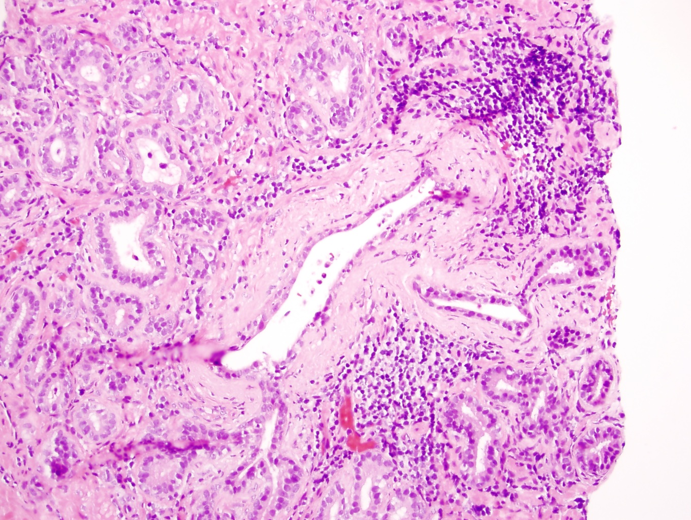
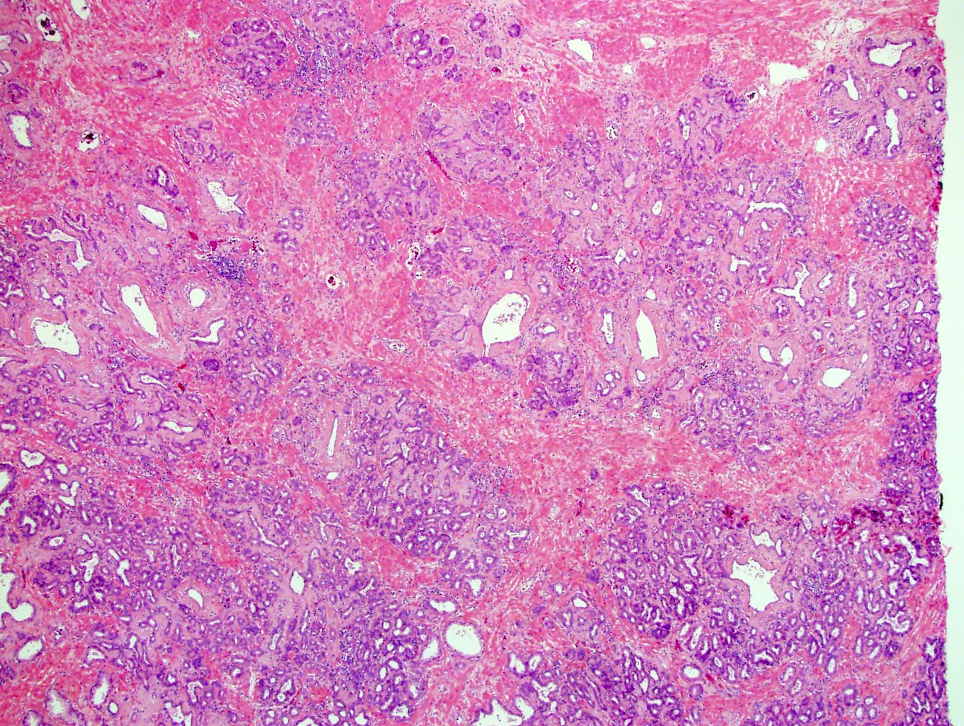
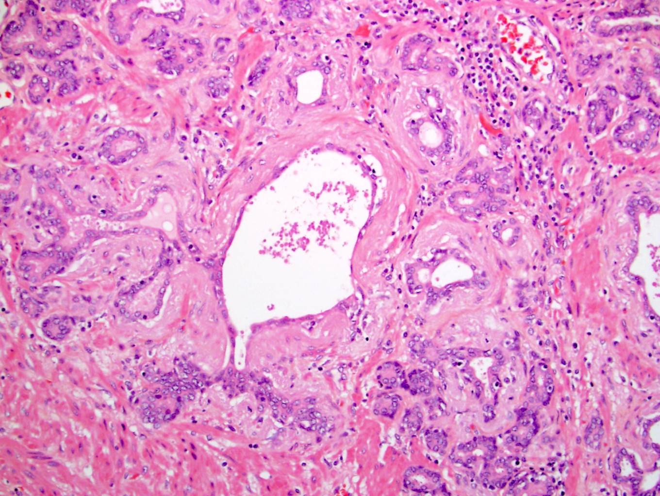
- Usually has a central, dilated atrophic gland surrounded by clustered smaller glands within a fibrotic / sclerotic stroma; each lobular unit of acini is circumscribed
- May have more prominent nucleoli than simple atrophy does and this heightens its ability to mimic cancer (Am J Surg Pathol 1998;22:1073)
Microscopic (histologic) images
Positive stains
- Basal cell stains (high molecular weight cytokeratin, p63) should be at least focally positive
Negative stains
- AMACR / P504S should be mainly negative, as with atrophy in general
Molecular / cytogenetics description
- Can have an elevated proliferation index that is greater than that of benign, nonatrophic acini and greater than that of simple atrophy (Am J Surg Pathol 1998;22:1073)
Sample pathology report
- Prostate, biopsies:
- Benign prostatic tissue (see comment)
- Comment: Prostatic basal cell cocktail / P504S immunostain rules out cancer in the small acini.
Differential diagnosis
- Carcinoma with an atrophic appearance or atrophic variant of prostatic adenocarcinoma:
- Has a frankly infiltrative appearance, in which individual small cancer acini are interspersed between larger benign acini
- Nuclear enlargement and prominent nucleoli are seen in a higher percent of cells than in atrophy
- There is often concomitant presence of nonatrophic carcinoma
- Can also have a sclerotic stroma
- p63-, HMWK-
- Simple atrophy:
- Far more common
- Has fewer prominent nucleoli (Am J Surg Pathol 1998;22:1073)
- Has a lower proliferation (Ki67) index
Board review style question #1
Prostate tissue is shown. Which histologic finding, if it were present in this image, would make this proliferation of small acini suspicious for cancer?
- Central dilated duct space
- Clustered or lobular arrangement of the small acini
- Irregular and uncircumscribed infiltration of the small acini among larger definitely benign ones
- Prominent nucleoli in only a few cells and no nuclear enlargement
- Sclerotic stroma
Board review style answer #1
C. Postatrophic hyperplasia is shown. Irregular and uncircumscribed infiltration of the small acini would raise the suspicion of cancer. The clustered, lobular and circumscribed arrangement of the small acini (B) favors a benign diagnosis. The presence of the central dilated duct space (A) is also very characteristic of postatrophic hyperplasia. Sclerotic stroma (E) is a feature of postatrophic hyperplasia that can also occur in cancer and causes it to mimic cancer. Finally, as long as prominent nucleoli are visible in only a few cells (D) but not most cells, without nuclear enlargement (compared with other benign acini), that also is not suspicious for cancer.
Comment Here
Reference: Postatrophic hyperplasia
Comment Here
Reference: Postatrophic hyperplasia


