
Prostate gland & seminal vesicles
Acinar / ductal adenocarcinomas
Acinar adenocarcinoma, histologic patterns
Microcystic adenocarcinoma
Author: Ankur Sangoi, M.D.
Editorial Board Member: Bonnie Choy, M.D.
Deputy Editor-in-Chief: Maria Tretiakova, M.D., Ph.D.

Last author update: 17 July 2023
Last staff update: 20 January 2025
Copyright: 2019-2025, PathologyOutlines.com, Inc.
PubMed Search: Microcystic adenocarcinoma


Table of Contents
Definition / general | Essential features | Terminology | ICD coding | Sites | Diagnosis | Case reports | Microscopic (histologic) description | Microscopic (histologic) images | Positive stains | Negative stains | Sample pathology report | Differential diagnosis | Additional references | Board review style question #1 | Board review style answer #1Cite this page: Sangoi A. Microcystic adenocarcinoma. PathologyOutlines.com website. https://www.pathologyoutlines.com/topic/prostatemicrocystic.html. Accessed April 1st, 2025.
Definition / general
- One of the unusual histological patterns of acinar adenocarcinoma
- While often easier to recognize at radical prostatectomy given the juxtaposition to usual pattern acinar adenocarcinoma (incidence of 11%), deceptive morphology can be challenging to recognize on prostate biopsy (incidence of 1%) (Am J Surg Pathol 2010;34:556, Int J Surg Pathol 2020;28:584)
- As these are graded in the usual fashion, formal designation as microcystic pattern is not indicated in the pathology report and is more important to recognize for accurate diagnosis
Essential features
- Cystic glands with marked dilatation (reminiscent of cystic atrophy) at low power magnification but often showing luminal crystalloid, blue intraluminal mucin and prominent nucleoli at higher power magnification
Terminology
- Sometimes synonymous with pseudohyperplastic pattern prostatic adenocarcinoma
ICD coding
- ICD-O: 8140/3 - acinar adenocarcinoma
- ICD-10: C61 - malignant neoplasm of the prostate
- ICD-11: 2C82.0 & XH4PB1 - adenocarcinoma of prostate & acinar adenocarcinoma of prostate
Sites
- Prostate
Diagnosis
- Core needle biopsies or transurethral resection of the prostate
- Immunohistochemistry may be used to confirm the absence of basal cells
Case reports
- Limited series of atrophic / microcystic prostatic adenocarcinoma (Int J Surg Pathol 2020;28:584)
Microscopic (histologic) description
- Cystic glands with marked dilatation (reminiscent of cystic atrophy; glands can become quite attenuated), approximately 10x the size of typical malignant acini (Am J Surg Pathol 2010;34:556)
- Sometimes crowded growth at low power magnification but often showing luminal crystalloid, blue intraluminal mucin and prominent nucleoli at higher power magnification
- Typically coexists with usual acinar pattern prostatic adenocarcinoma
- One of the important deceptively bland histological patterns of prostate cancer, alongside atrophic, pseudohyperplastic and foamy gland prostatic adenocarcinoma
Microscopic (histologic) images
Contributed by Ankur Sangoi, M.D.
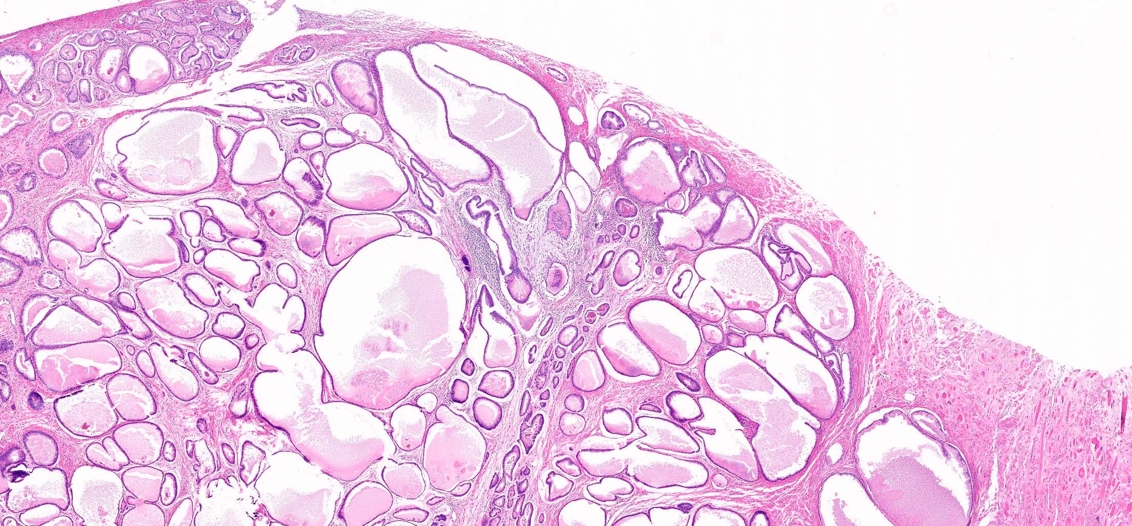
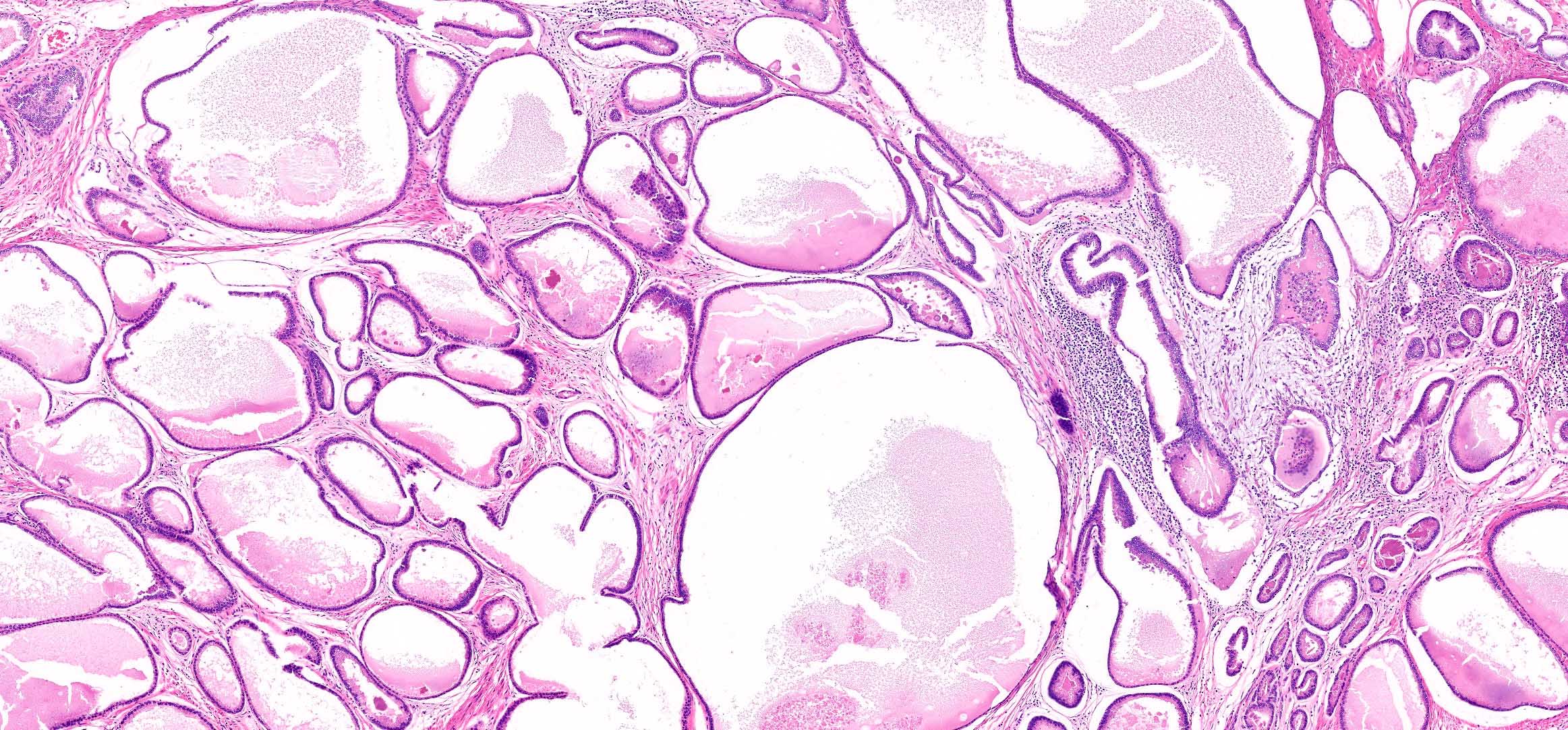
Compact dilated cysts
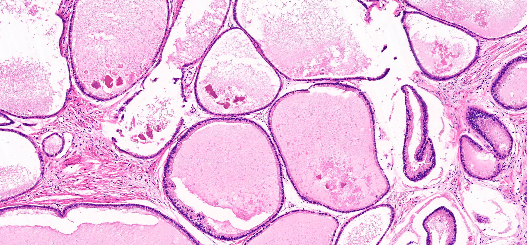
Basal nuclei with amphophilic cytoplasm
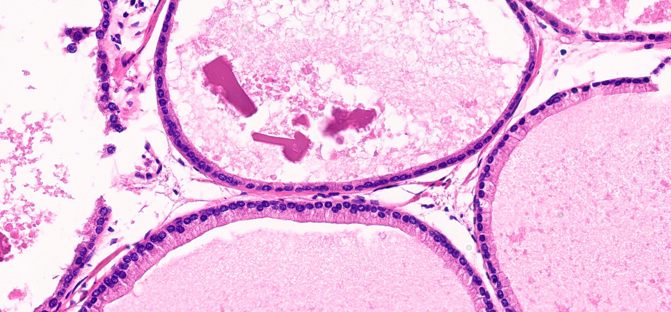
Prominent nucleoli and luminal crystalloid
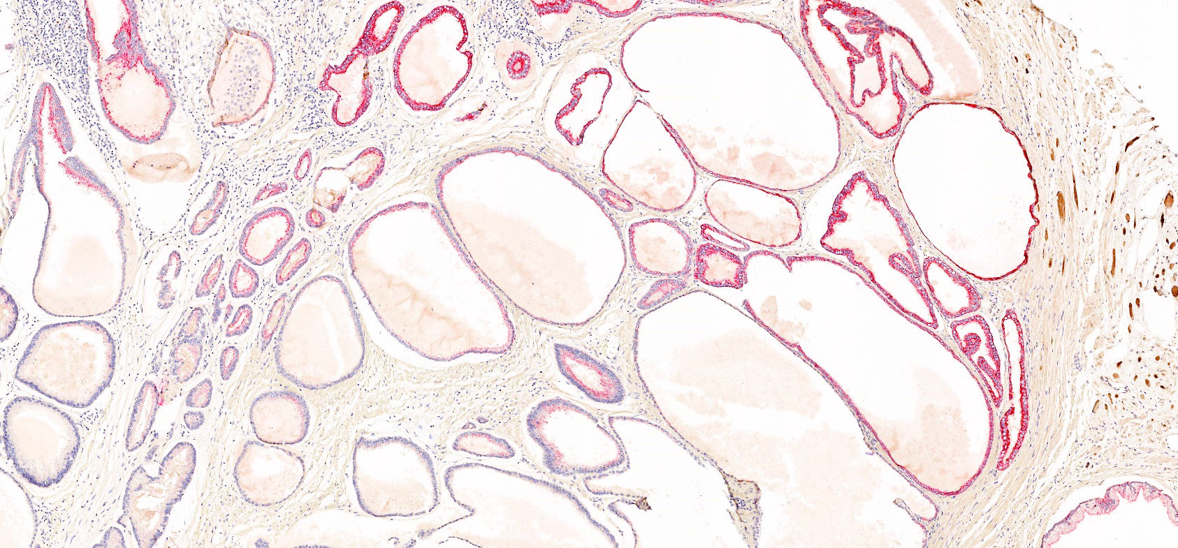
Cytoplasmic P504S
Positive stains
- AMACR (P504S) luminal expression in 96% of cases (Am J Surg Pathol 2010;34:556)
Negative stains
- Complete loss of basal cell reactivity (34 beta E12, p63) in 96% of cases (Am J Surg Pathol 2010;34:556)
Sample pathology report
- Same as typical acinar adenocarcinoma; it is not needed to report the presence of this histological pattern
- Prostate, radical prostatectomy:
- Prostatic adenocarcinoma, Gleason score 3 + 3 = 6, grade group 1 (see synoptic report)
Differential diagnosis
- Cystic atrophy:
- Lacks amphophilic cytoplasm, prominent nucleoli, luminal mucin / crystalloid
- While it may show patchy, cytoplasmic P504S staining, it shows retained basal cell reactivity (p63 / high molecular weight keratin)
- High grade prostatic intraepithelial neoplasia:
- While it may show luminal crystalloid, it typically lacks luminal mucin
- Basal cells are somewhat appreciable on H&E staining but immunohistochemistry shows retained basal cell reactivity (p63 / high molecular weight keratin)
- Atrophic pattern prostatic adenocarcinoma (including aberrant p63 positivity)
- Often shows reduced cytoplasm compared to microcystic pattern prostatic adenocarcinoma
- Can show aberrant nuclear staining for p63 in the tumor cells (absent p63 / high molecular weight keratin staining in basal cells)
Additional references
Board review style question #1
Which of the following statements is true regarding microcystic pattern prostatic adenocarcinoma?
- It is one of the patterns of prostatic adenocarcinoma that is excluded from grading
- It most closely mimics cystic atrophy but shows an immunoprofile of usual acinar adenocarcinoma (cytoplasmic P504S, loss of basal cell reactivity)
- It rarely shows luminal crystalloid or prominent nucleoli
- It shows atrophic glands with aberrant nuclear positivity for p63
Board review style answer #1
B. It most closely mimics cystic atrophy but shows an immunoprofile of usual acinar adenocarcinoma (cytoplasmic P504S, loss of basal cell reactivity). Answer A is incorrect because Gleason grading is applicable to microcystic pattern prostatic adenocarcinoma (graded on architecture, usually Gleason grade 3 + 3). Answer C is incorrect because it often shows luminal crystalloid with prominent nucleoli. Answer D is incorrect because it does not show features of p63 positive prostatic adenocarcinoma (atrophic glands with aberrant nuclear positivity for p63).
Comment Here
Reference: Microcystic adenocarcinoma
Comment Here
Reference: Microcystic adenocarcinoma
