Table of Contents
Definition / general | Essential features | Terminology | ICD coding | Epidemiology | Sites | Pathophysiology | Diagnosis | Prognostic factors | Case reports | Treatment | Gross description | Microscopic (histologic) description | Microscopic (histologic) images | Positive stains | Negative stains | Molecular / cytogenetics description | Sample pathology report | Differential diagnosis | Additional references | Board review style question #1 | Board review style answer #1 | Board review style question #2 | Board review style answer #2Cite this page: Samaddar A, Jimenez RE. Clear cell cribriform hyperplasia. PathologyOutlines.com website. https://www.pathologyoutlines.com/topic/prostateclearcellcribhyper.html. Accessed April 1st, 2025.
Definition / general
- Epithelial cribriform proliferation that is considered to be a histological variant of benign prostatic hyperplasia (BPH)
Essential features
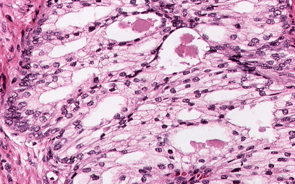
- Nodular clusters of cribriform acini lined by clear cells with small monotonous nuclei and inconspicuous nucleoli
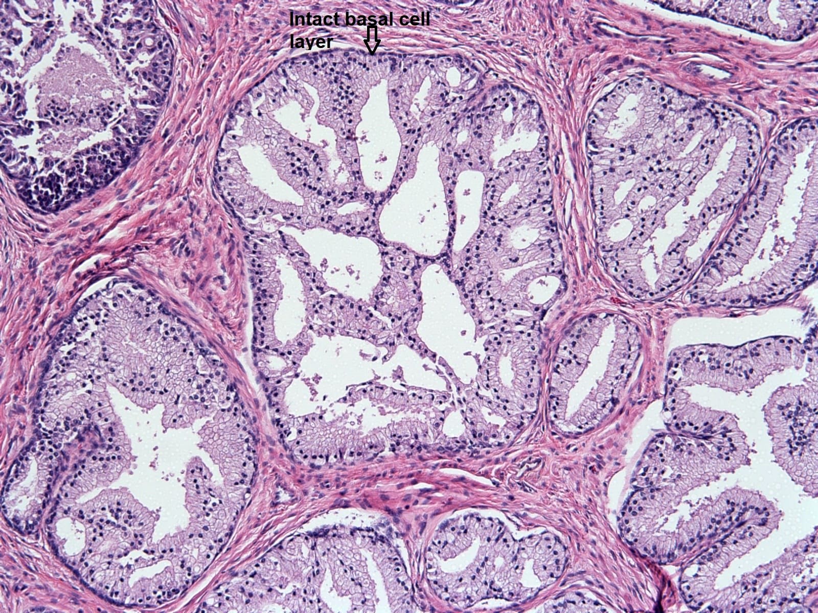
- Basal cell layer is intact
- Predominantly occurs in the transitional zone of the prostate
Terminology
- Synonym: cribriform hyperplasia (Virchows Arch 2009;454:1)
ICD coding
- ICD-11: GA90 - hyperplasia of prostate
Epidemiology
- As a morphologic variant of benign prostatic hyperplasia, clear cell cribriform hyperplasia shares the same epidemiology
Sites
- Transitional zone of the prostate
Pathophysiology
- Considered a variant of benign prostatic hyperplasia whose pathophysiology is incompletely understood
- Dominant role of androgens and androgen receptors is recognized and recently the role of prostatic inflammation and metabolic factors has been described (Gerontology 2019;65:458)
Diagnosis
- Mostly in transurethral resection of the prostate (TURP) specimens of men with lower urinary tract obstruction secondary to benign prostatic hyperplasia (Am J Surg Pathol 1986;10:665)
Prognostic factors
- Not a risk factor for prostate cancer
Case reports
- 58 - 88 year old men with clear cell cribriform hyperplasia of the prostate (Am J Clin Pathol 1991;95:446)
- 60 year old man with benign prostatic hyperplasia (Ann Int Med Den Res 2017;3:25)
- 62 - 87 year old men with clear cell cribriform hyperplasia of prostate (Am J Surg Pathol 1986;10:665)
Treatment
- Mostly found incidentally in TURP specimens treated for benign prostatic hyperplasia; no additional therapy is required
Gross description
- No specific gross features
Microscopic (histologic) description
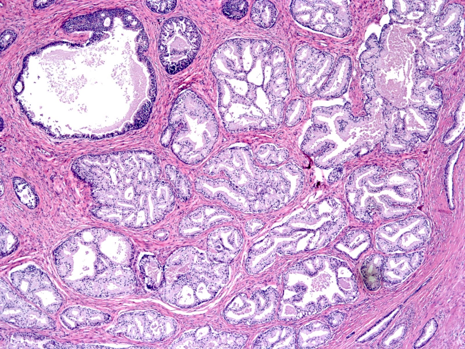
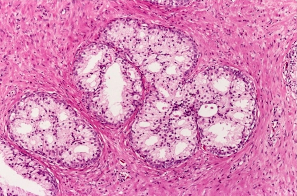
- Focal or diffuse nodular clusters of acini with cribriform morphology
- Involved ducts follow the architectural distribution of adjacent benign prostatic hyperplasia
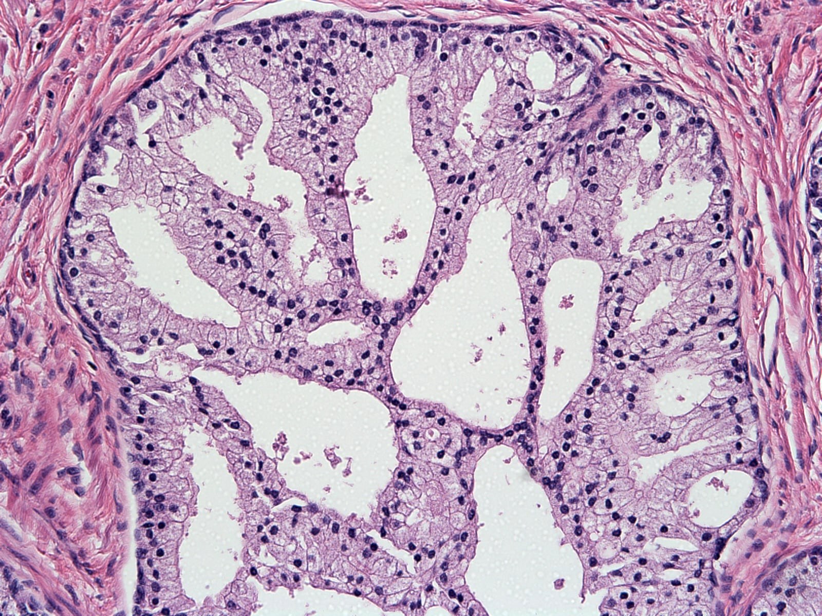
- Cuboidal to columnar cells with clear to pale eosinophilic granular cytoplasm and small, monotonous nuclei with inconspicuous nucleoli
- Basal cell layer is prominent and frequently discernible on light microscopic examination
- Stroma surrounding glands is cellular (benign prostatic hyperplasia-like)
- References: Am J Clin Pathol 1991;95:446, Surg Pathol Clin 2022;15:591
Microscopic (histologic) images
Positive stains
- High molecular weight cytokeratin, p63 and p40 highlight the basal cells, supporting a nonneoplastic diagnosis
Molecular / cytogenetics description
- Flow cytometry showed diploid DNA (n = 13) (Am J Clin Pathol 1991;95:446)
Sample pathology report
- Prostate, transurethral resection:
- Benign prostatic hyperplasia
- Note: it is not necessary to specify clear cell cribriform hyperplasia in the diagnosis as it has no clinical impact and the term may cause confusion
Differential diagnosis
- Central zone histology (Hum Pathol 2002;33:518, Cancers (Basel) 2022;14:3041):
- Central zone of the prostate near the base can have focal cribriform glands
- Also lacks cytological atypia and inconspicuous nucleoli
- Basal cell hyperplasia (cribriform / pseudocribriform pattern) (Hum Pathol 2005;36:480, Cancers (Basel) 2022;14:3041):
- Commonly found in the transition zone
- Clusters of proliferating basal cells within back to back acini that are growing in a solid or cribriform pattern
- Scant cytoplasm with hyperchromatic nuclei, rendering a basophilic appearance
- May have prominent nucleoli
- Positive for basal cell markers (p63, HMWCK)
- Atypical intraductal cribriform proliferation (AIDCP):
- Loose cribriform (lumen spanning) architecture
- Moderate cytological atypia (enlarged, hyperchromatic nuclei) and prominent nucleoli
- Previously classified as high grade prostatic intraepithelial neoplasia with cribriform architecture
- Intraductal carcinoma:
- Invasive acinar adenocarcinoma with cribriform Gleason pattern 4:
- Ductal type adenocarcinoma:
Additional references
Board review style question #1
Which of the following is true about the prostatic lesion shown above?
- Carries a risk of progressing to high grade prostatic intraepithelial neoplasia
- Commonly encountered in the peripheral zone
- Considered to be an architectural variant of nodular prostatic hyperplasia
- Cytologically atypical with prominent nucleoli
Board review style answer #1
C. Considered to be an architectural variant of nodular prostatic hyperplasia. The image shows clear cell cribriform hyperplasia, which is considered to be a histological variant of benign prostatic hyperplasia. Answer B is incorrect because clear cell cribriform hyperplasia is commonly encountered in the transitional zone of the prostate. Answer D is incorrect because clear cell cribriform hyperplasia is cytologically bland with monotonous nuclei and inconspicuous nucleoli. Answer A is incorrect because clear cell cribriform hyperplasia is not a premalignant condition.
Comment Here
Reference: Clear cell cribriform hyperplasia
Comment Here
Reference: Clear cell cribriform hyperplasia
Board review style question #2
Which of the following matches the immunohistochemical profile for clear cell cribriform hyperplasia of the prostate?
- HMWCK-, p63-, AMACR-
- HMWCK-, p63-, AMACR diffuse +
- HMWCK-, p63+, AMACR-
- HMWCK+, p63+, AMACR-
- HMWCK+, p63+, AMACR diffuse +
Board review style answer #2
D. HMWCK+, p63+, AMACR-. Acini are lined by an intact basal cell layer positive for HMWCK and p63. AMACR is usually negative but may be focally positive in some cases of this entity, a histological variant of benign prostatic hyperplasia. This immunohistochemical profile would also be seen in central zone histology and basal cell hyperplasia with a cribriform / pseudocribriform pattern. Answer E is incorrect because atypical intraductal cribriform proliferation and intraductal carcinoma will show an intact basal cell layer positive for HMWCK and p63 (may show discontinuous staining) with AMACR positivity. Answer B is incorrect because invasive acinar adenocarcinoma with cribriform Gleason pattern 4 and ductal type adenocarcinoma do not have an intact basal cell layer and will be negative for HMWCK and p63, while being positive for AMACR. Answers A and C are incorrect because these immunohistochemical profiles do not align with any other entities in the differential.
Comment Here
Reference: Clear cell cribriform hyperplasia
Comment Here
Reference: Clear cell cribriform hyperplasia


