Table of Contents
Definition / general | Terminology | Epidemiology | Sites | Etiology | Case reports | Treatment | Clinical images | Microscopic (histologic) description | Microscopic (histologic) images | Positive stains | Electron microscopy description | Differential diagnosis | Additional referencesCite this page: Chaux A, Cubilla AL. Granuloma inguinale. PathologyOutlines.com website. https://www.pathologyoutlines.com/topic/penscrotumgranulomainguinale.html. Accessed April 1st, 2025.
Definition / general
- Initially described in India by McLeod (1882) and Donovan (1905)
- Sexually transmitted disease caused by Klebsiella granulomatis, formerly Calymmatobacterium granulomatis, a gram negative rod
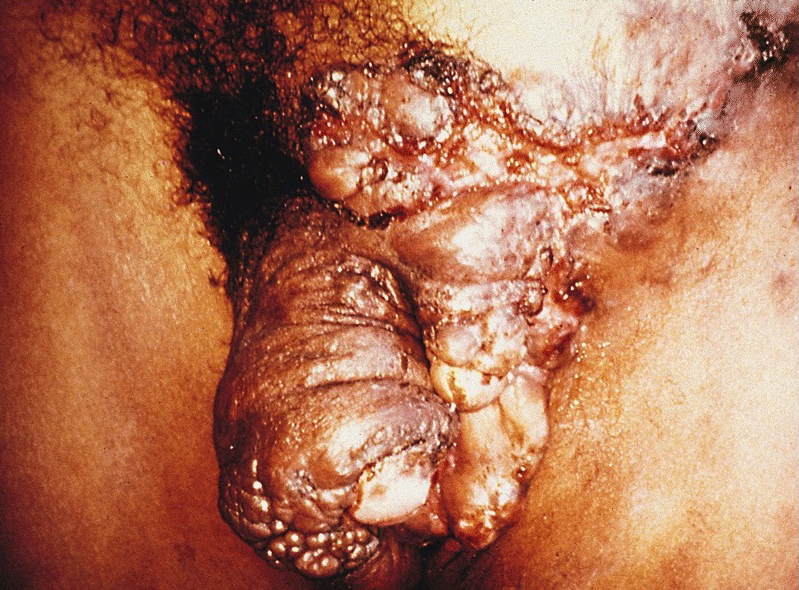
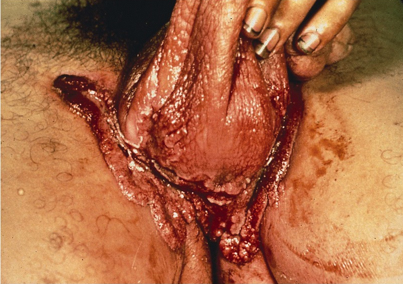
- Initially a small painful nodule at infection site that ulcerates; may have satellite lesions
Terminology
- Also called donovanosis
Epidemiology
- Rare in U.S. (100 cases/year)
- More common in African Americans, in individuals with a lower socioeconomic status and among those untrained in hygiene
- Endemic in tropical and subtropical climates such as Papua New Guinea, parts of South Africa, parts of India, Indonesia and Australian aborigines (Braz J Infect Dis 2008;12:521)
Sites
- Can affect foreskin, glans, penile shaft or scrotum
Etiology
- Caused by Klebsiella granulomatis, formerly Calymmatobacterium granulomatis, a gram negative rod (Int J Syst Bacteriol 1999;49:1695)
Case reports
- 21 year old HIV+ man with coexisting squamous cell carcinoma (Dermatol Online J 2008;14:8)
- 48 year old man (Dermatol Online J 2006;12:14, free full text)
Treatment
- Three weeks of treatment with erythromycin, streptomycin or tetracycline or 12 weeks of treatment with ampicillin
- Usually clinical improvement within 1 week
Clinical images
Microscopic (histologic) description
- Massive plasma cell infiltrate without lymphocytes in granulation tissue
- Diffuse infiltration by neutrophils forming microabscesses
- Large mononuclear cells (also called Pund cells) with Donovan bodies (large intracytoplasmic encapsulated bipolar bodies, highlighted with Warthin-Starry or Wright-Giemsa stain)
Microscopic (histologic) images
Positive stains
- Wright-Giemsa or Warthin-Starry stains show Donovan bodies in tissue sample
Electron microscopy description
- Bacteria residing inside phagosomes of macrophages
Differential diagnosis


