Table of Contents
Definition / general | Terminology | Epidemiology | Etiology | Clinical features | Treatment | Clinical images | Microscopic (histologic) description | Microscopic (histologic) imagesCite this page: Chaux A, Cubilla AL. Chancroid. PathologyOutlines.com website. https://www.pathologyoutlines.com/topic/penscrotumchancroid.html. Accessed April 1st, 2025.
Definition / general
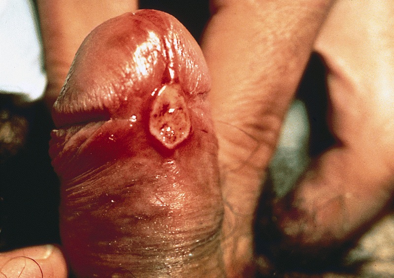
- Sexually transmitted disease caused by Haemophilus ducreyi which produces a painful genital ulcer and inguinal adenopathy
Terminology
- Dwarf chancroid: soft, painful, small ulcer
- Giant chancroid: may extend rapidly and be associated with ruptured inguinal abscess
- Phagedenic chancroid: may destroy external genitalia if superimposed Fusobacterium infection is present
- Do not confuse with chancre, a lesion typical of infection with syphilis
Epidemiology
- Mainly in developing countries, particularly Africa, Asia and Latin America
- Associated with commercial sex workers
Etiology
- Caused by Haemophilus ducreyi, a small gram negative rod
Clinical features
- Painful genital ulcer associated with tender suppurative inguinal adenopathy is suggestive
- Cofactor for HIV transmission (CDC: Sexually Transmitted Diseases Treatment Guidelines, 2006 [Accessed 28 March 2018])
- Often culture negative because Haemophilus ducreyi is very fragile in transport
- Molecular techniques are useful for diagnosis
- Must rule out Treponema pallidum (serology or darkfield examination) and HSV, which may coexist
Treatment
- Single oral dose of azithromycin or a single IM dose of ceftriaxone or oral erythromycin for seven days
Clinical images
Microscopic (histologic) description
- Zonation phenomenon at ulcer base
- Upper layer is ulcer base with fibrin, neutrophils and necrosis
- Middle layer has granulation tissue, palisading blood vessels and thrombosis
- Deep layer has marked lymphoplasmacytic infiltrate


