Table of Contents
Definition / general | Epidemiology | Sites | Etiology | Clinical features | Case reports | Gross description | Gross images | Microscopic (histologic) description | Microscopic (histologic) images | Positive stains | Differential diagnosisCite this page: Chaux A, Cubilla AL. Adenosquamous carcinoma. PathologyOutlines.com website. https://www.pathologyoutlines.com/topic/penscrotumadenosq.html. Accessed April 1st, 2025.
Definition / general
- Mixed tumor composed of neoplastic squamous nests intermingled with areas of glandular differentiation
- ICD-0: 8560 / 3
Epidemiology
- 1 - 2% of all penile carcinomas (Anal Quant Cytol Histol 2007;29:185)
- Mean age of 55 years (range 30 - 74 years)
Sites
- Most common is glans but extension to coronal sulcus and inner foreskin is also common
Etiology
- May originate in misplaced glandular cells in perimeatal region, in metaplastic goblet cells of foreskin mucosa or as aberrant differentiation of squamous epithelium
Clinical features
- Local recurrence in up to 25% and inguinal nodal metastases in 43 - 50% of cases
- Low mortality rate (0 - 14%)
Case reports
- 3 patients (ages 37, 72 and 74 years) with superficial tumors (Am J Surg Pathol 1996;20:156)
Gross description
- Firm, gray-white and granular tumor
Microscopic (histologic) description
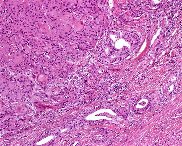
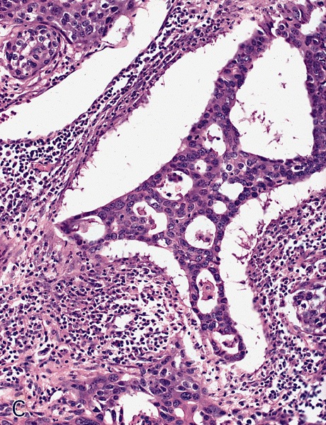
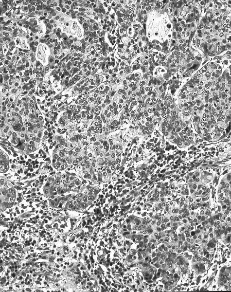
- Squamous cell and glandular patterns, with squamous cell pattern usually predominating
- Both components are usually discrete but mixtures can be found
- Glands produce intraluminal and intracellular mucin
- Frequent presence of penile intraepithelial neoplasia in adjacent mucosa
Microscopic (histologic) images
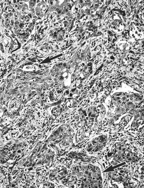
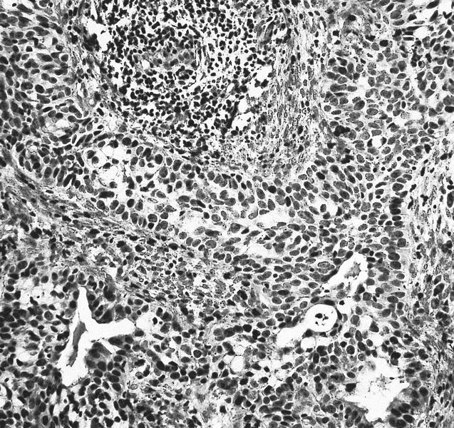
Contributed by Alcides Chaux, M.D. and Antonio Cubilla, M.D.
AFIP images
Images hosted on other servers:
Differential diagnosis
- Adenosquamous (mucoepidermoid) carcinoma of urethra: ventral in penis, restricted to periurethral tissue and corpus cavernosa
- Littré gland adenocarcinoma: ventral in penis, restricted to periurethral tissue and corpus cavernosa
- Metastatic disease: usually involves shaft, tumor emboli present (Int J Surg Pathol 2011;19:597)
- Mucoepidermoid carcinoma: mixed tumor with mucin but no glandular or ductal structures
- Pseudoglandular (acantholytic, adenoid) carcinoma: prominent acantholysis simulates glandular spaces but lining is composed of squamous epithelium; spaces contain necrotic debris and keratin, not mucin


