Table of Contents
Definition / general | Essential features | Terminology | ICD coding | Epidemiology | Sites | Pathophysiology | Etiology | Clinical features | Diagnosis | Laboratory | Radiology description | Radiology images | Prognostic factors | Case reports | Treatment | Gross description | Gross images | Microscopic (histologic) description | Microscopic (histologic) images | Virtual slides | Positive stains | Negative stains | Molecular / cytogenetics description | Sample pathology report | Differential diagnosis | Board review style question #1 | Board review style answer #1 | Board review style question #2 | Board review style answer #2Cite this page: Ma L. Metastases to ovary. PathologyOutlines.com website. https://www.pathologyoutlines.com/topic/ovarytumormetastatic.html. Accessed January 21st, 2025.
Definition / general
- Secondary involvement of ovary from malignant neoplasms of extraovarian primary sites
Essential features
- Ovary is the most common site of metastasis within the gynecologic tract
- Colorectal and breast carcinoma are the most common primary tumors to metastasize to the ovary
- Usually bilateral, small (< 10 cm), multinodular tumors with extraovarian spread
Terminology
- Krukenberg tumor
- Designation should be reserved for tumors with an appreciable component (> 10%) of signet ring cells (Gynecol Oncol 2006;101:152)
- Most commonly of gastric primary (Arch Pathol Lab Med 2006;130:1725)
ICD coding
Epidemiology
- ~15% of ovarian malignancies are of metastatic origin (range: 3 - 30%) (Int J Gynecol Cancer 2009;19:1160)
- Ovarian metastasis may present either synchronously or metachronously with primary tumor
- Ovary is the most common site of metastasis within the gynecologic tract (Int J Gynecol Pathol 2019;38:363)
- Colorectal carcinoma is the most common primary tumor to metastasize to the ovary, followed by breast carcinoma (Int J Gynecol Pathol 2023;42:414, Int J Gynecol Cancer 2009;19:1160)
Sites
- Ovary and primary organ site, with or without other organs
Pathophysiology
- Tumors can spread to the ovary by several pathways, depending on the primary
- Direct extension
- Lymphovascular / hematogeneous spread (Obstet Gynecol Int 2011;2011:612817)
- Transcoelomic / transperitoneal dissemination (e.g., Krukenberg tumors of gastric primary)
- Tumor cell exfoliation / transtubal spread (e.g., concurrent low grade endometrioid endometrial and ovarian carcinomas [FIGO stage IIIA endometrial carcinoma] simulating independent primary tumors) (Histopathology 2020;76:37)
Etiology
- Malignant tumor of extraovarian primary site
Clinical features
- Clinical features often relate to the primary tumor
- Pelvic (ovarian) mass may be the first manifestation of the disease from a clinically occult primary (Virchows Arch 2017;470:69)
Diagnosis
- Comprehensive medical history, physical evaluation, intraoperative evaluation, imaging, endoscopy
- Final diagnosis is rendered by histopathology and immunohistochemistry (with or without molecular studies)
Laboratory
- CA-125 is not a useful biomarker in the primary diagnostics of metastases to the ovary (Clin Exp Metastasis 2017;34:295)
- CA-125/CEA ratio may be of clinical use in distinguishing primary ovarian tumors from colorectal carcinoma metastases (Tumour Biol 1992;13:18)
Radiology description
- No specific imaging features differentiate primary ovarian malignancy from metastatic ovarian tumors (Magn Reson Imaging Clin N Am 2023;31:93)
- MRI appearances that suggest a mucinous lesion should trigger a search on the scan for an alternative primary mass in the gastrointestinal tract
Prognostic factors
- Ovarian metastases from colorectal carcinoma are often unresponsive to chemotherapy and are associated with poor survival (Cancer 2017;123:1134)
Case reports
- 37 year old woman with primary jejunal adenocarcinoma presenting as bilateral ovarian metastasis (Gastroenterology Res 2017;10:366)
- 57 year old Japanese woman with malignant phyllodes tumor of the breast metastasizing to the ovary (Int J Surg Pathol 2022;30:427)
- 61 year old woman with metastatic angiosarcoma to an ovarian Brenner tumor (Int J Gynecol Pathol 2023;42:176)
- 64 year old woman with isolated ovarian metastasis from pancreatic adenocarcinoma and the role of molecular analysis in establishing diagnosis (J Pancreat Cancer 2021;7:74)
- 66 year old woman with primary colorectal carcinoma and metachronous ovarian metastasis (Am J Case Rep 2019;20:1515)
Treatment
- Debulking surgery with or without chemotherapy
Gross description
- Bilateral ovarian involvement (overall occurs in 40 - 80% of cases)
- Primary mucinous ovarian tumors, uncommonly bilateral
- Size: usually < 10 cm
- Generally applied to metastatic mucinous tumors, though may be less reliable than previously reported (Gynecol Oncol 2006;101:152)
- Multinodular, solid and cystic, or friable
- Surface involvement
- Hilar involvement (common in hematogenous spread)
Microscopic (histologic) description
- Morphology varies with appearance of primary tumor
- General features
- Involvement of ovarian surface and superficial cortex
- Infiltrative growth pattern with stromal desmoplasia
- Heterogenous nodular invasive growth (Kurman: Blaustein's Pathology of the Female Genital Tract, 7th Edition, 2019)
- Metastatic tumors more often envelop preexisting normal ovarian structures than primary tumors
- Extensive lymphovascular invasion
- Colorectal adenocarcinoma
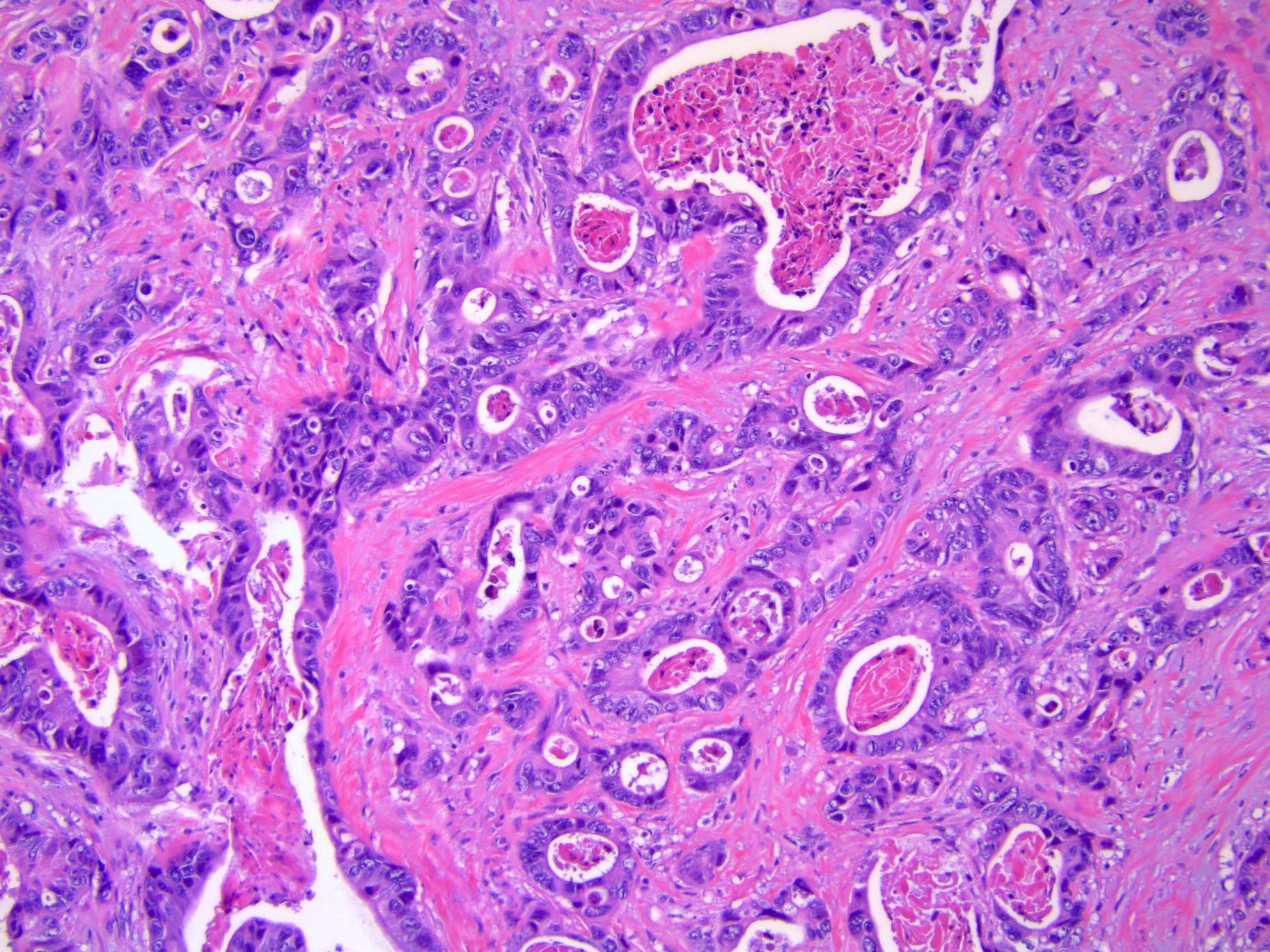
- Dilated glands with cribriform epithelium draped along the periphery of luminal dirty necrosis (garland pattern)
- Low grade appendiceal mucinous neoplasm
- Hypermucinous columnar epithelium with low grade cytologic atypia
- Retraction of epithelium from underlying stroma is commonly seen
- Abundant pseudomyxoma ovarii
- Gastric carcinoma
- Frequently presents as Krukenberg tumor (composed predominantly of signet ring cells)
- Stroma may alternate from hypercellular to hypocellular (edematous)
- Breast carcinoma
- Lobular or ductal differentiation
- Signet ring cells may be present
- Endocervical adenocarcinoma
- HPV associated
- Variety of glandular patterns
- Epithelium with hyperchromatic nuclei, apical mitoses, basal apoptoses
- HPV independent, gastric type
- Mucinous epithelium with clear / pale cytoplasm with distinct borders
- Cytologic atypia may range from minimal to severe
- HPV associated
- Endometrial adenocarcinoma
- Endometrioid histotype is more common than serous
- Synchronous endometrial and ovarian endometrioid carcinomas
- May apply Scully criteria, though recent studies suggest that these neoplasms are clonally related (J Natl Cancer Inst 2016;108:djv427, J Natl Cancer Inst 2016;108:djv428)
- Melanoma
- Follicle-like spaces are frequently seen
- Diffuse or nodular growth is common
- Intracytoplasmic melanin pigment
Microscopic (histologic) images
Virtual slides
Positive stains
- Depends on the primary tumor
- References: J Clin Pathol 2012;65:596, Int J Gynecol Pathol 2015;34:257
Negative stains
Molecular / cytogenetics description
- KRAS, SMAD4 and NTKR1 mutations are more frequent in cases of ovarian metastasis from colorectal carcinoma than in cases of colorectal carcinoma without ovarian metastasis (Cancer 2017;123:1134)
Sample pathology report
- Fallopian tube and ovary, right and left, bilateral salpingo-oophorectomy:
- Metastatic carcinoma, consistent with patient's known history of breast primary, involving bilateral ovaries (see comment)
- Bilateral fallopian tubes, negative for tumor
- Comment: On immunohistochemistry, the tumor cells are positive for GATA3, mammaglobin and GCDFP-15, while negative for PAX8. These findings support the diagnosis above.
Differential diagnosis
- Primary ovarian mucinous cystic tumor:
- Differential diagnosis for metastatic low grade appendiceal mucinous neoplasm
- Typically unilateral
- Rarely associated with pseudomyxoma peritonei
- May be associated with Brenner tumor or mature cystic teratoma
- CK7+, CK20-, SATB2-
- Primary ovarian endometrioid carcinoma:
- Differential diagnosis for metastatic colorectal adenocarcinoma
- Commonly associated with endometriosis or adenofibromatous component
- Squamous differentiation may be seen
- CK7+, ER+, PAX8+
- Differential diagnosis for metastatic endometrial endometrioid carcinoma in cases of synchronous endometrial and ovarian endometrioid carcinomas
- Scully criteria favoring primary ovarian endometrioid carcinoma (Scully: Tumors of the Ovary, Maldeveloped Gonads, Fallopian Tube, and Broad Ligament: Atlas of Tumor Pathology, 1st Edition, 1998)
- Associated with endometriosis or endometriotic lesions, a subset of which are clonal and demonstrate cancer associated mutations (J Pathol 2015;236:201)
- Low grade (FIGO grade 1 or 2)
- Histologic dissimilarity between ovarian and endometrial tumors
- No or superficial myometrial invasion of endometrial tumor
- No vascular space invasion, surface implants,or predominantly hilar location in ovary
- Ovarian tumor is unilateral
- Regardless of histopathologic criteria, genomic studies suggest that most cases are clonally related (J Natl Cancer Inst 2016;108:djv427, J Natl Cancer Inst 2016;108:djv428)
- Clonal relationship may not apply to Lynch syndrome patients (Mod Pathol 2021;34:994)
- Scully criteria favoring primary ovarian endometrioid carcinoma (Scully: Tumors of the Ovary, Maldeveloped Gonads, Fallopian Tube, and Broad Ligament: Atlas of Tumor Pathology, 1st Edition, 1998)
- Differential diagnosis for metastatic colorectal adenocarcinoma
- Signet ring stromal tumor of the ovary:
- Differential diagnosis for metastatic signet ring cell carcinoma
- Signet ring morphology in a background of fibromatous stroma
- Signet ring cells contain a single large nonmucinous intracytoplasmic vacuole
- Eosinophilic hyaline globules are common
- SF1+, calretinin+, EMA-
Board review style question #1
A 65 year old woman with an ovarian mass undergoes an oophorectomy. A representative section of the mass is shown above. Which of the following immunoprofiles best supports a diagnosis of metastatic colorectal adenocarcinoma?
- CK7+, CK20+, PAX8-, GATA3+, CDX2-, SATB2-
- CK7+, CK20-, PAX8+, GATA3-, CDX2+, SATB2-
- CK7-, CK20+, PAX8-, GATA3-, CDX2+, SATB2+
- CK7-, CK20-, PAX8-, GATA3-, CDX2-, SATB2-
Board review style answer #1
C. CK7-, CK20+, PAX8-, GATA3-, CDX2+, SATB2+. Compared with the other answer choices, this immunoprofile best supports a colorectal adenocarcinoma primary, which is typically CK7-, CK20+, PAX8-, GATA3-, CDX2+ and SATB2+. Answer A is incorrect as it fits the immunoprofile for a metastatic urothelial carcinoma. Answer B is incorrect as it is more consistent with a primary ovarian endometrioid carcinoma. Answer D is incorrect as it is not immunoreactive to any typical adenocarcinoma markers.
Comment Here
Reference: Metastases to ovary
Comment Here
Reference: Metastases to ovary
Board review style question #2
Which of the following organs is the most common site of metastasis within the gynecologic tract?
- Cervix
- Endometrium
- Fallopian tube
- Ovary
- Vulva
Board review style answer #2
D. Ovary. The ovary is the most common site of metastasis within the gynecologic tract. Answers A, B, C and E are incorrect because the vagina is the second most frequent site of metastasis within the gynecologic tract following the ovary (Cancer 1984;53:1978).
Comment Here
Reference: Metastases to ovary
Comment Here
Reference: Metastases to ovary


