Table of Contents
Definition / general | Microscopic (histologic) description | Microscopic (histologic) images | Differential diagnosisCite this page: Ehdaivand S. Endometrioid borderline tumor. PathologyOutlines.com website. https://www.pathologyoutlines.com/topic/ovarytumorendoborderline.html. Accessed April 3rd, 2025.
Definition / general
- Rare (< 200 cases reported)
- Mean age 55 years, range 28-86 years
- 97% present with stage I disease
- 36% associated with endometriosis
- Typically no recurrent disease or metastasis on follow-up, even if intraepithelial carcinoma or microinvasion (Am J Surg Pathol 2003;27:1253, Am J Surg Pathol 2000;24:1465)
Microscopic (histologic) description
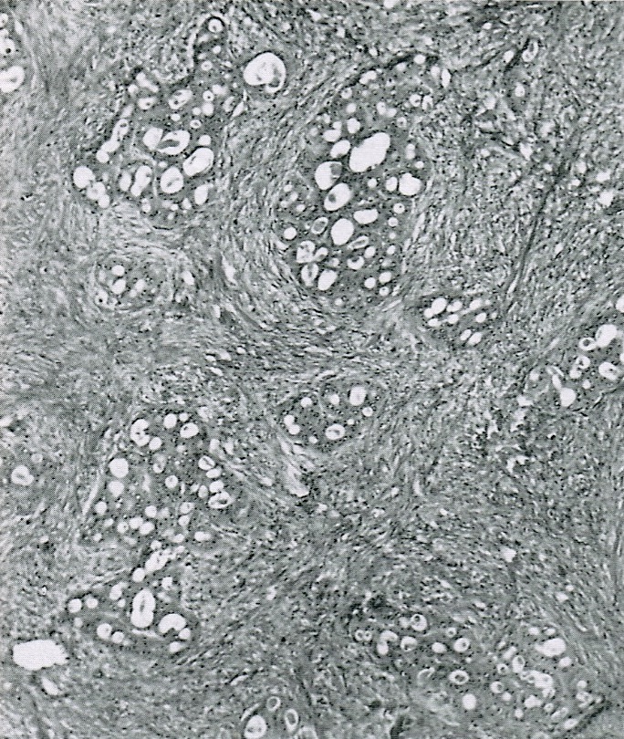
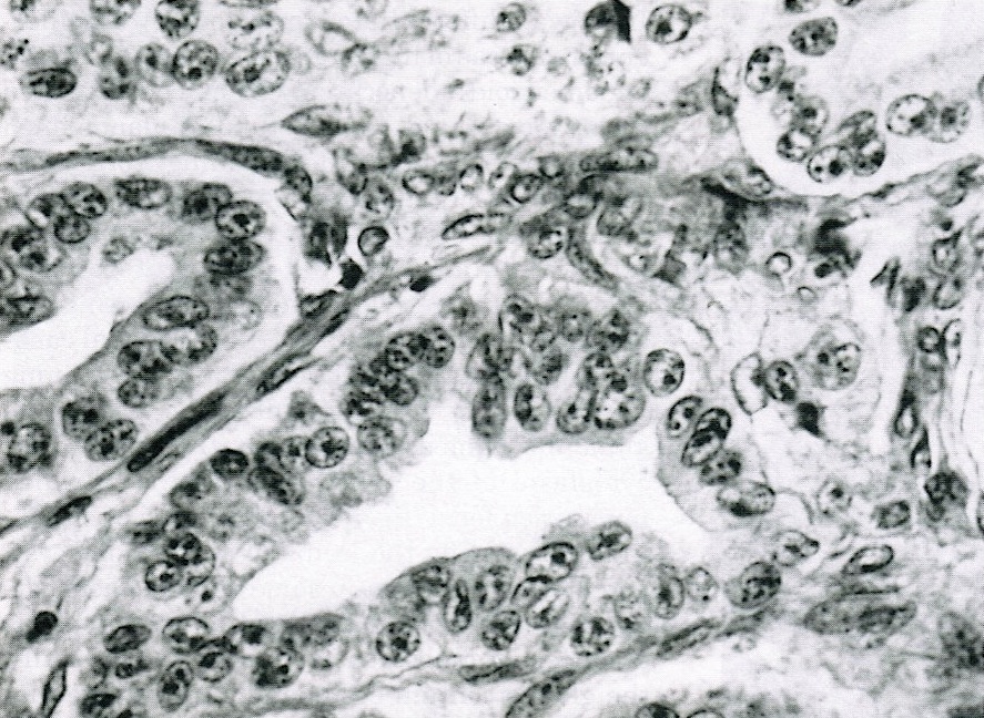
- Composed of aggregates, glands or cysts of endometrioid-type epithelium that is atypical or cytologically malignant
- May have architectural atypia including non-branching villous papillae, cribriform glands but lacks destructive stromal invasion, glandular confluence or stromal disappearance
- Often has adenofibromatous pattern (47%) or squamous differentiation (47%)
- May have foci of intraepithelial carcinoma (7%, high grade / grade 3 nuclei are large, pleomorphic with coarse chromatin or hypochromasia, large irregular nucleoli; also many mitotic figures; associated with villoglandular architecture or microinvasion)
- Microinvasion if areas of invasion are 10 mm2 or less (7%)
Microscopic (histologic) images
Differential diagnosis
- Atypical endometriosis: will have endometrial stroma


