Table of Contents
Definition / general | Epidemiology | Etiology | Clinical features | Laboratory | Case reports | Treatment | Gross description | Microscopic (histologic) description | Microscopic (histologic) images | Positive stains | Additional referencesCite this page: Ghofrani M. Granulomatous inflammation. PathologyOutlines.com website. https://www.pathologyoutlines.com/topic/ovarynontumorgranulomatous.html. Accessed December 25th, 2024.
Definition / general
- Necrotizing or nonnecrotizing granulomatous inflammation of the ovary
Epidemiology
- Cortical granulomas are common incidental findings with uncertain clinical significance
- Primary ovarian tuberculosis and other primary ovarian granulomas are uncommon
- Frequency of secondary ovarian granulomas depends on primary cause
Etiology
- Tuberculosis: hematogenous spread to ovary or secondary to tuberculous salpingitis (Arch Gynecol Obstet 2008;278:359); may clinically resemble malignancy (Arch Gynecol Obstet 2010;282:643)
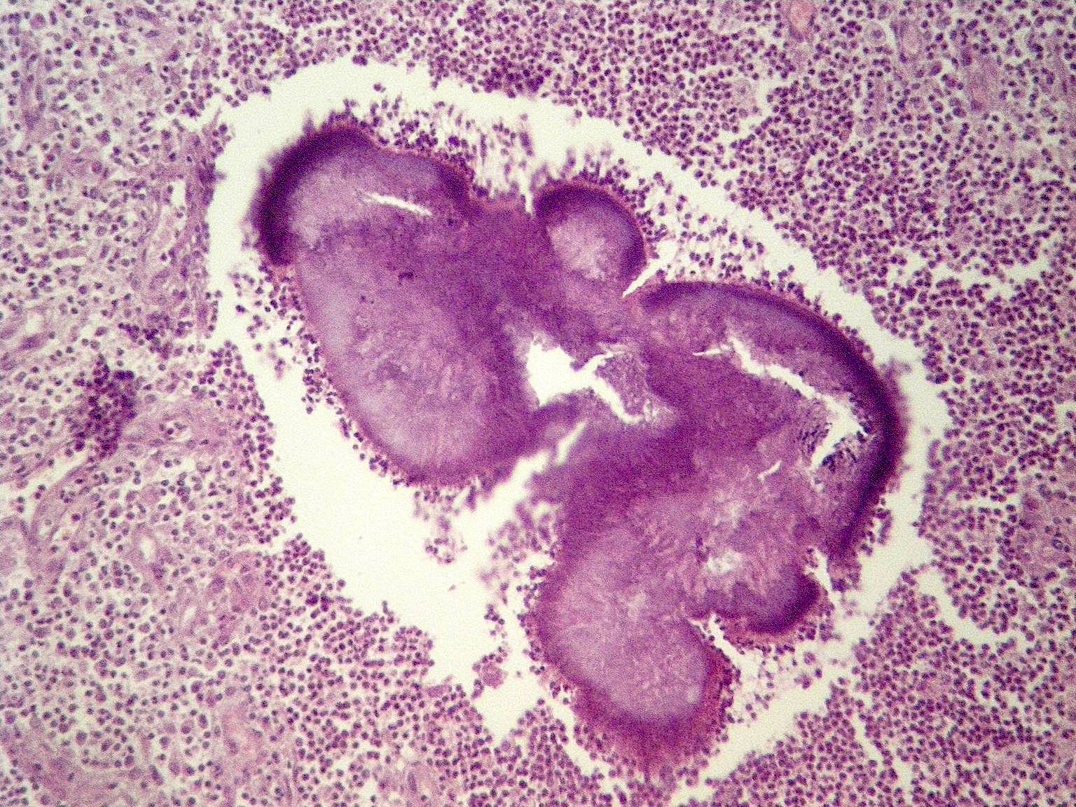
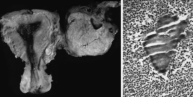
- Actinomyces: associated with IUD (Am J Clin Pathol 1977;68:622)
- Ascaris lumbricoides: J Coll Physicians Surg Pak 2009;19:663
- Bowel contents via colo-ovarian fistula: Obstet Gynecol 1987;69:533
- Crohn disease: usually extension from bowel (Am J Obstet Gynecol 1975;122:527)
- Dysgerminoma: granulomatous reaction
- Enterobius vermicularis: Eur J Gynaecol Oncol 2007;28:513
- Foreign material: often postsurgical, including suture, talc, starch, lubricant and carbon (Obstet Gynecol 1985;66:701, Hum Pathol 1996;27:1008)
- Keratin from cystic teratomas and squamous elements of endometrial and ovarian endometrioid adenocarcinomas
- Malakoplakia: Obstet Gynecol 1987;69:537
- Necrobiotic (palisading) granuloma: often with history of surgery or cauterization months to years previous
- Sarcoidosis: rare (Am J Obstet Gynecol 1990;162:1284)
- Schistosomiasis
Clinical features
- Often an incidental finding or part of a systemic granulomatous disorder
Laboratory
- May be associated with elevated CA 125, raising concern for cancer (Rom J Intern Med 2009;47:297)
Case reports
- Due to microfibrillar collagen hemostat (avitene) used to control bleeding (Arch Pathol Lab Med 1995;119:1161)
Treatment
- In secondary cases, treat primary cause
Gross description
- May present as a mass and simulate a tumor
Microscopic (histologic) description
- Tuberculosis is typically confined to the cortex
Microscopic (histologic) images
Positive stains
- AFB is typically positive in tuberculous oophoritis
Additional references


