Table of Contents
Definition / general | Essential features | Terminology | ICD coding | Epidemiology | Sites | Pathophysiology | Etiology | Clinical features | Diagnosis | Prognostic factors | Case reports | Treatment | Clinical images | Gross description | Gross images | Microscopic (histologic) description | Microscopic (histologic) images | Virtual slides | Positive stains | Negative stains | Videos | Sample pathology report | Differential diagnosis | Additional references | Board review style question #1 | Board review style answer #1 | Board review style question #2 | Board review style answer #2Cite this page: Smith MH. Pyogenic granuloma. PathologyOutlines.com website. https://www.pathologyoutlines.com/topic/oralcavitypyogenicgranuloma.html. Accessed March 27th, 2025.
Definition / general
- Benign and often reactive vascular proliferation of the oral mucosa, most commonly found on the gingiva
Essential features
- Benign red / vascular soft tissue proliferation, most commonly found on the anterior maxillary gingiva
- Associated with poor oral hygiene, chronic trauma, pregnancy, medications
- Form of capillary hemangioma that is most often considered reactive and may demonstrate a lobular architecture histopathologically
- May demonstrate rapid growth and clinically mimic malignancy (Clin Case Rep 2018;6:690, Case Rep Dent 2021;2021:5575896)
- Constitutes 3.8 - 7% of oral biopsy diagnoses (J Oral Maxillofac Surg 2010;68:2185)
Terminology
- Lobular capillary hemangioma, subtype
- Pregnancy tumor
- Epulis gravidarum / granuloma gravidarum: pyogenic granuloma occurring in a setting of pregnancy
- Epulis granulomatosum: pyogenic granuloma located in an extraction socket
ICD coding
Epidemiology
- Large age range; most common in the second and third decades of life (J Oral Maxillofac Surg 2010;68:2185, Oral Maxillofac Surg 2012;16:305)
- Female predilection (J Oral Maxillofac Surg 2010;68:2185, Pathol Int 2005;55:391, Oral Maxillofac Surg 2012;16:305)
Sites
- Gingiva affected most often (maxillary anterior) (J Oral Maxillofac Surg 2010;68:2185, Head Neck Pathol 2019;13:4)
- May also affect lips, buccal mucosa, tongue or palate
Pathophysiology
- Largely unknown, although in pregnancy, estrogen enhances vascular endothelial growth factor (VEGF) production in macrophages, likely contributing to the development of pyogenic granulomas (J Dermatol Sci 2005;38:1)
- Also in pregnancy, progesterone may function as an immunosuppressant in gingiva, preventing an acute inflammatory reaction against oral bacteria and resulting in proliferative gingival inflammation (J Clin Periodontol 1991;18:262)
- Increased estrogen and progesterone in pregnancy increases concentrations of Prevotella intermedia in the subgingival biofilm, decreases the host response to the bacteria and increases the vascular permeability and infiltration of fluids into the gingival tissues, contributing to formation of pyogenic granulomas (J Appl Oral Sci 2013;21:215)
- 2018 International Society for the Study of Vascular Anomalies (ISSVA) Classification of Vascular Anomalies lists BRAF, RAS and GNA14 as causal genes for the lobular capillary hemangioma (pyogenic granuloma) (ISSVA: ISSVA Classification for Vascular Anomalies [Accessed 11 August 2021])
Etiology
- Associated with chronic irritation (J Oral Maxillofac Surg 2010;68:2185, Head Neck Pathol 2019;13:4)
- Poor oral hygiene
- Ill fitting oral appliances or overhang margins of dental restorations
- Persistent bite trauma
- May be seen in patients with drug induced gingival overgrowth from use of calcium channel blockers, calcineurin inhibitors, antiseizure medications or anticancer medications (e.g. TNF alpha antagonists, BRAF inhibitors, tyrosine kinase inhibitors, epidermal growth factor receptor inhibitors, mTOR inhibitors, taxanes, pyrimidine analogs) (Case Rep Dent 2021;2021:5575896, Pediatr Transplant 2020;e13947)
Clinical features
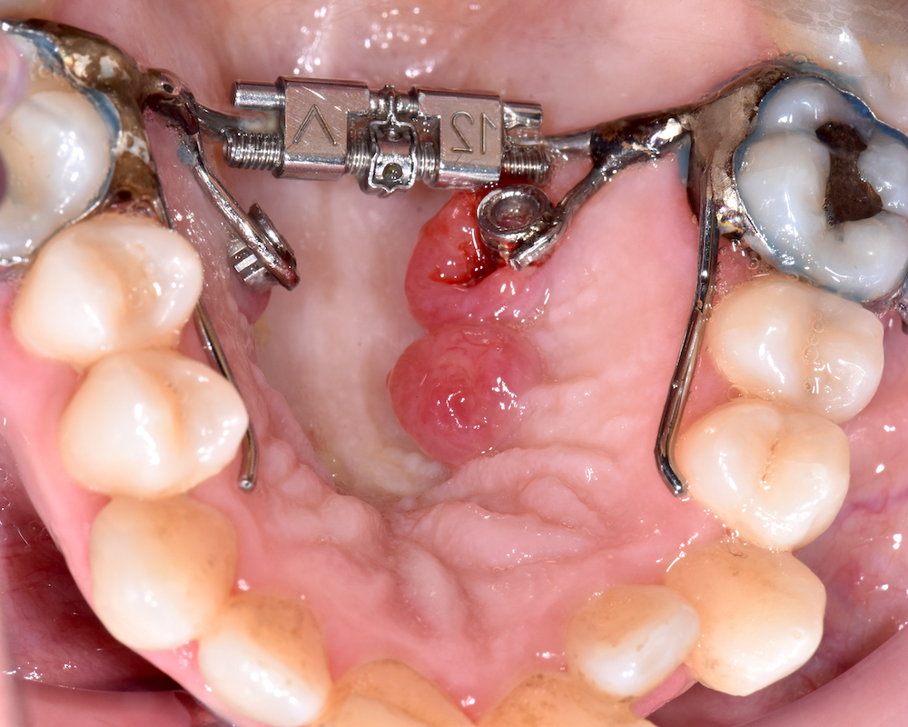
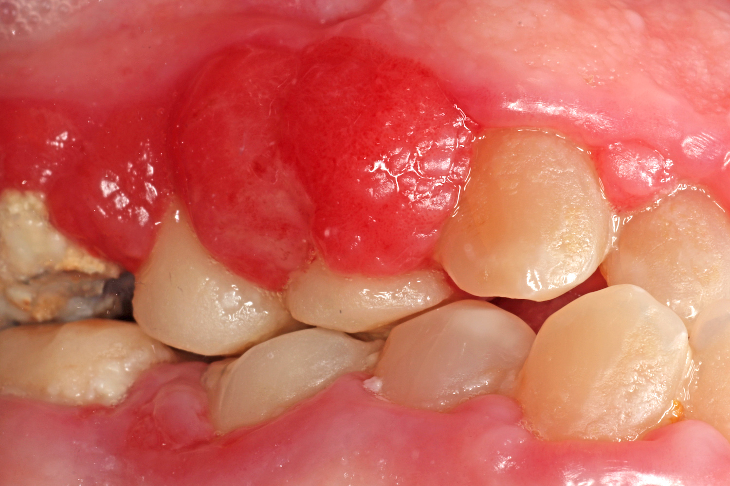
- Smooth or lobulated exophytic mass
- Ranges from red / pink to deep red / blue / purple in color (Head Neck Pathol 2019;13:4)
- Frequently ulcerated or crusted (Pathol Int 2005;55:391)
- Often pedunculated, although the lobular capillary hemangioma variant is more commonly sessile (Pathol Int 2005;55:391)
- May bleed easily upon gentle manipulation
- May exhibit rapid growth (Case Rep Dent 2021;2021:5575896)
- 1 mm to several centimeters in diameter
- Lesions tend to become more fibrotic / pink / rubbery over time and with elimination of irritating factors (termed sclerosed pyogenic granuloma or inflammatory fibrous hyperplasia) (J Oral Maxillofac Surg 2010;68:2185)
Diagnosis
- Made upon histopathological examination of the excisional specimen
Prognostic factors
- Good prognosis with low recurrence rate (8.2%) (J Oral Maxillofac Surg 2010;68:2185)
Case reports
- 15 and 26 year old women with large, progressive pyogenic granulomas (Clin Case Rep 2018;6:690)
- 51 year old woman with pyogenic granuloma treated with diode laser (Saudi Med J 2016;37:1395)
- 57 year old man taking amlodipine with rapid development of an epulis granulomatosum (Case Rep Dent 2021;2021:5575896)
Treatment
- Conservative excision, extending to periosteum, with subsequent histopathologic examination (J Oral Maxillofac Surg 2010;68:2185, Neville: Oral and Maxillofacial Pathology, 4th Edition, 2015)
- Elimination of precipitating factors
- Granuloma gravidarum (pyogenic granuloma) should be excised after pregnancy, unless significant esthetic or functional concerns are present (J Appl Oral Sci 2013;21:215)
- In pregnancy, pyogenic granulomas may spontaneously regress after removal of local irritants or after childbirth (J Appl Oral Sci 2013;21:215)
Clinical images
Contributed by Brittany Camenisch, D.M.D., Nehal Almehmadi, B.D.S., Sana Naheed, B.D.S., Evan Lynch, M.D., Ph.D.,
Justin Kolasa, M.D., D.M.D., Molly Housley Smith, D.M.D. and Susanna Goggin, D.M.D.
Gross description
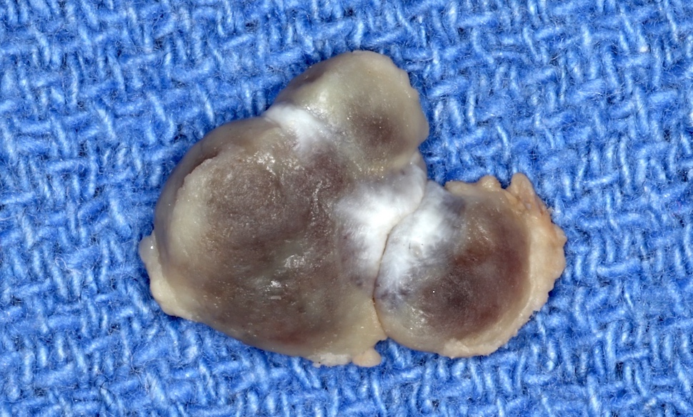
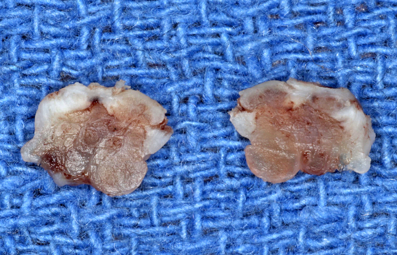
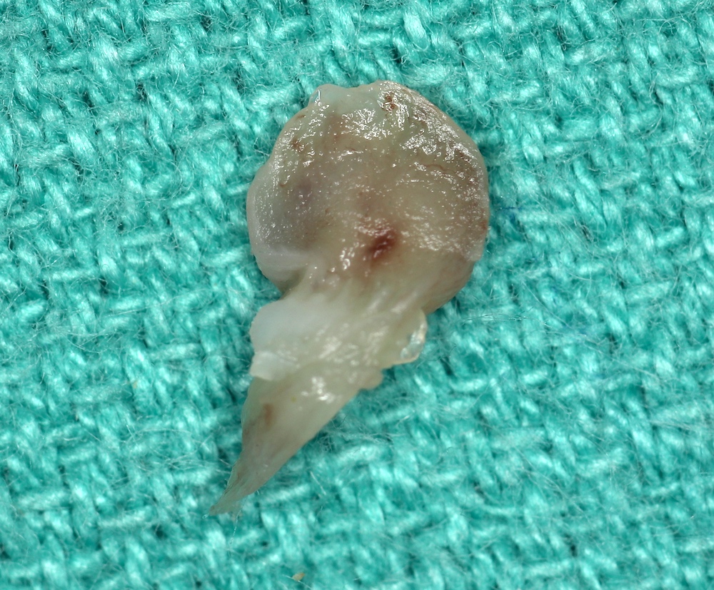
- Tan / brown mass, often pedunculated or lobulated
- May demonstrate hemorrhagic foci on cut surface (Case Rep Dent 2016;2016:1323798)
- Often exhibits ulcerated surface
Gross images
Microscopic (histologic) description
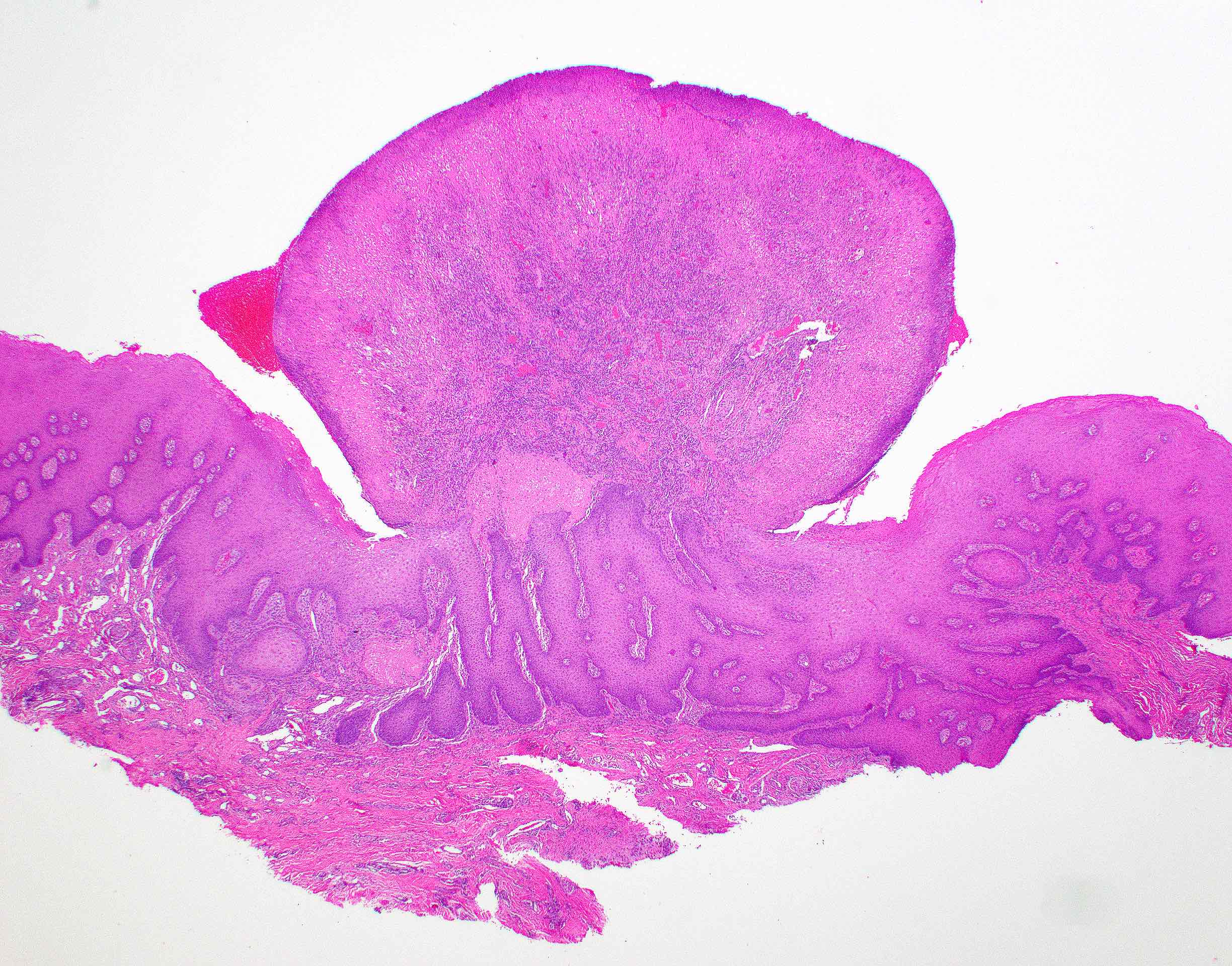
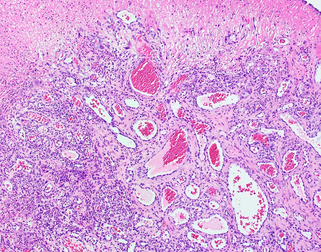
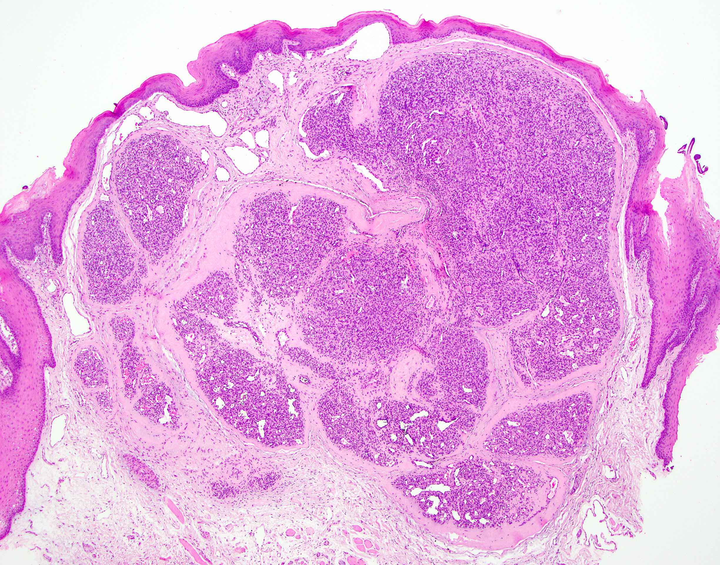
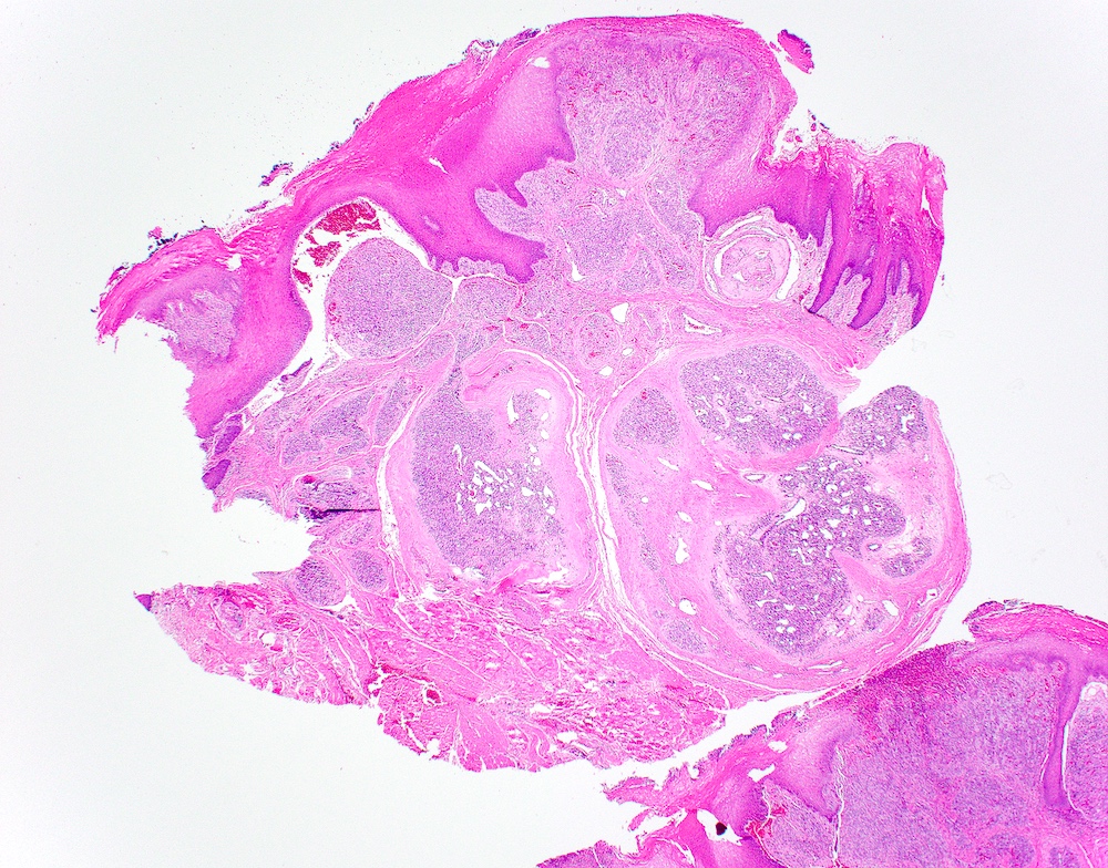
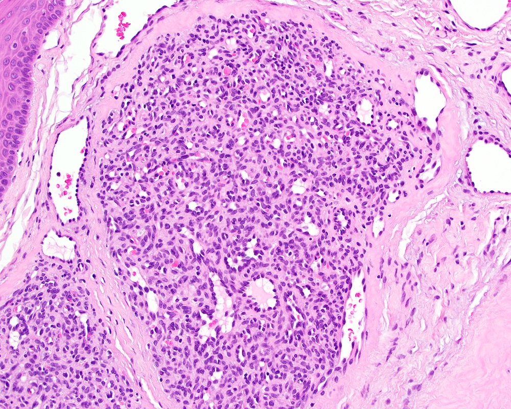
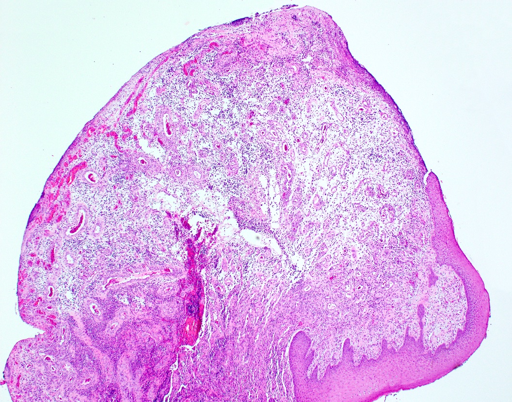
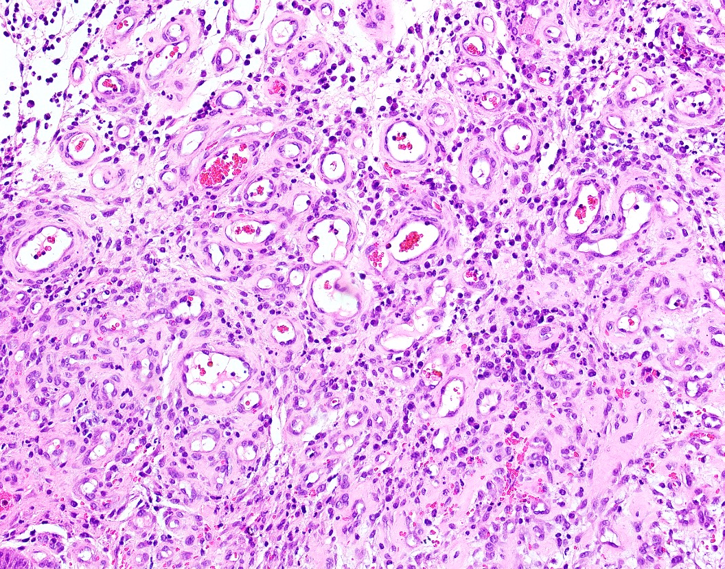
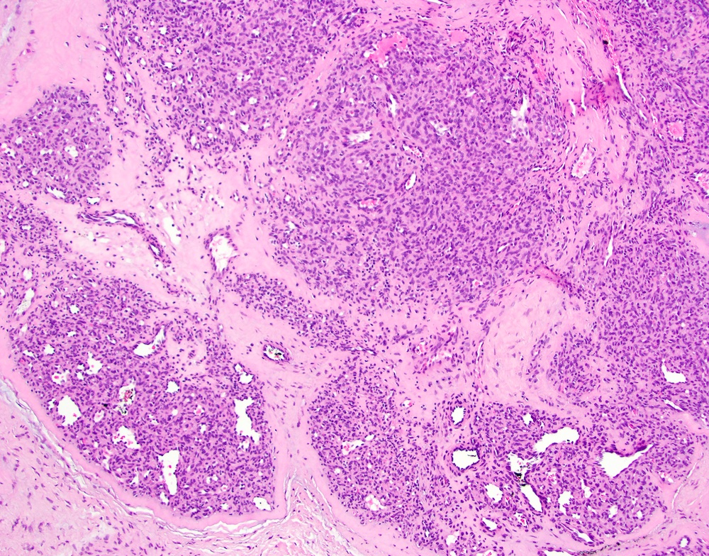
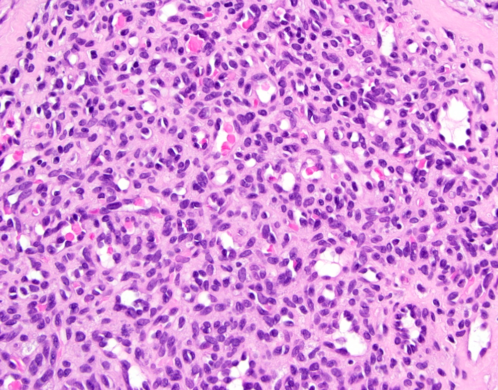
- Highly vascularized proliferation of granulation tissue (J Oral Maxillofac Surg 2010;68:2185)
- Often demonstrates surface ulceration and a subacute inflammatory cell infiltrate comprised of neutrophils, lymphocytes and plasma cells
- May demonstrate a lobular arrangement of capillary vessels and proliferating endothelial cells delineated by fibrous septae (termed lobular capillary hemangioma)
- May be pedunculated
- May show brisk mitotic rate (up to 10 mitotic figures per high power field); however, lacks pleomorphism (Ann Diagn Pathol 2020;46:151506)
Microscopic (histologic) images
Positive stains
- Immunohistochemistry is not routinely performed; however, CD34, CD31, erythroblast transformation specific related gene (ERG), alpha smooth muscle actin (SMA) and muscle specific actin (MSA) highlight the prominent vasculature throughout the mass
- WT1 (Ann Diagn Pathol 2020;46:151506)
Negative stains
Videos
Histopathology of pyogenic granuloma
Sample pathology report
- Gingiva, excision:
- Pyogenic granuloma
Differential diagnosis
- Peripheral ossifying fibroma:
- May be clinically identical
- Only on the gingiva / alveolar ridge
- Differentiated based on histopathologic examination
- Peripheral ossifying fibroma demonstrates the presence of bone, at least focally, within the mesenchymal stroma
- Peripheral giant cell granuloma:
- May be clinically identical
- Only on the gingiva / alveolar ridge
- Differentiated based on histopathologic examination
- Presence of osteoclast type multinucleated giant cells
- Parulis:
- May be clinically identical, although may express red-yellow fluid on palpation
- Associated with an adjacent nonvital tooth or inflamed periodontal pocket
- Can be traced using gutta percha to reveal the inflammatory origin
- Differentiated based on histopathologic examination
- Accumulations of neutrophils arranged in a linear fashion
- Metastatic disease:
- May be clinically identical
- Differentiated based on histopathologic examination
- Gingival squamous cell carcinoma:
- May be clinically identical, although often demonstrates a granular or papillary surface architecture (Oral Surg Oral Med Oral Pathol Oral Radiol Endod 2009;107:92)
- Differentiated based on histopathologic examination
- Invasive islands of squamous epithelium arising from surface
- Spongiotic gingival hyperplasia / localized juvenile spongiotic gingival hyperplasia:
- Papillary / pebbly surface architecture
- Recalcitrant to oral hygiene measures
- < 1 cm in size (J Cutan Pathol 2019;46:839)
- Traditionally localized, although multifocal examples have been noted
- Histopathologic examination reveals papillary proliferation of spongiotic, nonkeratinized squamous epithelium with elongation of rete ridges, intermixed capillary vessels and neutrophilic exocytosis
- Angiosarcoma:
- Rare in the oral cavity (Int J Oral Maxillofac Surg 2014;43:917)
- Differentiated based on histopathologic examination
- Infiltrative
- May demonstrate rounded, polygonal or spindled cells, often with an epithelioid morphology (Lancet Oncol 2010;11:983)
- Brisk mitotic rate with atypical mitotic figures or pleomorphism
- Forms vascular sinusoids and less clearly defined vascular spaces (Lancet Oncol 2010;11:983)
- May demonstrate keratin or EMA positivity
- Kaposi sarcoma:
- Often multifocal or associated with diffuse violaceous gingival enlargement (JAMA Dermatol 2019;155:370)
- Associated with AIDS (JAMA Dermatol 2019;155:370)
- Differentiated based on histopathologic examination
- HHV8 positive
Additional references
Board review style question #1
The above mass is found on the gingiva. What is true regarding this diagnosis?
- This entity is more commonly found in pregnant women
- This entity is more commonly found in men
- This entity is more commonly found in patients who are taking long term steroids
- No treatment is indicated for this particular entity; clinical diagnosis is sufficient
Board review style answer #1
Board review style question #2
Board review style answer #2


