
Mandible & maxilla
Other benign tumors / uncertain malignant potential tumors / tumor-like conditions
Melanotic neuroectodermal tumor of infancy
Last author update: 1 September 2017
Last staff update: 6 October 2022
Copyright: 2004-2025, PathologyOutlines.com, Inc.
PubMed Search: Melanotic neuroectodermal tumor of infancy [title]


Table of Contents
Definition / general | Essential features | Terminology | Epidemiology | Sites | Etiology | Clinical features | Diagnosis | Laboratory | Radiology images | Prognostic factors | Case reports | Treatment | Clinical images | Gross description | Gross images | Microscopic (histologic) description | Microscopic (histologic) images | Positive stains | Negative stains | Molecular / cytogenetics description | Differential diagnosis | Board review style question #1 | Board review style answer #1Cite this page: Magliocca K, and Martinez A. Melanotic neuroectodermal tumor of infancy. PathologyOutlines.com website. https://www.pathologyoutlines.com/topic/mandiblemaxillaPigNEtumor.html. Accessed April 2nd, 2025.
Definition / general
- A rare, locally aggressive biphasic tumor composed of a small round blue cell (neuroblast-like) component along with larger, melanin producing epithelioid cells
Essential features
- A rapidly growing lesion in infants (usually in the first year of life) that can be associated with elevated VMA
- Histologically is a biphasic tumor composed of a small round blue cell neuroblast-like component and larger, melanin producing epithelioid cells
Terminology
- Melanotic neuroectodermal tumor of infancy (MNTI)
- Other synonyms the current WHO no longer recommends:
- Melanotic progonoma
- Retinal anlage tumor
Epidemiology
- Rare, > 90% are in infants
- Median age 5 months
- Slightly more common in males (J Oral Maxillofac Surg 2015 Oct;73:1946)
Sites
- Most cases occur in the craniofacial region, most commonly the maxilla ( > 60% of cases)
Etiology
- Thought to be neural crest origin, based on:
- Secretion of vanillylmandelic acid (VMA), characteristic of other neural crest tumors such as pheochromocytoma and neuroblastoma
- VMA levels generally return to normal when excised
Clinical features
- Usually presents as a pigmented, rapidly expansile mass
Diagnosis
- Based on clinical, radiographic and pathologic features, but most important is biphasic components with histology
Laboratory
- Can show elevated vanillylmandelic acid (VMA) levels
Radiology images
Images hosted on other servers:

Presence of bone erosion

Heterogenously enhancing soft tissue mass lesion

Radiolucent expansile, solid lesion
Prognostic factors
- Wide range of recurrence rates:
- One large study found local recurrence rates after resection of 10 - 15% (Oral Surg Oral Med Oral Pathol Oral Radiol Endod 2006;102:204)
- Others indicate recurrence rate is > 35% (Fetal Pediatr Pathol 2006;25:59)
- A systematic review found a metastatic rate of 6.5% (Oral Surg Oral Med Oral Pathol Oral Radiol Endod 2006;102:204)
Case reports
- 2 month old female with mass arising in fibula (BMC Cancer 2016;16:629)
- 3 month old girl with swelling of the upper gums (J Clin Diagn Res 2016;10:ZJ07)
- 3 month old with MNTI involving anterior maxilla (Ann Maxillofac Surg 2015;5:234)
- 10 month old with soft tissue mass near right elbow (Int J Clin Exp Pathol 2015;8:13584)
Treatment
- Complete local excision with clear margins
- Adjuvant therapy for recurrent or residual tumor
Clinical images
Images hosted on other servers:

Swelling on maxillary alveolus

Upper vestibule, alveolar ridge and anterior hard palate
Gross description
- Lesions are usually gray to blue, firm and lobulated
Gross images
Images hosted on other servers:

Excised tumor
Microscopic (histologic) description
- Biphasic population of cells composed of:
- Nodules of round blue cells that are small and hyperchromatic with scant cytoplasm, often termed “neuroblastoma-like”
- A second population of larger epithelioid cells arranged in
cords and nests composed of abundant pale cytoplasm and round nuclei with vesicular
chromatin
- Within the cytoplasm is melanin pigment, although it can be focal and difficult to identify
- The background consists of dense fibrosis creating the appearance of third component.
Microscopic (histologic) images
Contributed by Kelly Magliocca, D.D.S., M.P.H. and Karen Fritchie, M.D.
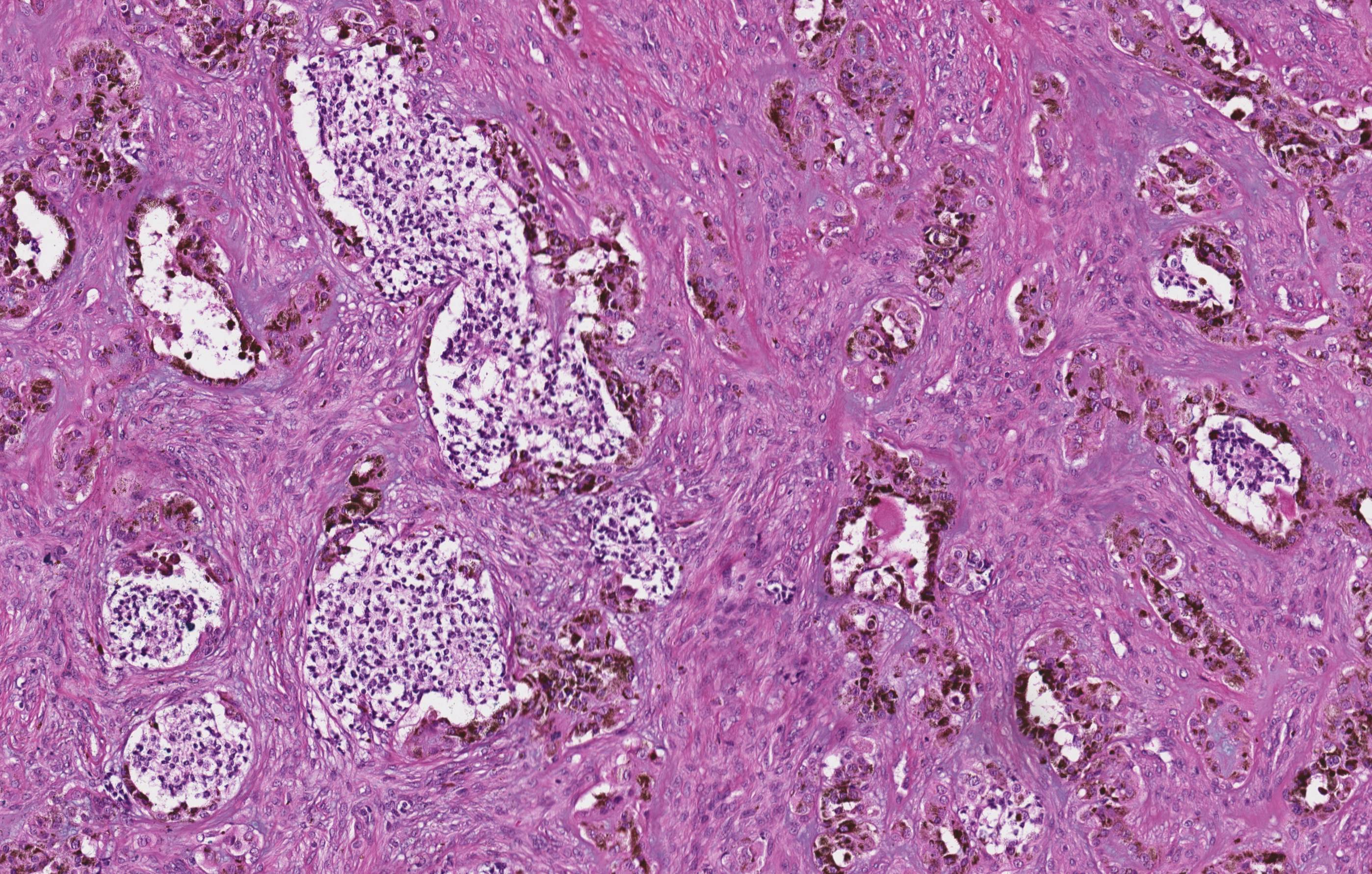
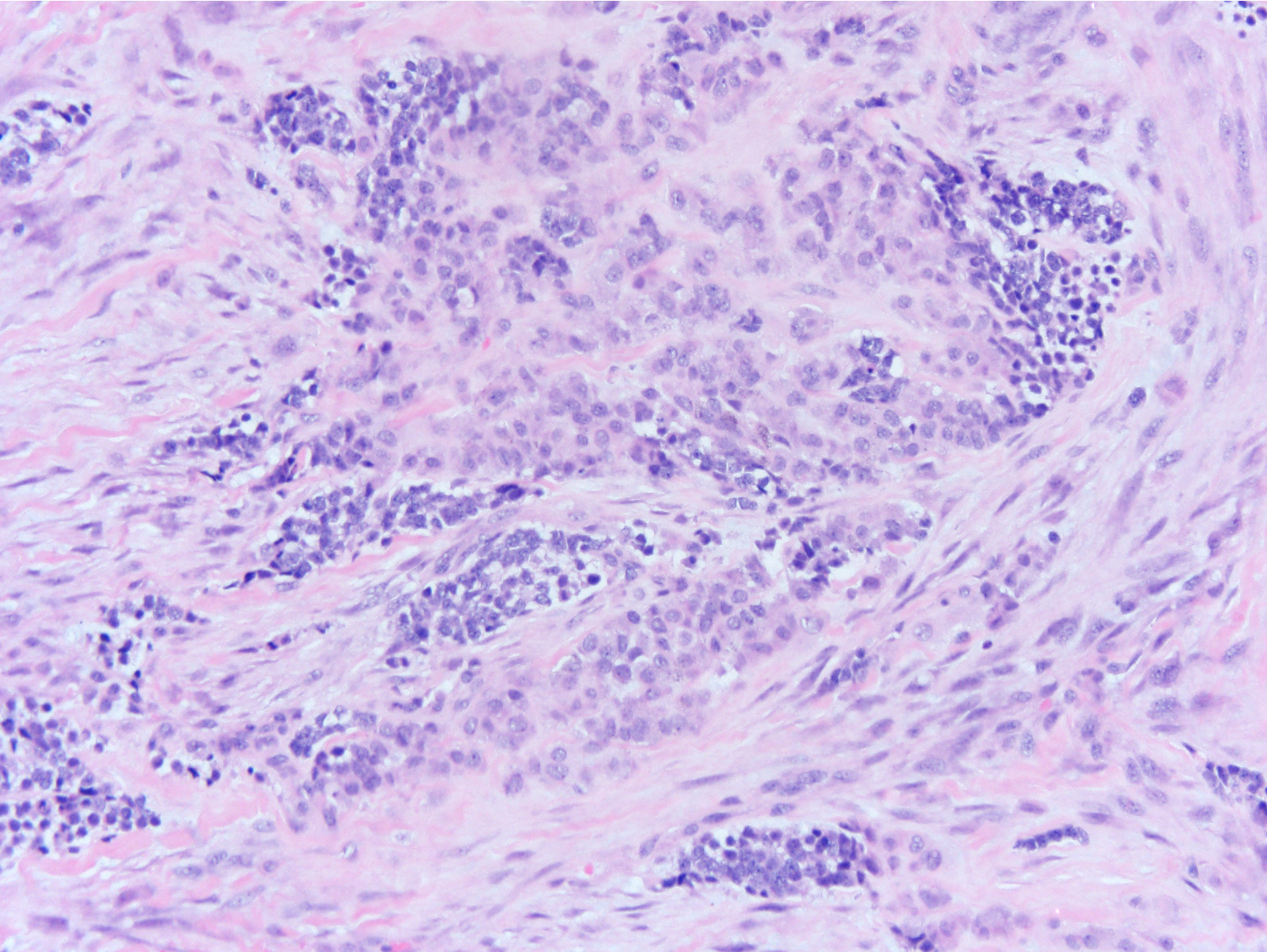
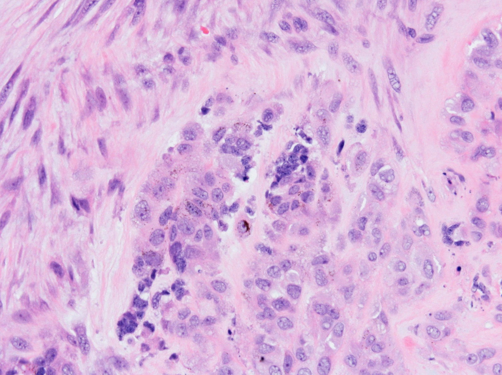
Melanotic neuroectodermal tumor of infancy
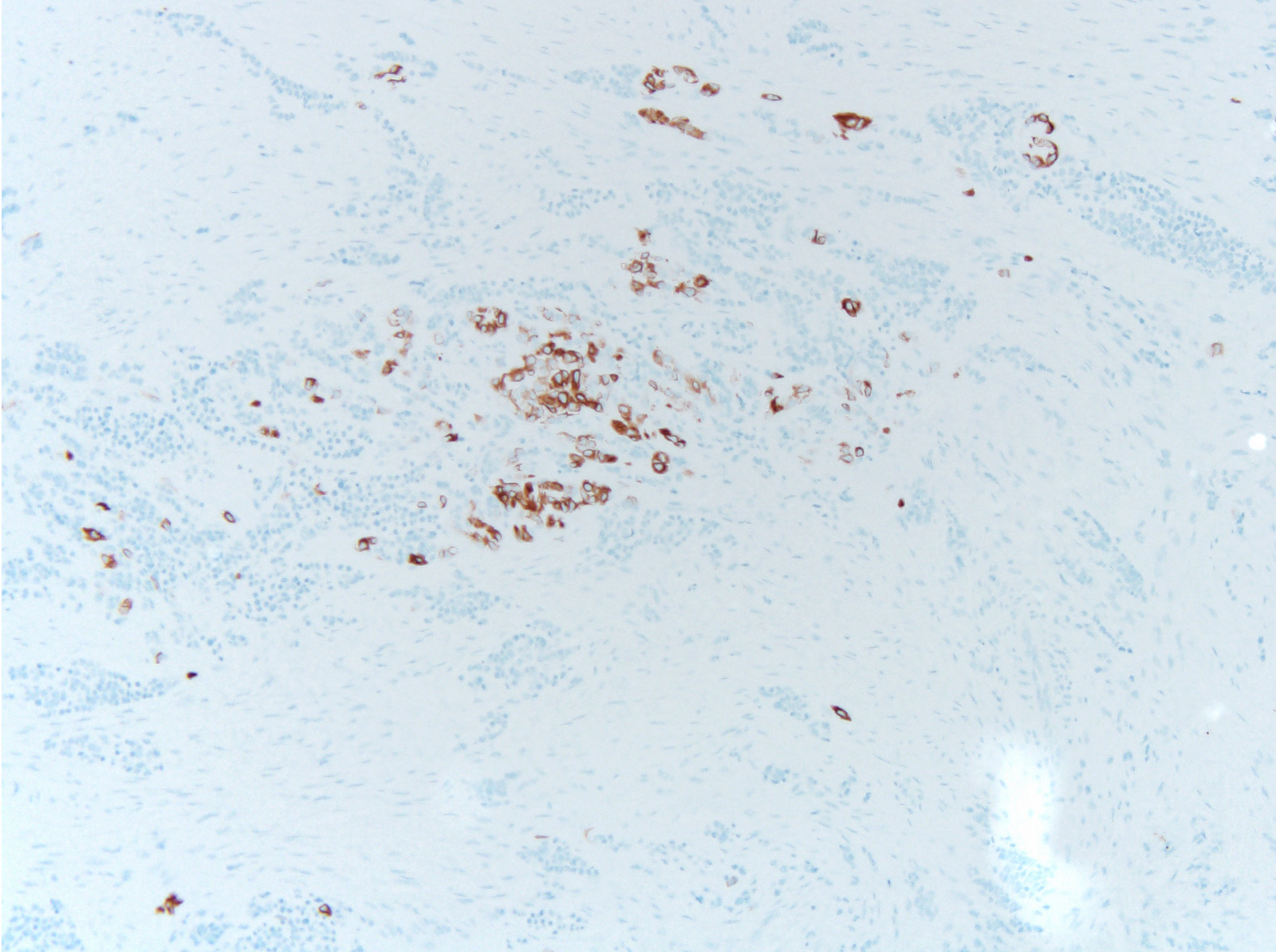
AE1 / AE3
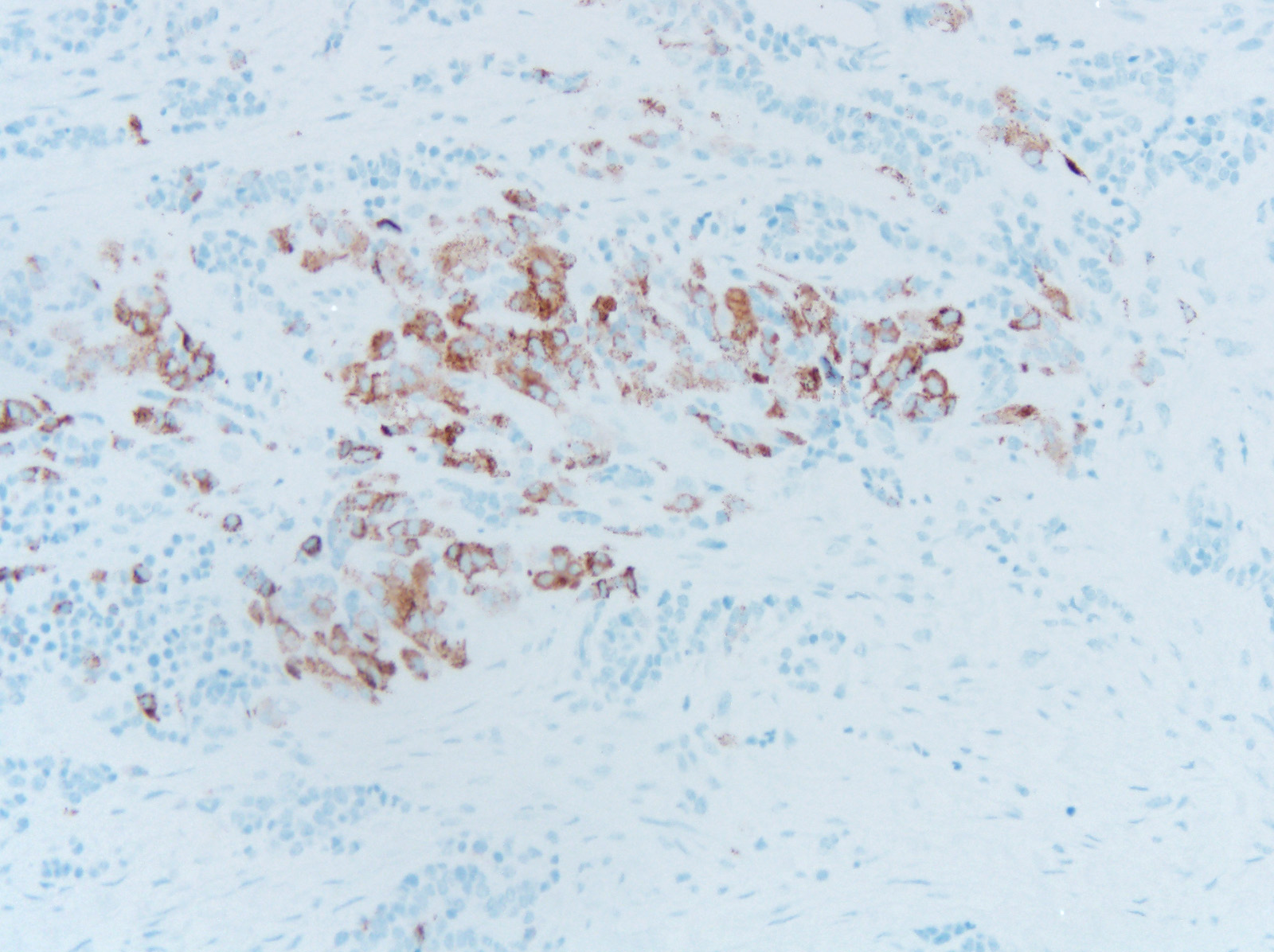
HMB45
Positive stains
Negative stains
- Usually negative for S100, chromogranin
Molecular / cytogenetics description
- Limited data but occasional mutations identified:
- Germline mutation of CDKN2A and a novel RPLP1- C19MC fusion identified in 1 case (BMC Cancer 2016;16:629)
- A BRAFV600E mutation was found in 1 case (Pediatrics 2015;136:e267)
Differential diagnosis
- Ewing sarcoma
- Only 2 - 10% develop in the head and neck
- Small round blue cell tumor that grows in sheets and nests
- Can have similar appearance to “neuroblast-like” component of MNTI, but lacks the epithelioid component
- Should have rearrangements of EWSR1, unlike MNTI
- Olfactory neuroblastoma
- Most common during fifth and sixth decades
- Typically involves the cribriform plate, nasal concha and septum
- Also composed of small round blue cells
- Can have Homer-Wright rosettes imparting biphasic
morphology, but they are all the same population of cells
- These will also stain with NSE and synaptophysin like the primitive component of MNTI
- No epithelioid component with melanin pigment
- Can have Homer-Wright rosettes imparting biphasic
morphology, but they are all the same population of cells
- Desmoplastic small round cell tumor
- Rare, aggressive tumor, usually in abdomen of adolescents
and young adults
- More commonly affects men
- Composed of nests of round blue cells with variable amounts of cytoplasm and hyperchromatic nuclei surrounded by desmoplastic stroma
- Should not have the epithelioid component with melanin pigment
- Also shows EWSR1 rearrangement as in Ewing sarcoma
- Rare, aggressive tumor, usually in abdomen of adolescents
and young adults
- Rhabdomyosarcoma
- Individuals tend to be older at presentation than MNTI
- More commonly involves sinonasal tract
- Also composed of small primitive round blue cells but with scattered rhabdomyoblasts
- Embryonal most common subtype in younger children
- Should be positive for myogenic markers such as myogenin and myoD1
- Should not have the epithelioid component with melanin pigment of MNTI
- Lymphoma
- Approximately 16% of NHLs involve the jawbones
- 2/3 are diffuse large B cell lymphoma (DLBCL) which usually occur in an older age group
- Main lymphoma mimics for MNTI are lymphoblastic lymphoma or Burkitt lymphoma, which feature monotonous, small to medium size round cells, without a biphasic component
- Flow cytometry, immunohistochemistry (such as Tdt) and molecular studies would help classify and risk stratify the patient
- Approximately 16% of NHLs involve the jawbones
Board review style question #1
What rearrangement has been described in melanotic neuroectodermal
tumor of infancy?
A. EWRS1-FLI1
B. EWSR1-WT1
C. PAX3-FOXO1
D. RPLP1-C19MC
A. EWRS1-FLI1
B. EWSR1-WT1
C. PAX3-FOXO1
D. RPLP1-C19MC
Board review style answer #1
D. RPLP1-C19MC has been described in one case of melanotic neuroectodermal tumor of infancy. EWRS1-FLI1 is seen in Ewing sarcoma, EWSR1-WT1 is seen in desmoplastic small round cell tumor and PAX3-FOXO1 is seen in alveolar rhabdomyosarcoma
Comment Here
Reference: Melanotic neuroectodermal tumor of infancy
Comment Here
Reference: Melanotic neuroectodermal tumor of infancy
