Table of Contents
Definition / general | Essential features | Terminology | ICD coding | Epidemiology | Sites | Pathophysiology | Etiology | Clinical features | Diagnosis | Radiology images | Prognostic factors | Case reports | Treatment | Clinical images | Gross description | Gross images | Microscopic (histologic) description | Microscopic (histologic) images | Virtual slides | Cytology description | Cytology images | Positive stains | Negative stains | Differential diagnosis | Additional references | Board review style question #1 | Board review style answer #1Cite this page: Wu R, Jain D. Pleomorphic carcinoma. PathologyOutlines.com website. https://www.pathologyoutlines.com/topic/lungtumorpleomorphic.html. Accessed April 3rd, 2025.
Definition / general
- Pleomorphic carcinoma:
- Subtype of sarcomatoid carcinoma; usually aggressive, malignant epithelial neoplasm composed of cells with significant cytologic atypia and nuclear pleomorphism
- Contains at least 10% spindle cells and / or giant cells
- Included under recent WHO classification of "carcinomas with pleomorphic, sarcomatoid or sarcomatous elements"
- Giant cell carcinoma:
- Subtype of sarcomatoid carcinoma consisting of purely giant, pleomorphic tumor cells
- Should not show differentiated non small cell components
- Tumor stains for cytokeratins
- Spindle cell carcinoma:
- Subtype of sarcomatoid carcinoma (WHO)
- < 1% of all primary lung carcinomas
Essential features
- Giant cell carcinoma:
- Classified as a subtype of sarcomatoid carcinoma that is composed almost entirely of tumor giant cells with no differentiated carcinomatous elements
- Tumor giant cells may or may not stain for human chorionic gonadotrophin
- Metastatic sarcoma or germ cell tumor should be excluded
Terminology
- Giant cell carcinoma:
- First described and characterized in 1958 (Cancer 1958;11:369)
- Use specific term giant cell carcinoma rather than general term sarcomatoid carcinoma whenever possible to avoid confusion (J Thorac Oncol 2015;10:1243)
- Giant cells can be a component of pleomorphic carcinoma; if tumor shows giant cells along with differentiated non small cell component, classify as pleomorphic carcinoma (Arch Pathol Lab Med 2010;134:1645)
- Large cell carcinoma is a different entity
ICD coding
- Giant cell carcinoma:
- Use code specific for location of tumor
- ICD-10: C34.90 - malignant neoplasm of unspecified part of unspecified bronchus or lung
Epidemiology
- Pure giant cell carcinoma is rare, 0.3 - 2% of lung cancers
- Mean age: 65 years, range: 42 - 81 years
- > 90% men, 92% smokers
Sites
- Giant cell carcinoma:
- May show predilection for upper lobes
- Giant cell tumors frequently metastasize to small intestine
Pathophysiology
- Giant cell carcinoma: may have increased incidence of gastrointestinal tract involvement (Cancer 1992;70:606)
Etiology
- Giant cell carcinoma: appears to be a morphologic phenotype expressed by a heterogeneous group of tumors (Histopathology 1998;32:225)
Clinical features
- Pleomorphic carcinoma:
- < 1% of all carcinomas
- Presumed epithelial origin, although epithelial and sarcomatous components express common markers differently (Am J Surg Pathol 2003;27:1203)
- Classified as carcinomas despite presence of sarcomatoid features (Arch Pathol Lab Med 2010;134:1645)
- Nodal metastases common
- Giant cell carcinoma:
- Cough, chest pain, dyspnea, malaise (varies by location)
Diagnosis
- Giant cell carcinoma: cannot be made on small biopsies or cytology; definite diagnosis only on resected tumor
Prognostic factors
- Giant cell carcinoma:
- Related to stage
- Extent of giant cells or trophoblastic differentiation does not affect prognosis (Histopathology 1998;32:225)
- Pleomorphic carcinoma:
- Stage 1 tumors have same prognosis as other stage 1 non small cell carcinomas; at higher stages, may have worse prognosis than other non small cell carcinomas of similar stage (Am J Surg Pathol 2003;27:311)
Case reports
- 51 year old man with small intestinal metastases (World J Surg Oncol 2005;3:32)
- 55 year old woman with jejunal intussusception from metastatic giant cell carcinoma of lung (BMJ Case Rep 2016;2016:bcr2016216030)
- 56 year old man with hemoptysis (J Cancer Res Ther 2011;7:363)
- 60 year old woman with pulmonary giant cell carcinoma associated with pseudomyxoma peritonei (J Bronchology Interv Pulmonol 2012;19:50)
- 66 year old man with giant cell lung carcinoma and HIV (Med Oncol 2009;26:167)
Treatment
- Giant cell carcinoma: not usually surgical since metastatic at diagnosis but resection and radiation may prolong survival
Gross description
- Pleomorphic carcinoma:
- 2 - 17 cm, necrosis and hemorrhage common
- Giant cell carcinoma:
- Solid, well demarcated, tan, peripheral mass with areas of necrosis and hemorrhage
Microscopic (histologic) description
- Pleomorphic carcinoma:
- Non small cell lung carcinoma with at least 10% neoplastic spindle or giant cells, usually with epithelial cells
- Epithelial component 10 - 85%, usually adenocarcinoma or large cell carcinoma, also squamous cell carcinoma (Am J Surg Pathol 2008;32:1727)
- Usually poorly differentiated
- Spindle cells resemble MPNST, MFH or fibrosarcoma
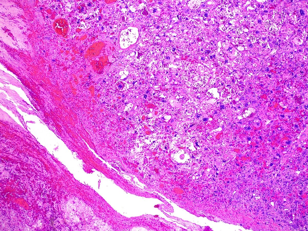
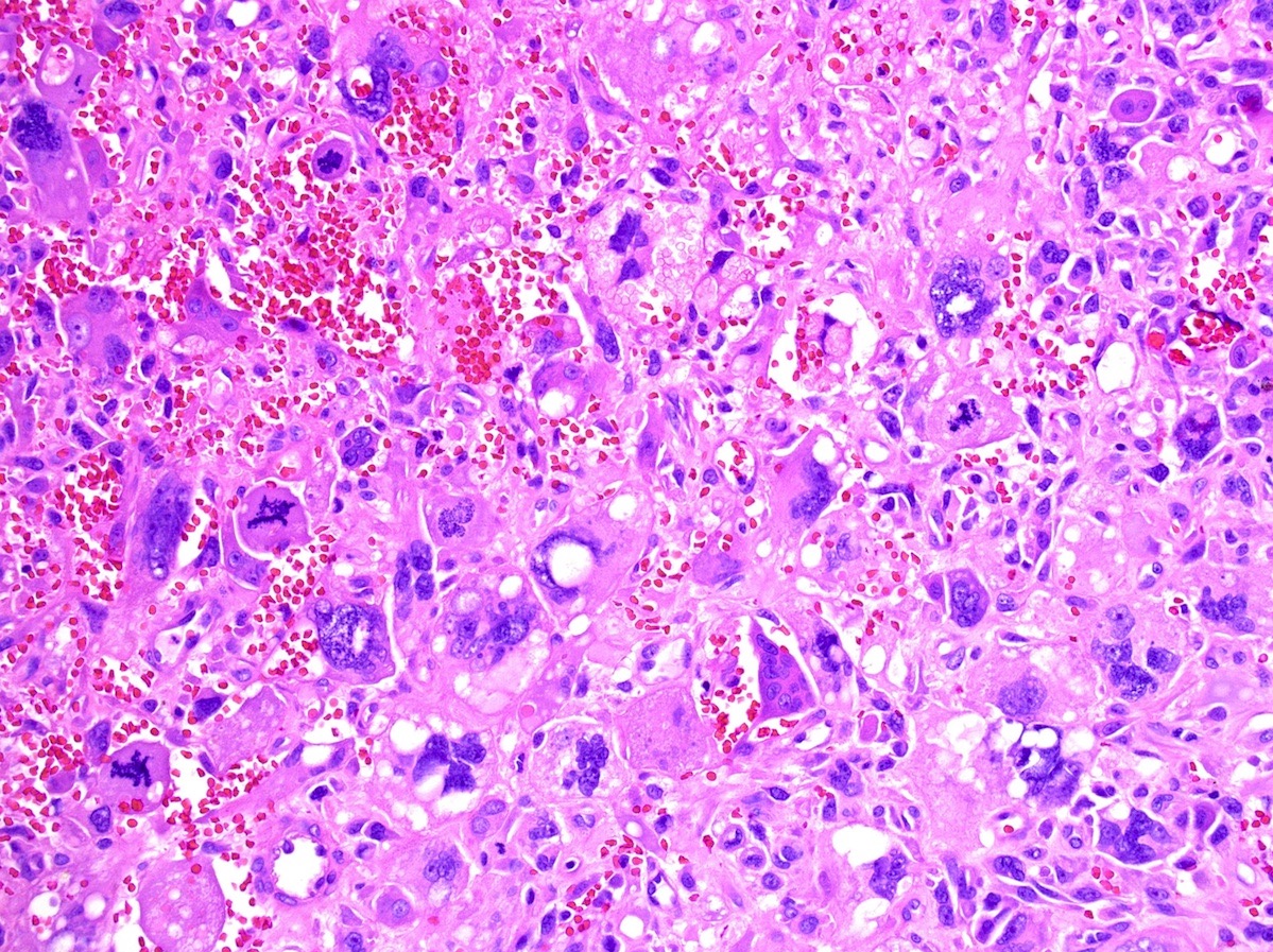
- Giant cells usually bizarre with multilobulated nuclei, abundant eosinophilic cytoplasm accompanied by heavy neutrophilic infiltrate with occasional ingested white blood cells
- Stroma often myxoid, frequent inflammatory infiltrate, collagen fibers
- Numerous mitotic figures
- Massive necrosis common
- Vascular invasion in 58%
- Giant cell carcinoma:
- Dyscohesive, polygonal, mono and multinucleated giant cells with abundant cytoplasm and prominent nucleoli
- Neoplastic, highly pleomorphic giant cells, often in inflammatory stroma with emperipolesis (Arch Pathol Lab Med 2010;134:1645)
- Giant cells are multinucleated, may resemble syncytiotrophoblasts and produce human chorionic gonadotropin but are usually fewer than in primary choriocarcinoma of lung (Histopathology 2000;36:17)
- 2 types of giant cells: βHCG positive syncytiotrophoblast-like giant cells with smudged nuclei and coarse chromatin with nuclear molding and βHCG negative giant cells with admixed neutrophils and cell in cell features (Histopathology 2016;68:680)
- Spindle cell carcinoma:
- Carcinoma composed exclusively of spindle shaped tumor cells
- Tumor cells often obliterate vessels
Microscopic (histologic) images
Cytology description
- Giant cell carcinoma:
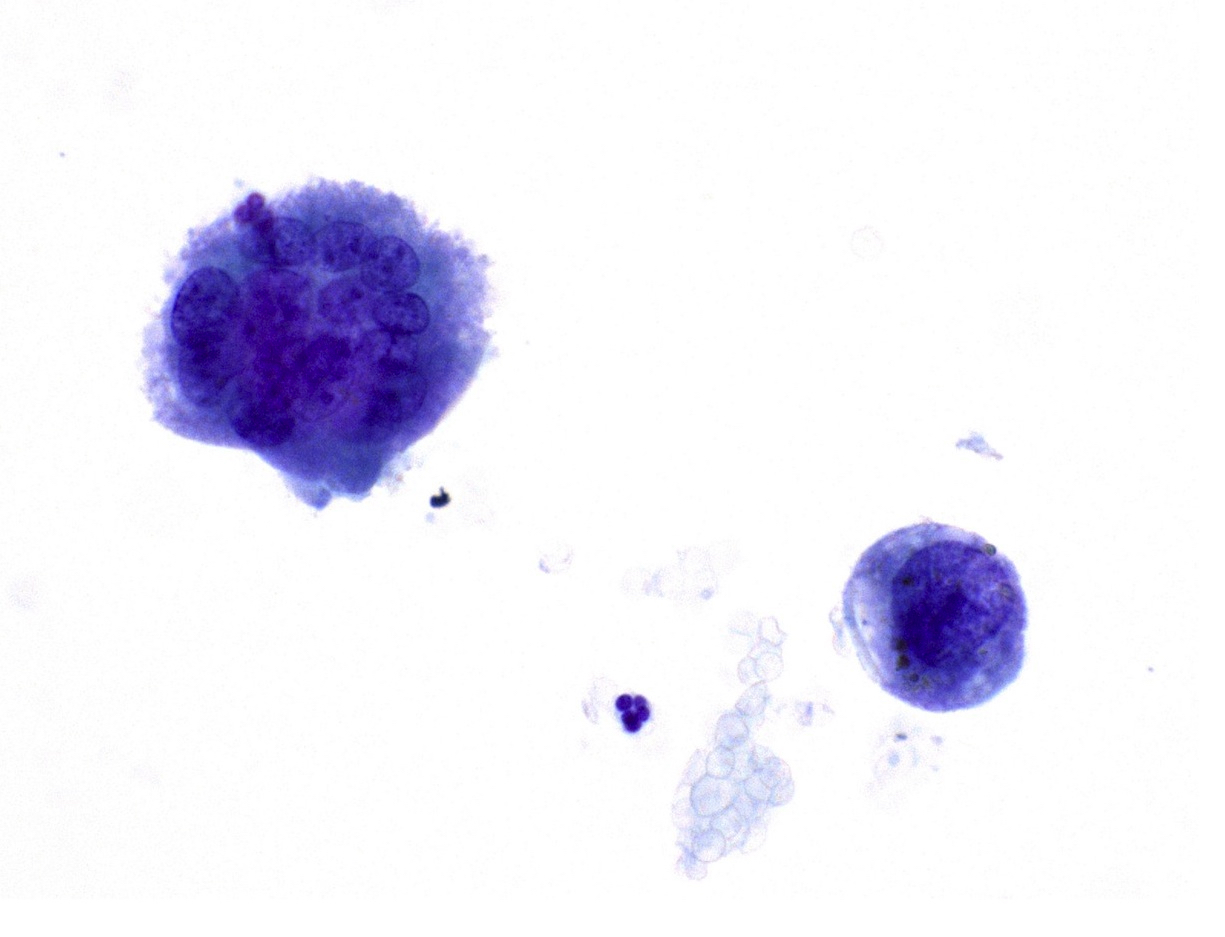
- Pleomorphic cells in flat loose clusters, with abundant, thick, well demarcated cytoplasm, multinucleation (Acta Cytol 2011;55:173)
- Sharp clear nuclear membranes, acidophilic cytoplasm with finely granular or foamy appearance, irregular coarse chromatin, bizarre mitoses, neutrophil engulfment (Diagn Cytopathol 2007;35:555)
Positive stains
- Pleomorphic carcinoma:
- Giant cell carcinoma:
- Calretinin (67%)
- Pancytokeratin, TTF1 variable
Negative stains
- Pleomorphic carcinoma: CK20
- Giant cell carcinoma: chromogranin, synaptophysin, CD68, desmin
- Spindle cell carcinoma: surfactant apoprotein A
Differential diagnosis
- Metastatic germ cell tumor (e.g., choriocarcinoma)
- Other subtypes of sarcomatoid carcinoma
- True sarcomas, such as undifferentiated pleomorphic sarcoma
Additional references
Board review style question #1
Giant cell carcinoma of the lung is most commonly associated with which epithelial component resembling non small cell carcinoma?
- Adenocarcinoma
- Adenosquamous carcinoma
- Large cell carcinoma
- Squamous cell carcinoma
- None of the above
Board review style answer #1
E. None of the above. Giant cell carcinoma is a subtype of sarcomatoid carcinoma composed almost entirely of tumor giant cells with no differentiated carcinomatous elements.
Comment Here
Reference: Pleomorphic carcinoma
Comment Here
Reference: Pleomorphic carcinoma


