Table of Contents
Definition / general | Essential features | Terminology | ICD coding | Epidemiology | Sites | Pathophysiology | Etiology | Clinical features | Diagnosis | Radiology images | Prognostic factors | Case reports | Treatment | Gross description | Gross images | Microscopic (histologic) description | Microscopic (histologic) images | Virtual slides | Cytology description | Cytology images | Positive stains | Negative stains | Molecular / cytogenetics description | Differential diagnosis | Additional references | Board review style question #1 | Board review style answer #1Cite this page: Wu R. Carcinosarcoma. PathologyOutlines.com website. https://www.pathologyoutlines.com/topic/lungtumorcarcinosarcoma.html. Accessed December 29th, 2024.
Definition / general
- Subtype of sarcomatoid carcinoma, consisting of a mixture of nonsmall cell lung cancer (typically squamous cell carcinoma or adenocarcinoma) and sarcomatous heterologous elements
- Monoclonal tumor with divergent lines of differentiation, leading to mixture of carcinomatous and sarcomatous elements (Am J Surg Pathol 2002;26:510)
Essential features
- Biphasic tumor consisting of a nonsmall cell carcinoma with heterologous sarcomatoid differentiation
- Rare tumor with poor prognosis
- Recommend molecular testing according to associated histologic components; i.e. tumors with adenocarcinoma component should be tested for EGFR and ALK (J Thorac Oncol 2015;10:1243)
Terminology
- Use specific term "carcinosarcoma" rather than general term "sarcomatoid carcinoma" whenever possible to avoid confusion (J Thorac Oncol 2015;10:1243)
- "Biphasic sarcomatoid carcinoma" has been proposed as replacement term
ICD coding
- Use code specific for location of tumor
- C34.90 malignant neoplasm of unspecified part of unspecified bronchus or lung
Epidemiology
- Rare, 0.1% of all lung cancers
- M > F, most with smoking history, average age 60
Sites
- Large airways and peripheral lung
Pathophysiology
- Primary lung carcinoma undergoes sarcomatoid change
Etiology
- Epithelial malignancy with divergent differentiation
Clinical features
- Dependent on location: endobronchial lesions have pneumonia, cough, shortness of breath, hemoptysis; peripheral lesions may have no symptoms
Diagnosis
- Difficult to diagnose on small biopsy / cytology but possible
Radiology images
Prognostic factors
- Poor prognosis overall; increased tumor size appears related to reduced survival (Am J Surg Pathol 1999;23:1514)
- Better prognosis for smaller or endobronchial lesions
Case reports
- 49 year old woman with a SBO secondary to small bowel metastases (Acta Chir Belg 2015;115:49)
- 50 year old woman with a 45 pack year smoking history (Surg Case Rep 2017;3:52)
- 50 year old man with complaint of pain in back radiating to right lower limb and chest pain for 3 months (J Cytol 2015r;32:28)
- 64 year old man with a seven-month history of shortness of breath (Contemp Oncol (Pozn) 2015;19:163)
- 65 year old man with bronchial carcinosarcoma (J Radiol Case Rep 2012;6:26)
- 65 year old man with tumor including chest wall invasion (Int J Clin Oncol 2010;15:319)
- 75 year old woman with diabetes, hypertension, chronic kidney disease, and coronary disease (Ochsner J 2015;15:196)
- 77 year old man with pulmonary carcinosarcoma and idiopathic pulmonary fibrosis (Lung 2016;194:171)
- 83 year old woman with glandular and lipoblastic carcinosarcoma (Case Rep Pulmonol 2016;2016:2020146)
- Cytology case report (Diagn Cytopathol 1988;4:239)
Treatment
- Surgical resection in majority of cases (Surg Today 2014;44:175), followed by radiation
- Chemotherapy for cases with metastases
- Tumors with EGFR mutation can be considered for targeted therapy (Oncotarget 2017;8:25323)
Gross description
- May show central necrosis and hemorrhage if larger tumor
- Irregular borders, tan to yellow
Microscopic (histologic) description
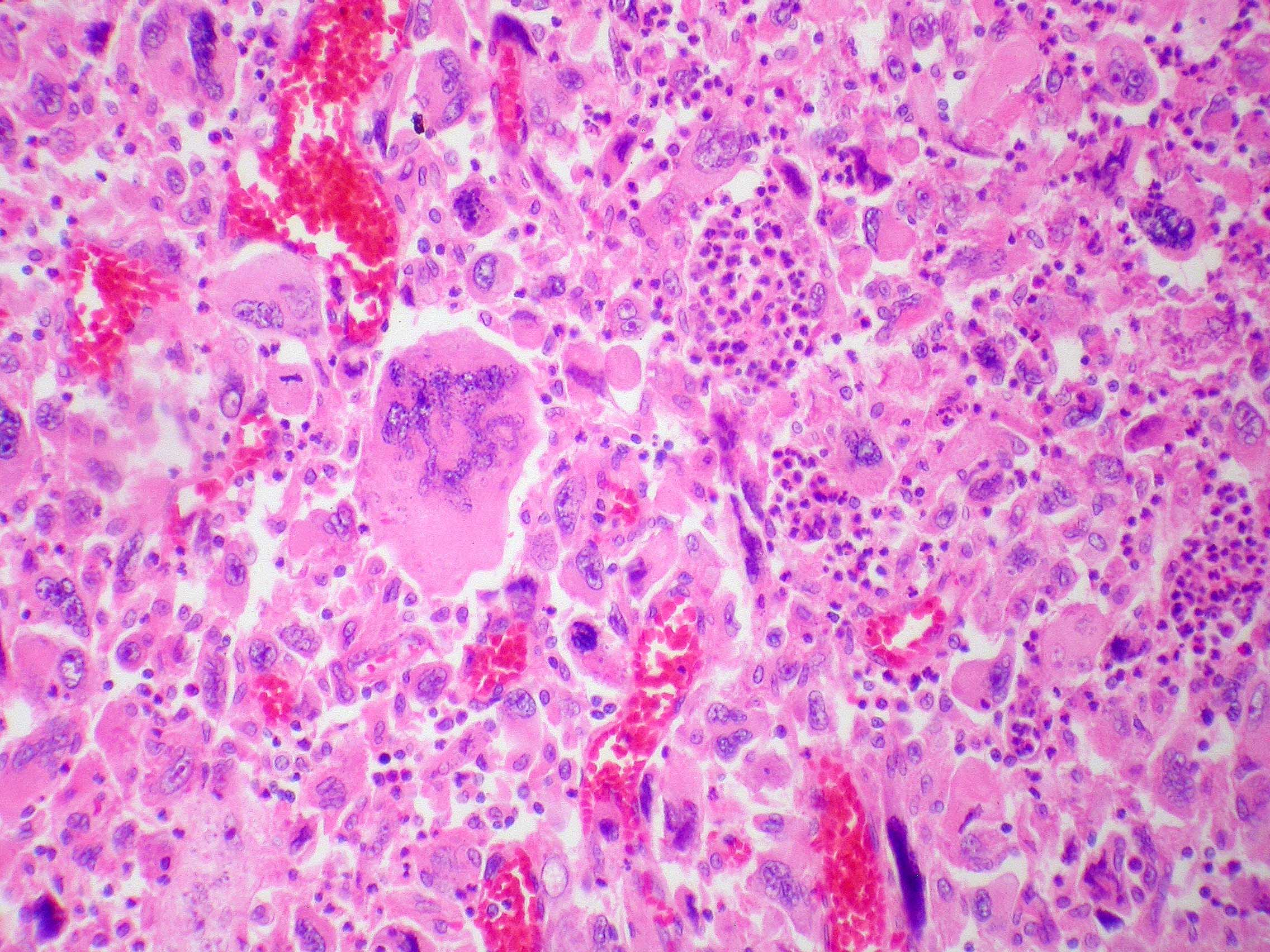
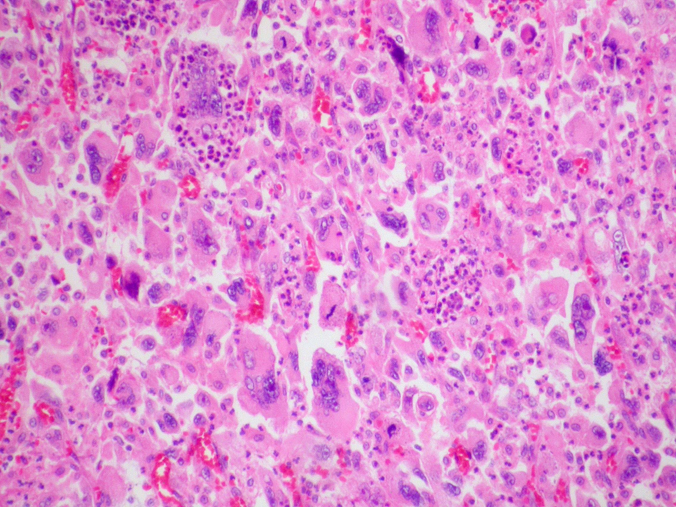
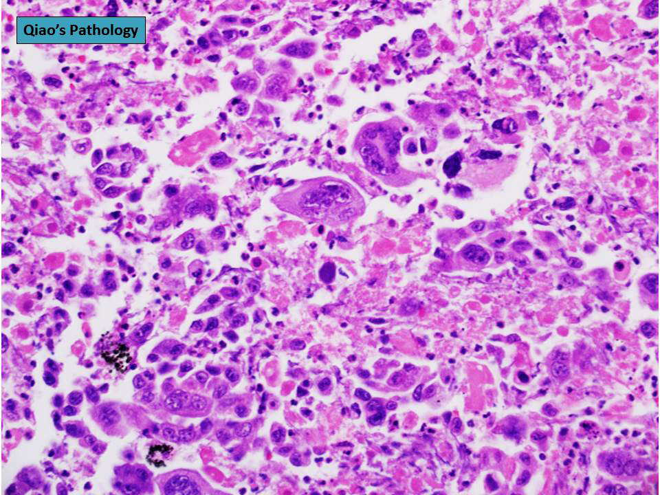
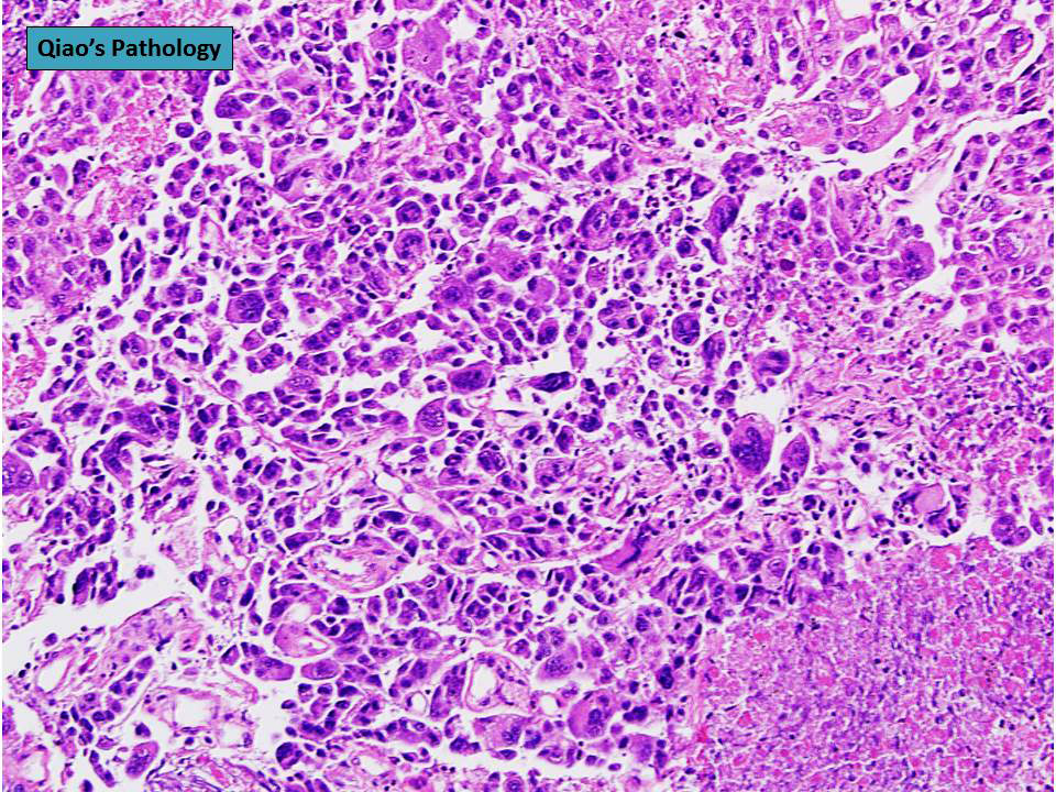
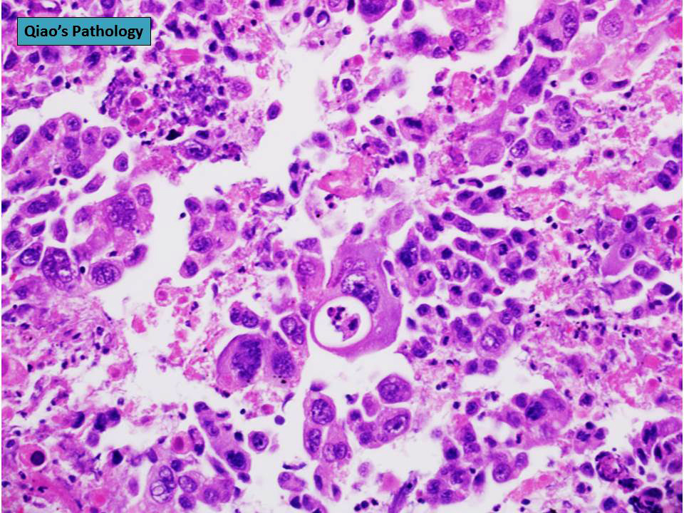
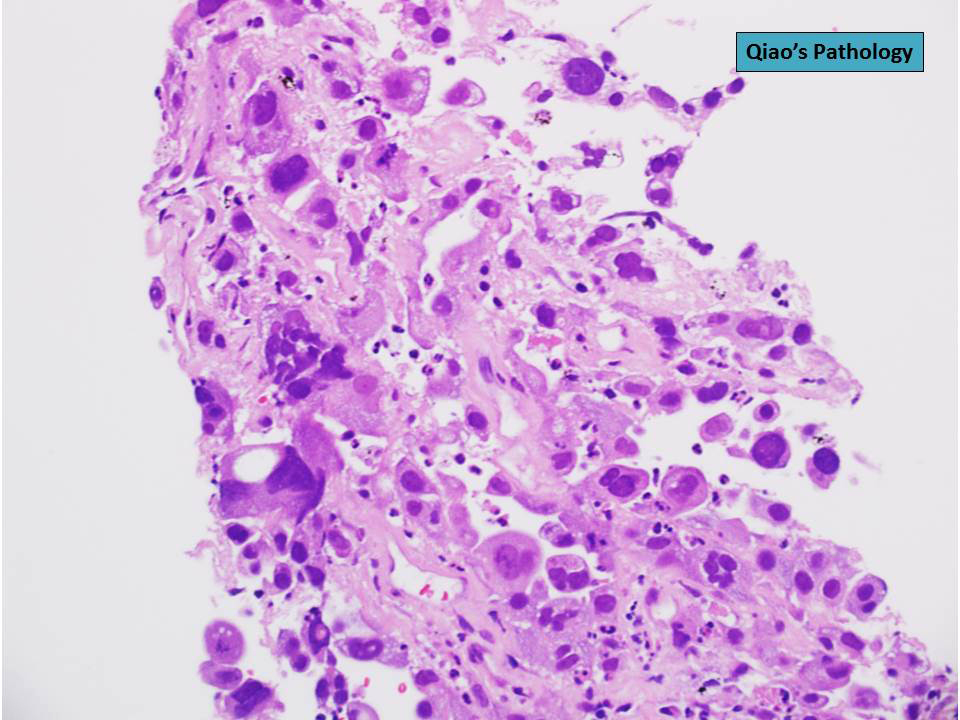
- Mixture of carcinomatous and sarcomatous elements
- Carcinoma component usually squamous but may be glandular or neuroendocrine
- Heterologous sarcomatoid component, such as rhabdomyosarcoma, osteosarcoma, chondrosarcoma
- Metastases may show carcinomatous or sarcomatous component or both
Microscopic (histologic) images
Scroll to see all images
Contributed by Yale Rosen, M.D. and Jian-Hua Qiao, M.D.
Images hosted on other servers:
Contributed by Yale Rosen, M.D. and Jian-Hua Qiao, M.D.
Images hosted on other servers:
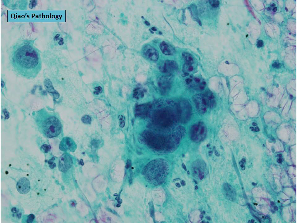
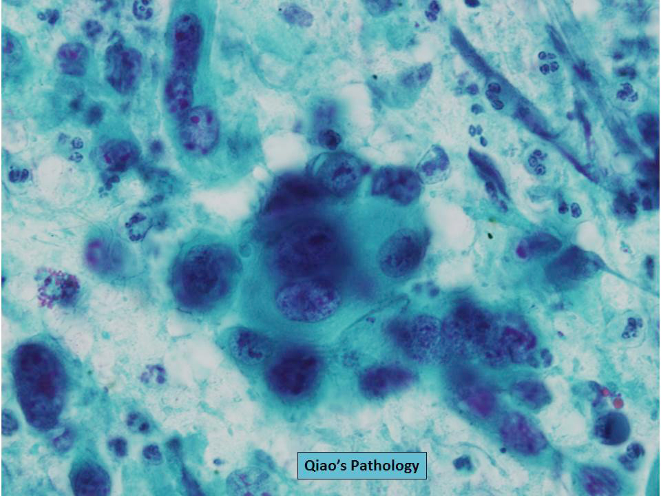
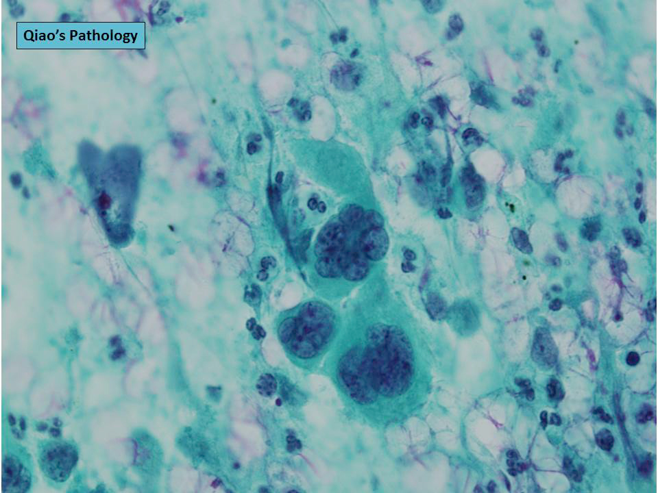
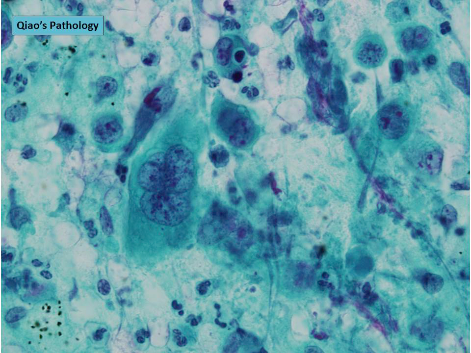
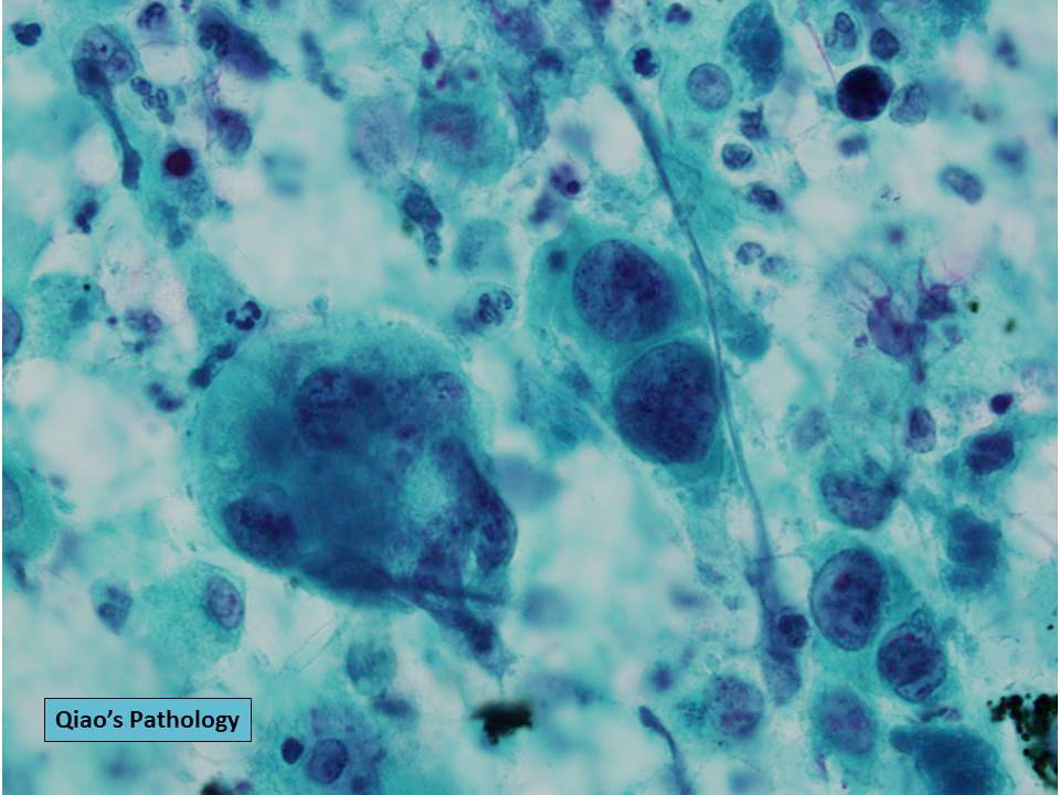
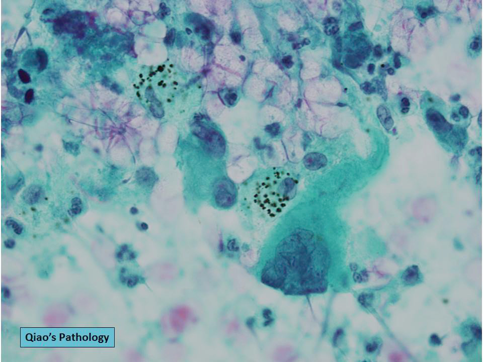
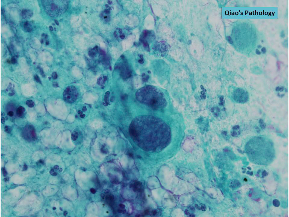
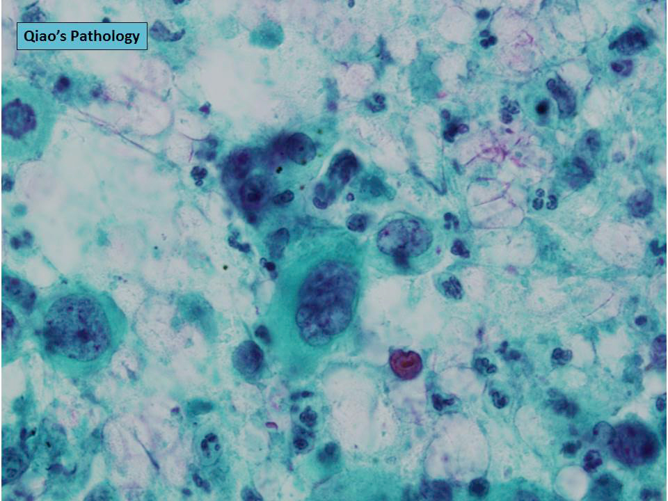
Cytology description
- May only show epithelial component, i.e. squamous cell carcinoma
Cytology images
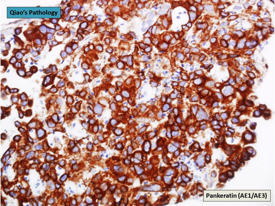
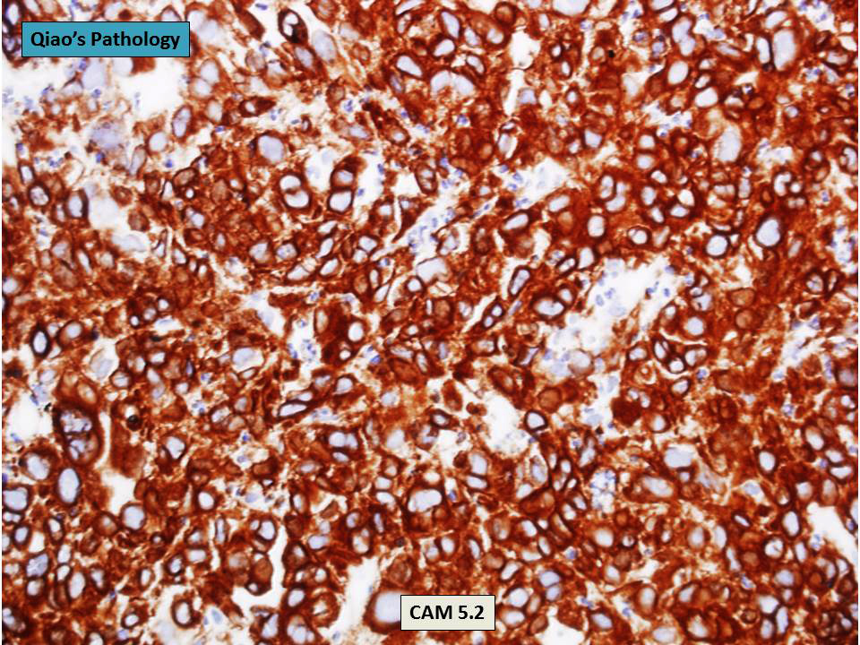
Positive stains
- Dependent on specific components of tumor
Negative stains
- Dependent on specific components of tumor
Molecular / cytogenetics description
- TP53 mutations often seen, Kras less frequent, EGFR mutations very uncommon (J Thorac Oncol 2015;10:1243)
- Equivalent allelic loss between the two components, supporting monoclonal origin (Am J Surg Pathol 2002;26:510)
Differential diagnosis
- Other subtypes of sarcomatoid carcinoma
- Sarcomatoid mesothelioma
- True sarcomas, such as synovial sarcoma
- Other types of sarcomatoid carcinoma such as pulmonary blastoma (younger ages)
Additional references
- Review of 6 patients with pulmonary carcinosarcoma (ScientificWorldJournal 2012;2012:167317)
Board review style question #1
The most common epithelial component of a primary pulmonary carcinosarcoma is:
- Adenocarcinoma
- Adenosquamous carcinoma
- Large cell carcinoma
- Small cell carcinoma
- Squamous cell carcinoma
Board review style answer #1


