Table of Contents
Definition / general | Essential features | Terminology | ICD coding | Epidemiology | Sites | Pathophysiology | Etiology | Clinical features | Diagnosis | Laboratory | Radiology description | Radiology images | Prognostic factors | Case reports | Treatment | Gross description | Gross images | Microscopic (histologic) description | Microscopic (histologic) images | Cytology description | Positive stains | Negative stains | Molecular / cytogenetics description | Differential diagnosis | Board review style question #1 | Board review style answer #1Cite this page: Wu R. Lymphoepithelioma-like. PathologyOutlines.com website. https://www.pathologyoutlines.com/topic/lungtumorLEL.html. Accessed March 31st, 2025.
Definition / general
- Morphologically similar to undifferentiated nasopharyngeal carcinoma
- Rare tumor in lung; more common in Asians
- Associated with Epstein-Barr virus (EBV) in Asian populations (Cancer 1998;82:2334)
- In Caucasians, usually EBV-
Essential features
- Pulmonary lymphoepithelioma-like carcinoma (LELC) is an epithelial malignancy characterized by large, syncytial cells with vesicular nuclei and prominent nucleoli, with a prominent lymphocytic infiltrate
- LELC has a strong association with Epstein-Barr virus (EBV) and is more common in Asian populations
- LELC shows similar morphologic features to nasopharyngeal carcinoma and may be mistaken for lymphoma due to prominent inflammatory infiltrate
Terminology
- Lymphoepithelioma-like carcinoma (LELC) primary to lung first described in 1987 by Bégin et al. (J Surg Oncol 1987;36:280)
- Previously considered subtype of large cell undifferentiated carcinoma
- By 2015 WHO, now classified under epithelial tumor, "other and unclassified carcinomas" (J Thorac Oncol 2015;10:1243)
ICD coding
- Use code specific for location of tumor
- C34.90 Malignant neoplasm of unspecified part of unspecified bronchus or lung
Epidemiology
- Very rare, mostly seen in younger adults
- In Asians, usually women, nonsmokers, with EBV+ tumors (Am J Surg Pathol 2002;26:715)
Sites
- Intrapulmonary, either peripheral or central
Pathophysiology
- Better prognosis than other non small cell carcinomas of lung (Cancer 2012;118:4748, Ann Thorac Surg 2014;98:1013)
Etiology
- Some association with EBV infection (Mod Pathol 1991;4:264)
Clinical features
- Nonspecific: cough, dyspnea, chest pain, weight loss
Diagnosis
- Exclude metastatic nasopharyngeal carcinoma and other non small cell carcinomas
- Presence of prominent inflammatory infiltrate helpful
- Ancillary studies for EBV infection
Laboratory
- EBV serology often reveals prior infection, with higher titers associated with larger tumor size and higher stage (Am J Surg Pathol 2002;26:715)
Radiology description
- Similar to imaging characteristics of other primary lung cancers
- Solitary, peripheral pulmonary nodule with direct contact with pleura (J Thorac Imaging 2014;29:246)
- FDG PET / CT with strong FDG uptake
Prognostic factors
- Good prognosis: early tumor stage, normal serum lactate dehydrogenase level, normal serum albumin level, no lymph node metastasis
- Those who underwent complete resection had better overall survival (Cancer 2012;118:4748)
- Tumor recurrence and necrosis (5% or more of tumor) associated with poor prognosis (Am J Clin Pathol 2001;115:841)
Case reports
- 25 year old Italian man with EBV+ tumor (Hum Pathol 2003;34:623)
- 53 year old woman with thin walled cavity (Ann Thorac Surg 2013;96:1857)
- 58 year old woman initially diagnosed as squamous metaplasia (Oncol Lett 2015;9:1767)
- 60 year old woman initially diagnosed as adenocarcinoma (Tuberc Respir Dis (Seoul) 2013;75:170)
- 70 year old man with partially regressed tumor (Ann Thorac Surg 2015;99:2197)
- 83 year old man with FDG avid lung lesion (Clin Nucl Med 2015;40:134)
Treatment
- Complete surgical resection for early stage disease, with adjuvant chemotherapy and radiotherapy in advanced cases (J Thorac Dis 2017;9:123)
Gross description
- Variable size, can be > 10 cm
- Well circumscribed, lobulated, solid nodule
- Necrosis common
Microscopic (histologic) description
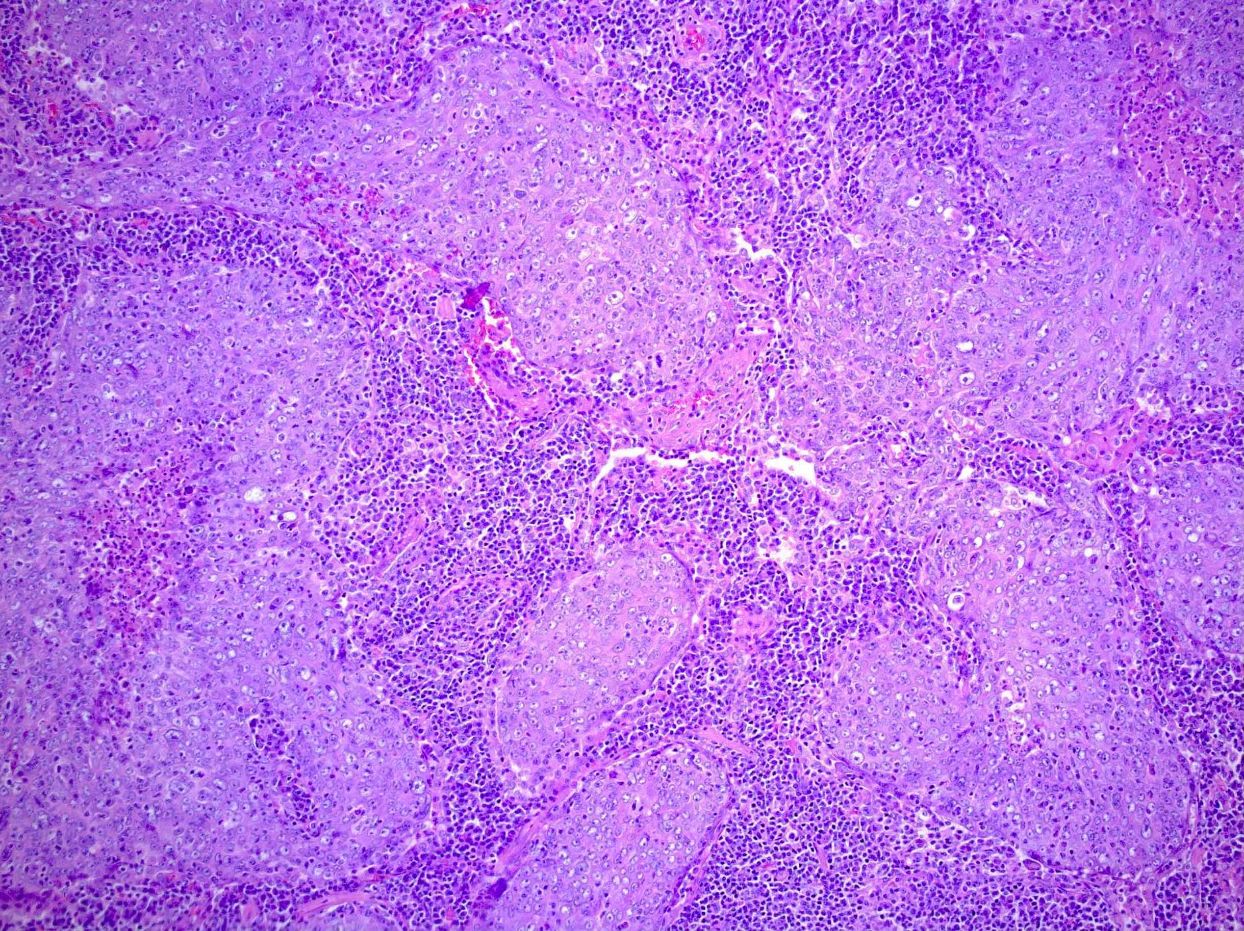
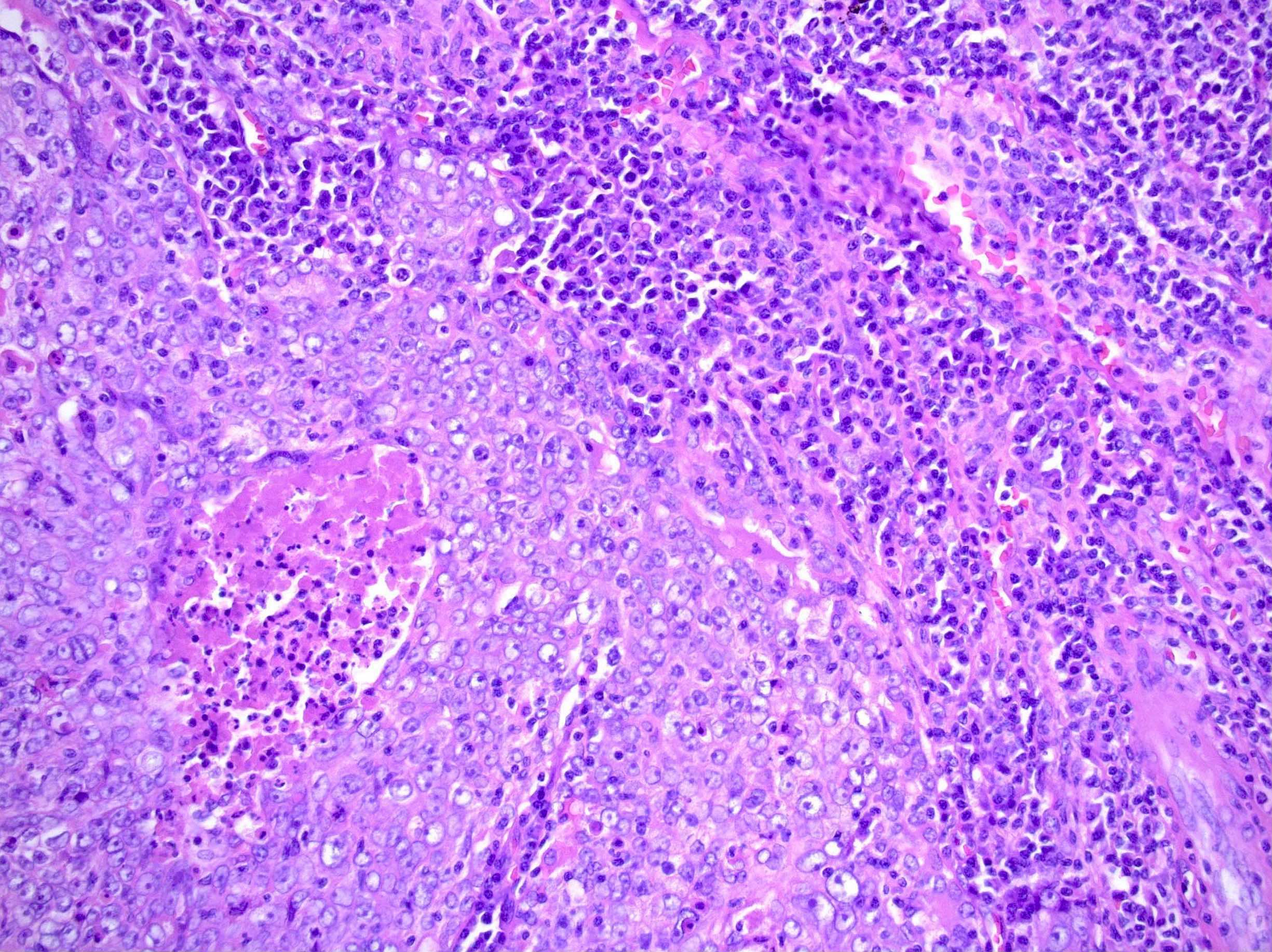
- Anastamosing islands, nests, cords or diffuse sheets of tumor cells
- Syncytial growth of monomorphous, polygonal epithelial cells with large vesicular nuclei, prominent eosinophilic nucleoli, accompanied by marked CD8+ lymphocytic infiltration
- Admixed lymphocytes, plasma cells, histiocytes, occasional neutrophils or eosinophils
- Predominantly pushing borders, permeative interface with adjacent lung
- Intratumoral amyloid deposition in a few cases (Am J Surg Pathol 2002;26:715)
Microscopic (histologic) images
Cytology description
- Large clusters of neoplastic cells with scant cytoplasm, large hyperchromatic nuclei, irregular nuclear contours, prominent nucleoli, brisk mitotic figures and prominent intratumoral lymphoid infiltration (Diagn Cytopathol 2012;40:820)
- Stripped, naked tumor nuclei
- Interspersed lymphocytes, some with crush artifact
Positive stains
Negative stains
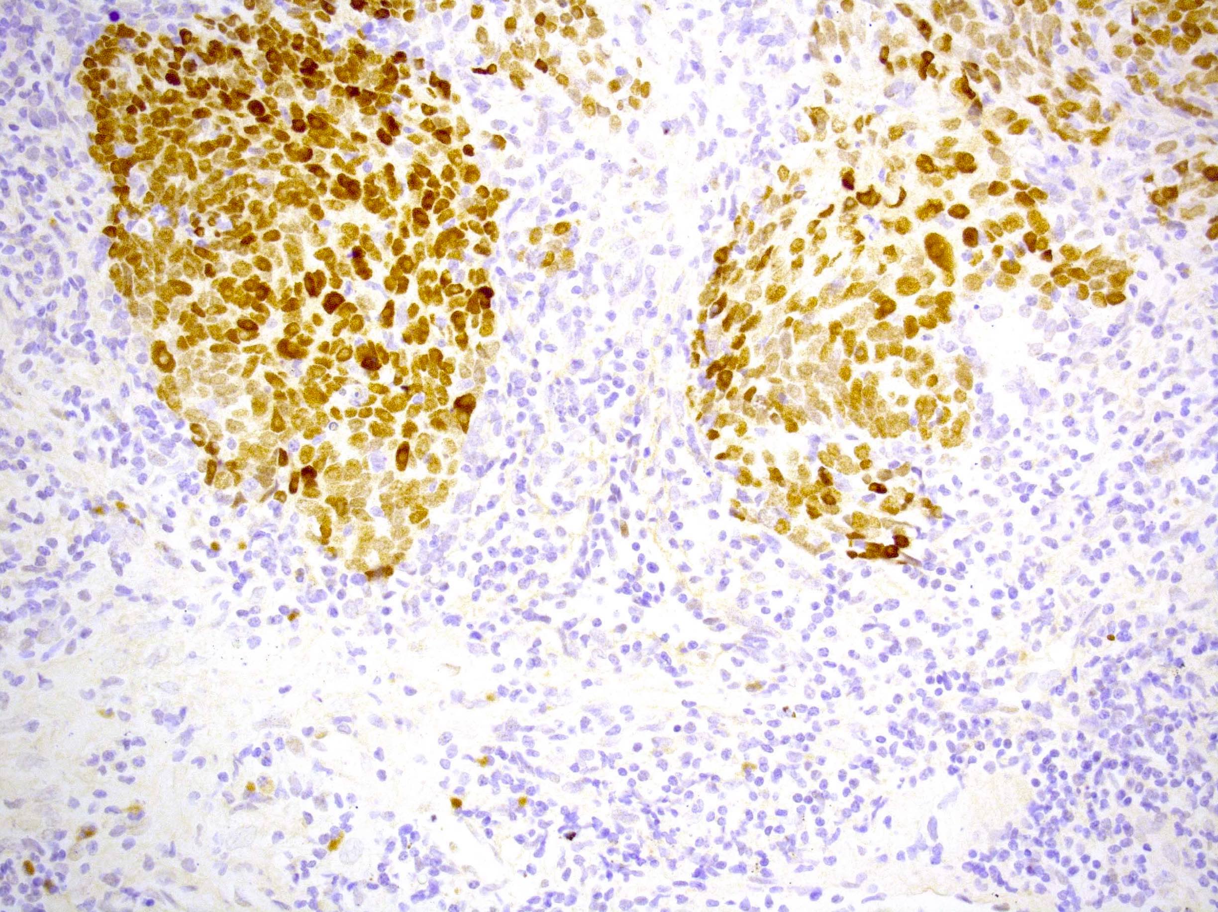
- Mostly TTF1 negative
- Napsin, neuroendocrine markers
- CD45 / LCA (except in stromal lymphocytes)
Molecular / cytogenetics description
- High PDL1 expression and infrequent driver mutations (Oncotarget 2015;6:33019, Lung Cancer 2015;88:254)
- Microsatellite instability is uncommon (Am J Clin Pathol 2007;127:282)
Differential diagnosis
- Inflammatory pseudotumor
- Lymphoma
- Melanoma
- Metastatic nasopharyngeal carcinoma
- Other non small cell lung carcinomas
Board review style question #1
Lymphoepithelioma-like carcinoma of the lung has been linked to which pathogen, particularly in Asian populations?
- Epstein-Barr virus (EBV)
- Human immunodeficiency virus (HIV)
- Human papillomavirus (HPV)
- Human T lymphotropic virus (HTLV)
- Respiratory syncytial virus (RSV)
Board review style answer #1


